| Product Includes | Product # | Quantity | Mol. Wt | Isotype/Source |
|---|---|---|---|---|
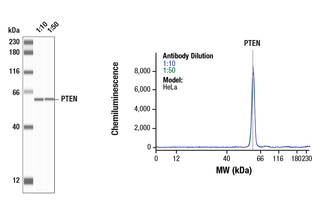
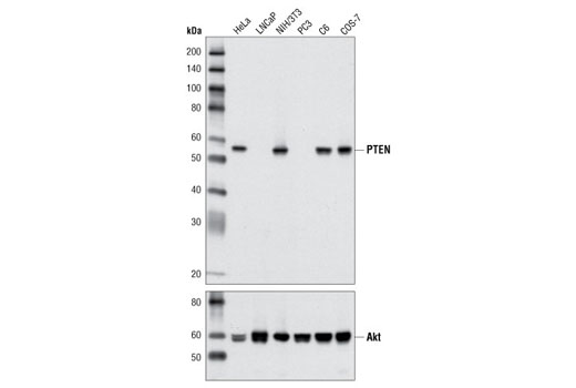
| PTEN (D4.3) XP® Rabbit mAb | 9188 | 20 µl | 54 kDa | Rabbit IgG |
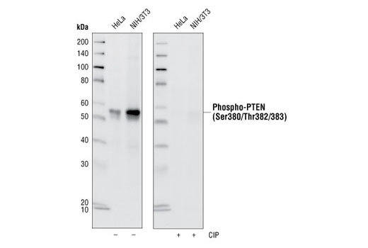
| Phospho-PTEN (Ser380/Thr382/383) (44A7) Rabbit mAb | 9549 | 20 µl | 54 kDa | Rabbit IgG |
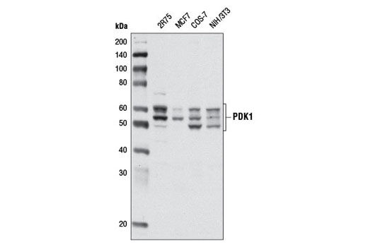
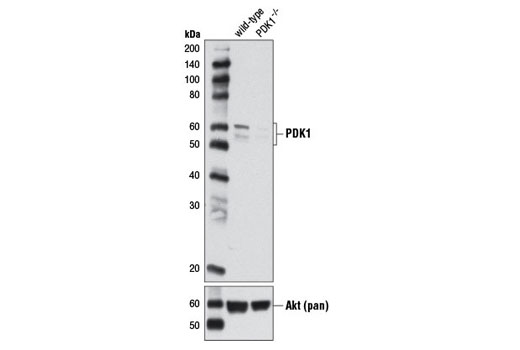
| PDK1 (D37A7) Rabbit mAb | 5662 | 20 µl | 58-68 kDa | Rabbit IgG |
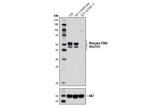
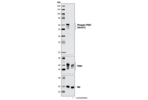
| Phospho-PDK1 (Ser241) (C49H2) Rabbit mAb | 3438 | 20 µl | 58 to 68 kDa | Rabbit IgG |
| Anti-rabbit IgG, HRP-linked Antibody | 7074 | 100 µl | Goat |
Please visit cellsignal.com for individual component applications, species cross-reactivity, dilutions, protocols, and additional product information.
Description
The PTEN and PDK1 Antibody Sampler Kit II provides an economical means to evaluate two key enzymes that regulate multiple signaling pathways. The kit includes enough antibodies to perform two western blot experiments with each primary antibody.
Storage
Background
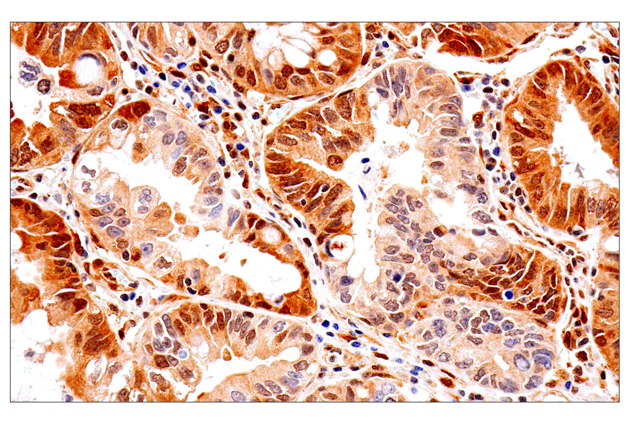
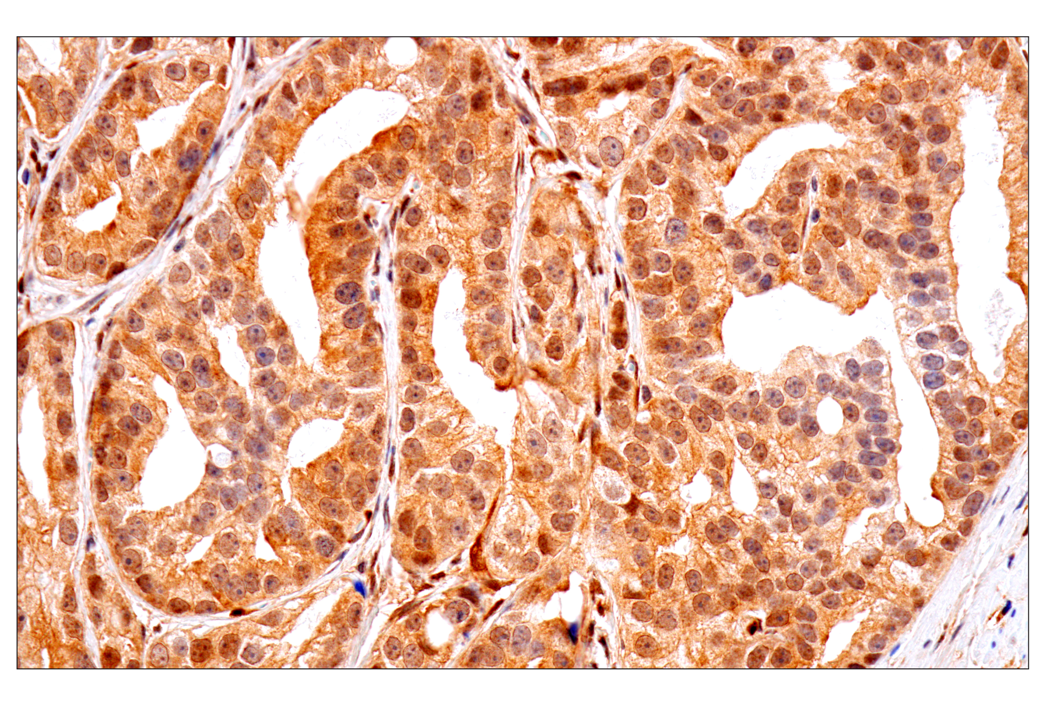
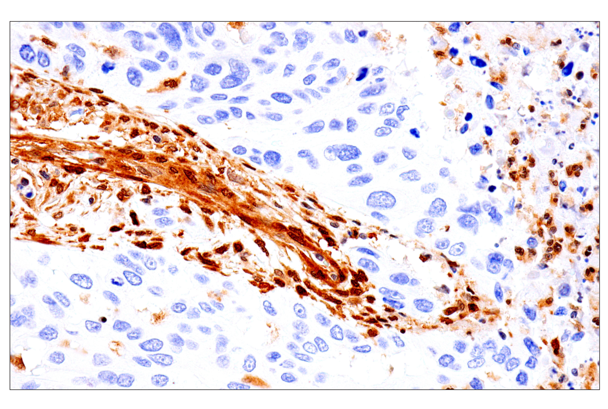
PTEN (phosphatase and tensin homologue deleted on chromosome ten), also referred to as MMAC (mutated in multiple advanced cancers) phosphatase, is a tumor suppressor implicated in a wide variety of human cancers (1). PTEN encodes a 403 amino acid polypeptide originally described as a dual-specificity protein phosphatase (2). The main substrates of PTEN are inositol phospholipids generated by the activation of the phosphoinositide 3-kinase (PI3K) (3). PTEN is a major negative regulator of the PI3K/Akt signaling pathway (1,4,5). PTEN possesses a carboxy-terminal, noncatalytic regulatory domain with three phosphorylation sites (Ser380, Thr382, and Thr383) that regulate PTEN stability and may affect its biological activity (6,7). PTEN regulates p53 protein levels and activity (8) and is involved in G protein-coupled signaling during chemotaxis (9,10).
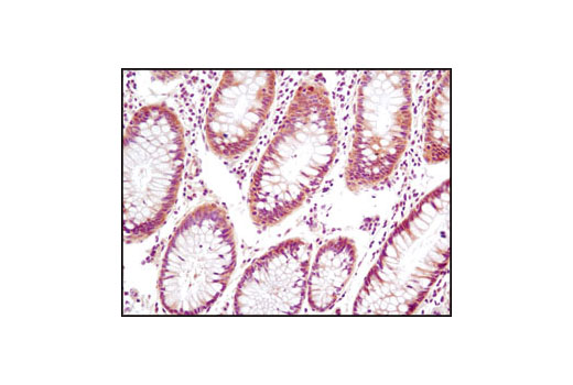
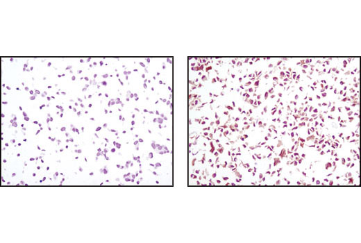
Phosphoinositide-dependent protein kinase 1 (PDK1) plays a central role in many signal transduction pathways (11,12), including the activation of Akt and the PKC isoenzymes p70 S6 kinase and RSK (13). Through its effects on these kinases, PDK1 is involved in the regulation of a wide variety of processes, including cell proliferation, differentiation, and apoptosis.
- Cantley, L.C. and Neel, B.G. (1999) Proc Natl Acad Sci USA 96, 4240-5.
- Myers, M.P. et al. (1997) Proc Natl Acad Sci USA 94, 9052-7.
- Myers, M.P. et al. (1998) Proc Natl Acad Sci USA 95, 13513-8.
- Wan, X. and Helman, L.J. (2003) Oncogene 22, 8205-11.
- Wu, X. et al. (1998) Proc Natl Acad Sci USA 95, 15587-91.
- Vazquez, F. et al. (2000) Mol Cell Biol 20, 5010-8.
- Torres, J. and Pulido, R. (2001) J Biol Chem 276, 993-8.
- Freeman, D.J. et al. (2003) Cancer Cell 3, 117-30.
- Funamoto, S. et al. (2002) Cell 109, 611-23.
- Iijima, M. and Devreotes, P. (2002) Cell 109, 599-610.
- Belham, C. et al. (1999) Curr Biol 9, R93-6.
- Toker, A. and Newton, A.C. (2000) Cell 103, 185-8.
- Williams, M.R. et al. (2000) Curr Biol 10, 439-48.
Background References
Trademarks and Patents
使用に関する制限
法的な権限を与えられたCSTの担当者が署名した書面によって別途明示的に合意された場合を除き、 CST、その関連会社または代理店が提供する製品には以下の条件が適用されます。お客様が定める条件でここに定められた条件に含まれるものを超えるもの、 または、ここに定められた条件と異なるものは、法的な権限を与えられたCSTの担当者が別途書面にて受諾した場合を除き、拒絶され、 いかなる効力も効果も有しません。
研究専用 (For Research Use Only) またはこれに類似する表示がされた製品は、 いかなる目的についても FDA または外国もしくは国内のその他の規制機関により承認、認可または許可を受けていません。 お客様は製品を診断もしくは治療目的で使用してはならず、また、製品に表示された内容に違反する方法で使用してはなりません。 CST が販売または使用許諾する製品は、エンドユーザーであるお客様に対し、使途を研究および開発のみに限定して提供されるものです。 診断、予防もしくは治療目的で製品を使用することまたは製品を再販売 (単独であるか他の製品等の一部であるかを問いません) もしくはその他の商業的利用の目的で購入することについては、CST から別途許諾を得る必要があります。 お客様は以下の事項を遵守しなければなりません。(a) CST の製品 (単独であるか他の資材と一緒であるかを問いません) を販売、使用許諾、貸与、寄付もしくはその他の態様で第三者に譲渡したり使用させたりしてはなりません。また、商用の製品を製造するために CST の製品を使用してはなりません。(b) 複製、改変、リバースエンジニアリング、逆コンパイル、 分解または他の方法により製品の構造または技術を解明しようとしてはなりません。また、 CST の製品またはサービスと競合する製品またはサービスを開発する目的で CST の製品を使用してはなりません。(c) CST の製品の商標、商号、ロゴ、特許または著作権に関する通知または表示を除去したり改変したりしてはなりません。(d) CST の製品をCST 製品販売条件(CST’s Product Terms of Sale) および該当する書面のみに従って使用しなければなりません。(e) CST の製品に関連してお客様が使用する第三者の製品またはサービスに関する使用許諾条件、 サービス提供条件またはこれに類する合意事項を遵守しなければなりません。