| Product Includes | Product # | Quantity | Mol. Wt | Isotype/Source |
|---|---|---|---|---|
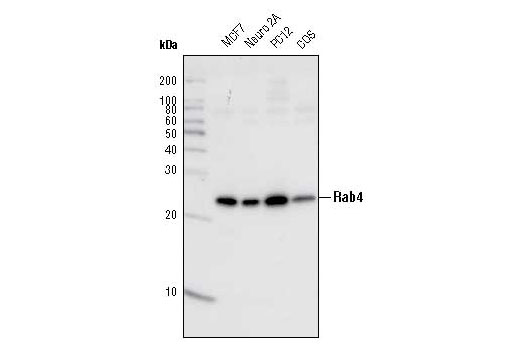
| Rab4 Antibody | 2167 | 20 µl | 25 kDa | Rabbit |
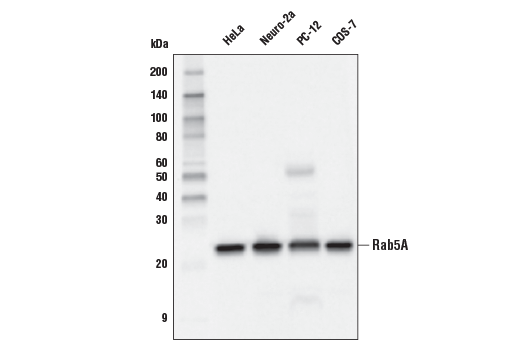
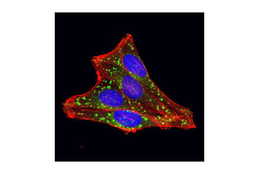
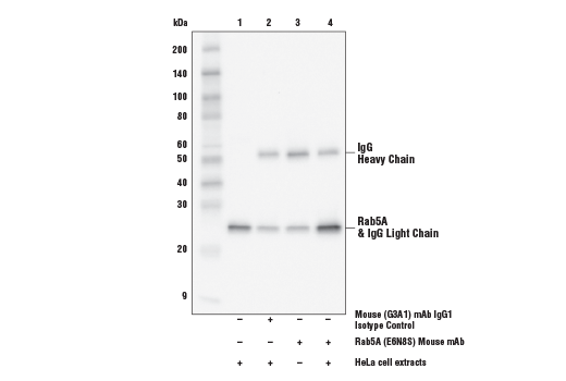
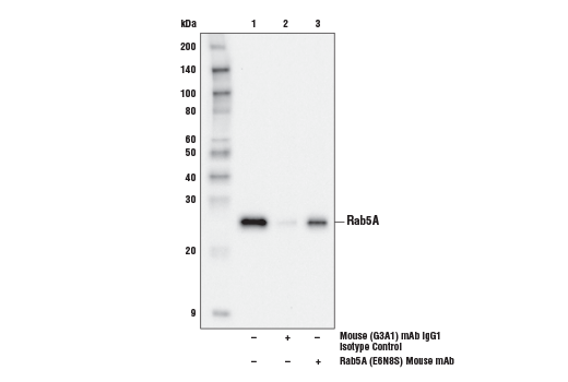
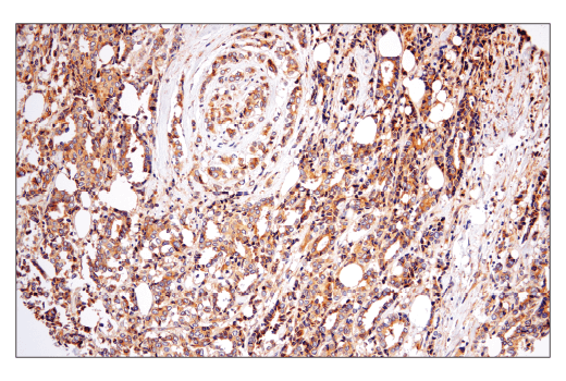
| Rab5A (E6N8S) Mouse mAb | 46449 | 20 µl | 25 kDa | Mouse IgG1 |
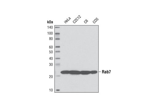
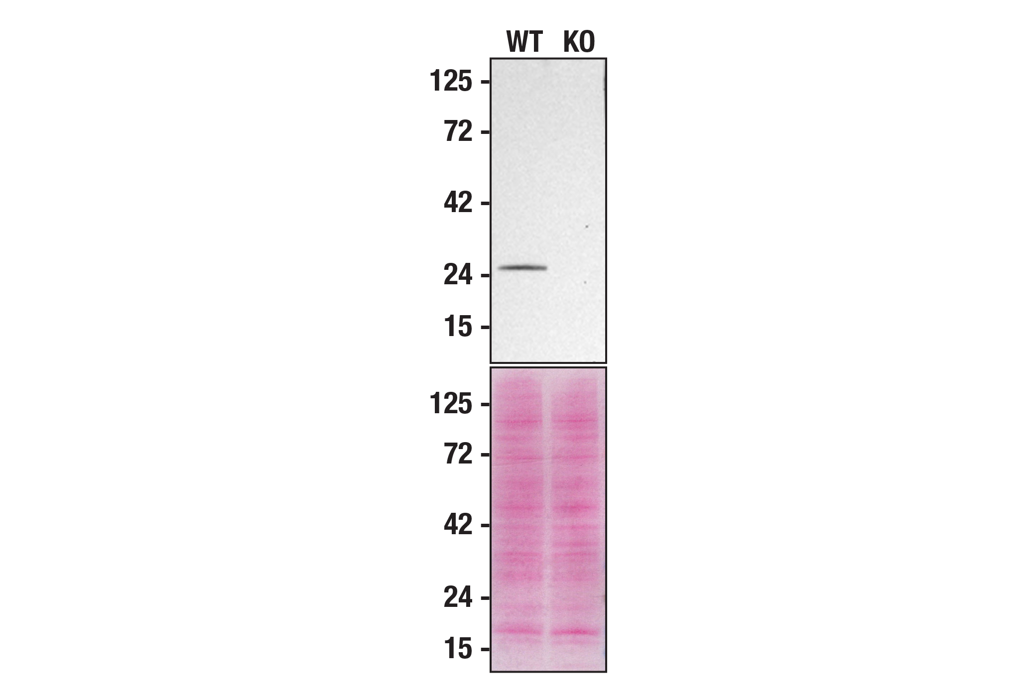
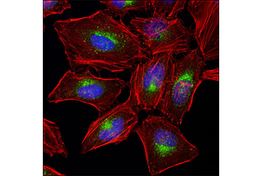
| Rab7 (D95F2) XP® Rabbit mAb | 9367 | 20 µl | 23 kDa | Rabbit IgG |
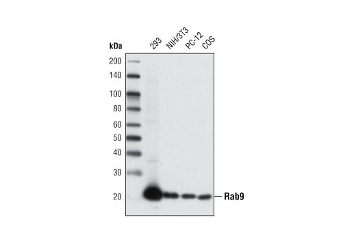
| Rab9A (D52G8) XP® Rabbit mAb | 5118 | 20 µl | 23 kDa | Rabbit IgG |
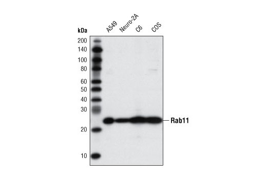
| Rab11 (D4F5) XP® Rabbit mAb | 5589 | 20 µl | 25 kDa | Rabbit IgG |
| Anti-rabbit IgG, HRP-linked Antibody | 7074 | 100 µl | Goat | |
| Anti-mouse IgG, HRP-linked Antibody | 7076 | 100 µl | Horse |
Please visit cellsignal.com for individual component applications, species cross-reactivity, dilutions, protocols, and additional product information.
Description
The Rab Family Antibody Sampler Kit provides an economical means to evaluate the presence and status of Rab proteins in cells. This kit provides enough primary and secondary antibodies to perform two Western blot experiments per primary antibody.
Storage
Background
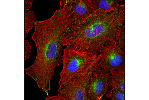
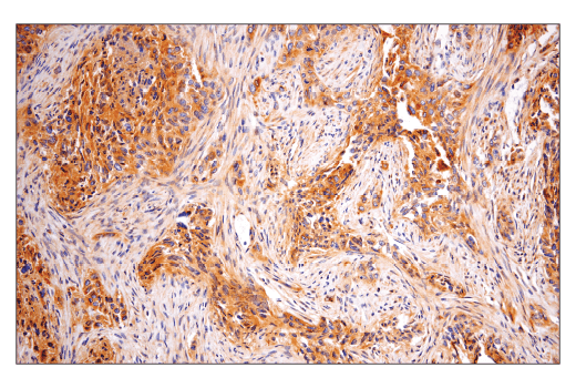
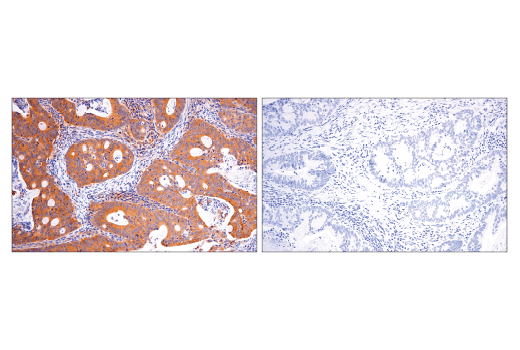
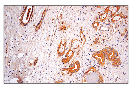
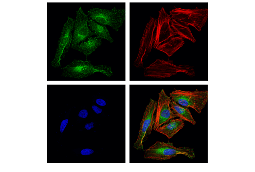
Rab family proteins are GTPases and members of the Ras superfamily of monomeric G proteins. These membrane-associated proteins are involved in many aspects of vesicle-mediated transport, taking part in the initial vesicle formation, transport of vesicles along the cytoskeleton, and eventual fusion of vesicle and target membranes. Rab4 is localized at early endosomes/recycling endosomes and functions as a key regulator for sorting/recycling of membrane and proteins (1,2). Both Rab4A and Rab4B isoforms are localized to similar cellular compartments and are believed to have similar functions (4). Rab4 interacts with several Rab4 effectors in a complex on a special endosome site to promote membrane/protein recycling (1,3). Rab5 is localized to the plasma membrane and early endosome and functions as a key regulator of vesicle trafficking during early endocytosis (1). The conformational change between Rab5-GTP and Rab5-GDP is essential for its biological function as a rate-limiting regulator at multiple steps during endocytosis (1,5). Similar to Rab4, Rab5 also interacts with specific Rab5 effectors on a specialized endosomal Rab domain to promote recycling between endosome and the plasma membrane (1,5,6). Both Rab7 and Rab9 are located in late endosomes but exert different functions. Rab7 associates with the RIPL effector protein to control membrane trafficking from early to late endosome and to lysosomes (7,8). Rab7 also helps to regulate growth receptor endocytic trafficking and degradation, and maturation of phagosome and autophagic vacuoles (8-11). Rab9 interacts with its effector proteins p40 and TIP47 (12,13) to promote the MPR (mannose 6-phosphate receptor)-associated lysosomal enzyme transport between late endosomes and the trans Golgi network (14,15). Rab11 (isoforms Rab11a and Rab11b) functions as a key regulator in the recycling of perinuclear, plasma membrane and Golgi compartment endosomes (16,17). Despite some overlap, distinct differences exist between Rab11a and Rab11b in both their cellular distribution and functional roles. Rab11a is ubiquitously expressed while Rab11b is found mainly in the heart and brain (18,19). Like other Rab proteins, Rab11 functions when associated with Rab11 family interacting proteins (FIPs). The three distinct classes of Rab11 FIPs all share a conserved carboxy-terminal Rab-binding domain that allows Rab-FIP protein interaction. When bound together, these proteins are thought to regulate membrane-associated protein sorting (20,21).
- Zerial, M. and McBride, H. (2001) Nat Rev Mol Cell Biol 2, 107-17.
- van der Sluijs, P. et al. (1992) Cell 70, 729-40.
- Deneka, M. et al. (2003) EMBO J 22, 2645-57.
- Krawczyk, M. et al. (2007) Nucleic Acids Res 35, 595-605.
- van der Bliek, A.M. (2005) Nat Cell Biol 7, 548-50.
- Haas, A.K. et al. (2005) Nat Cell Biol 7, 887-93.
- Feng, Y. et al. (1995) J Cell Biol 131, 1435-52.
- Méresse, S. et al. (1995) J Cell Sci 108 ( Pt 11), 3349-58.
- Ceresa, B.P. and Bahr, S.J. (2006) J Biol Chem 281, 1099-106.
- Jäger, S. et al. (2004) J Cell Sci 117, 4837-48.
- Méresse, S. et al. (1999) EMBO J 18, 4394-403.
- Díaz, E. et al. (1997) J Cell Biol 138, 283-90.
- Barbero, P. et al. (2002) J Cell Biol 156, 511-8.
- Lombardi, D. et al. (1993) EMBO J 12, 677-82.
- Riederer, M.A. et al. (1994) J Cell Biol 125, 573-82.
- Ullrich, O. et al. (1996) J Cell Biol 135, 913-24.
- Chen, W. et al. (1998) Mol Biol Cell 9, 3241-57.
- Lapierre, L.A. et al. (2003) Exp Cell Res 290, 322-31.
- Khvotchev, M.V. et al. (2003) J Neurosci 23, 10531-9.
- Junutula, J.R. et al. (2004) J Biol Chem 279, 33430-7.
- Hales, C.M. et al. (2001) J Biol Chem 276, 39067-75.
Background References
Trademarks and Patents
使用に関する制限
法的な権限を与えられたCSTの担当者が署名した書面によって別途明示的に合意された場合を除き、 CST、その関連会社または代理店が提供する製品には以下の条件が適用されます。お客様が定める条件でここに定められた条件に含まれるものを超えるもの、 または、ここに定められた条件と異なるものは、法的な権限を与えられたCSTの担当者が別途書面にて受諾した場合を除き、拒絶され、 いかなる効力も効果も有しません。
研究専用 (For Research Use Only) またはこれに類似する表示がされた製品は、 いかなる目的についても FDA または外国もしくは国内のその他の規制機関により承認、認可または許可を受けていません。 お客様は製品を診断もしくは治療目的で使用してはならず、また、製品に表示された内容に違反する方法で使用してはなりません。 CST が販売または使用許諾する製品は、エンドユーザーであるお客様に対し、使途を研究および開発のみに限定して提供されるものです。 診断、予防もしくは治療目的で製品を使用することまたは製品を再販売 (単独であるか他の製品等の一部であるかを問いません) もしくはその他の商業的利用の目的で購入することについては、CST から別途許諾を得る必要があります。 お客様は以下の事項を遵守しなければなりません。(a) CST の製品 (単独であるか他の資材と一緒であるかを問いません) を販売、使用許諾、貸与、寄付もしくはその他の態様で第三者に譲渡したり使用させたりしてはなりません。また、商用の製品を製造するために CST の製品を使用してはなりません。(b) 複製、改変、リバースエンジニアリング、逆コンパイル、 分解または他の方法により製品の構造または技術を解明しようとしてはなりません。また、 CST の製品またはサービスと競合する製品またはサービスを開発する目的で CST の製品を使用してはなりません。(c) CST の製品の商標、商号、ロゴ、特許または著作権に関する通知または表示を除去したり改変したりしてはなりません。(d) CST の製品をCST 製品販売条件(CST’s Product Terms of Sale) および該当する書面のみに従って使用しなければなりません。(e) CST の製品に関連してお客様が使用する第三者の製品またはサービスに関する使用許諾条件、 サービス提供条件またはこれに類する合意事項を遵守しなければなりません。