| Product Includes | Product # | Quantity | Mol. Wt | Isotype/Source |
|---|---|---|---|---|
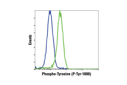
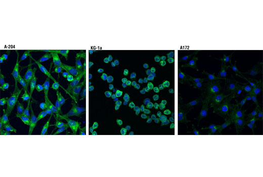
| Phospho-Tyrosine (P-Tyr-1000) MultiMab® Rabbit mAb mix | 8954 | 20 µl | N/A kDa | Rabbit IgG |
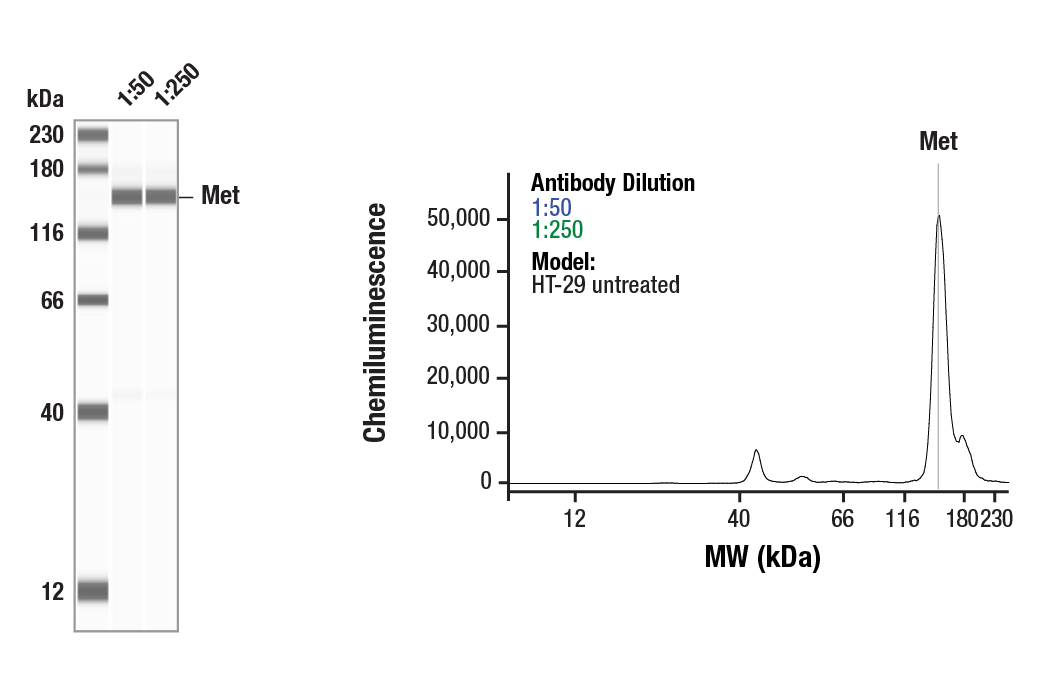
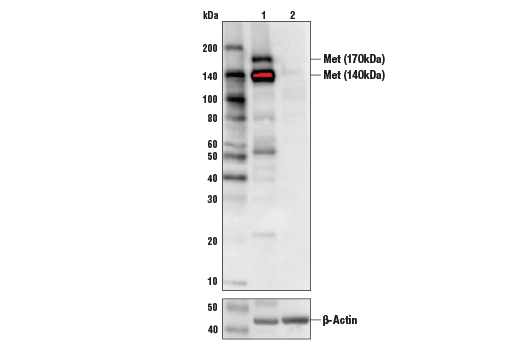
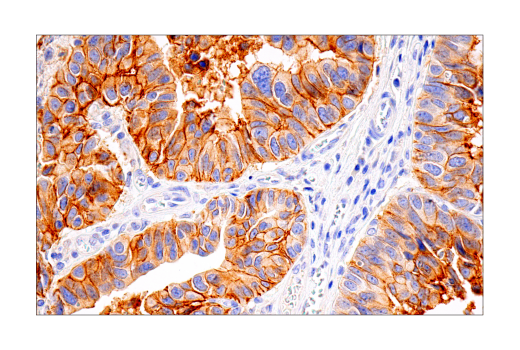
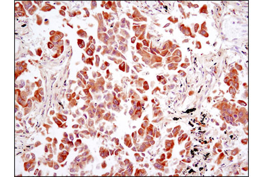
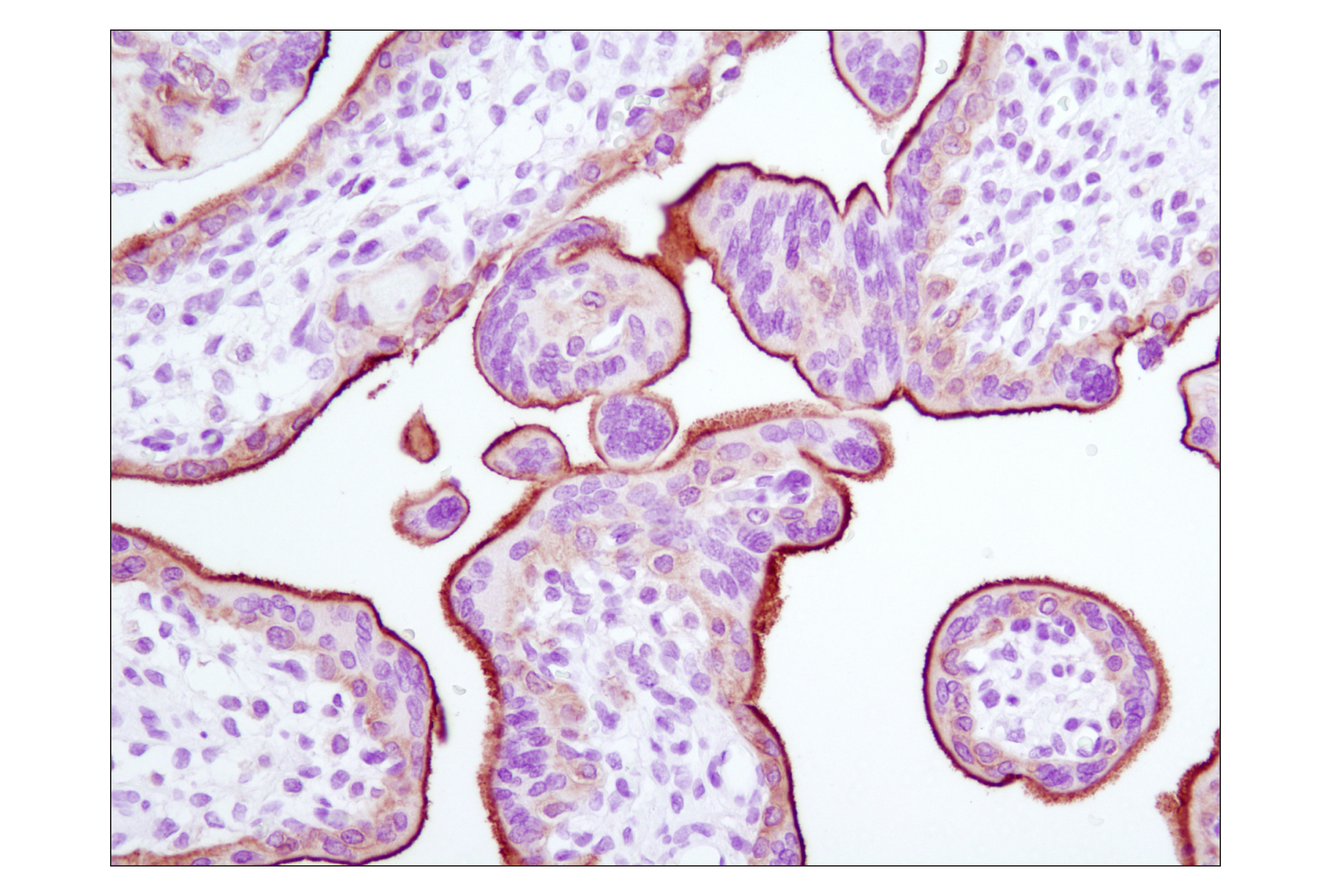
| Met (D1C2) XP® Rabbit mAb | 8198 | 20 µl | 140, 170 kDa | Rabbit IgG |
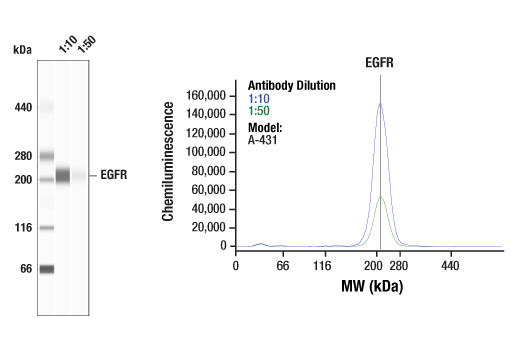
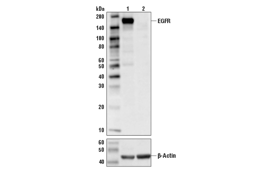
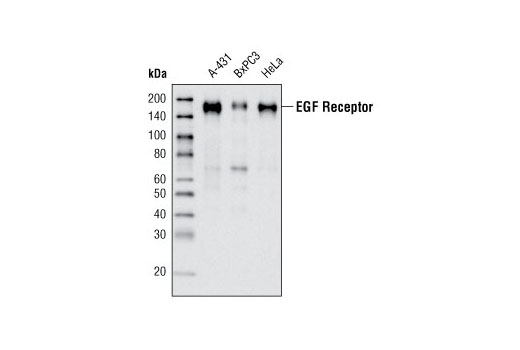
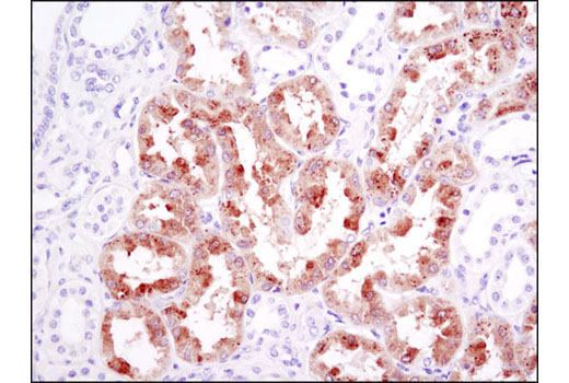
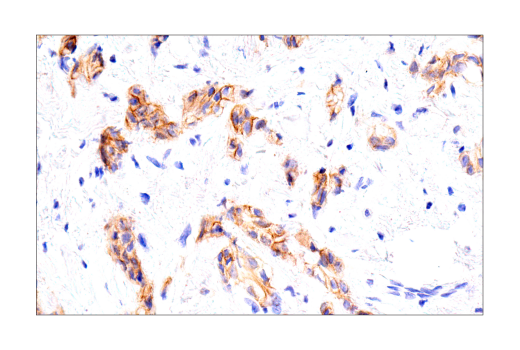
| EGF Receptor (D38B1) XP® Rabbit mAb | 4267 | 20 µl | 175 kDa | Rabbit IgG |
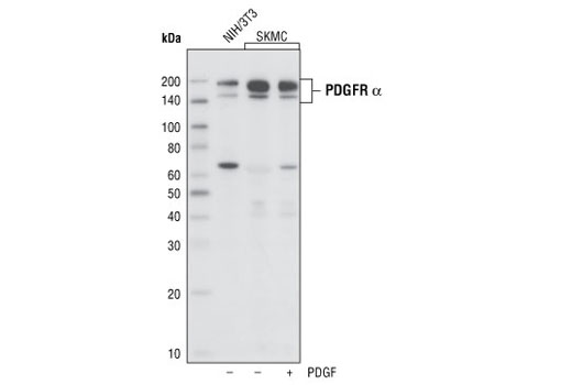
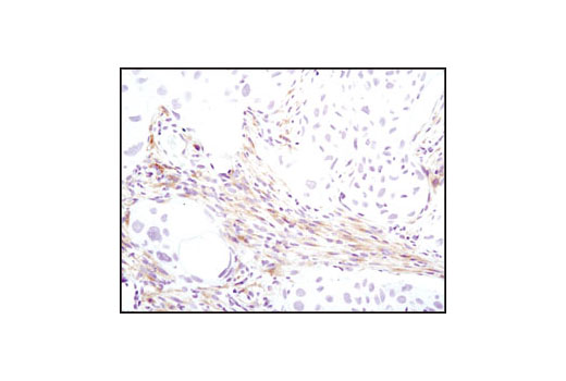
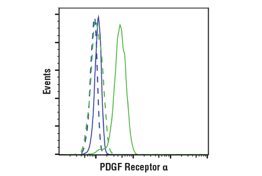
| PDGF Receptor α (D1E1E) XP® Rabbit mAb | 3174 | 20 µl | 190 kDa | Rabbit IgG |
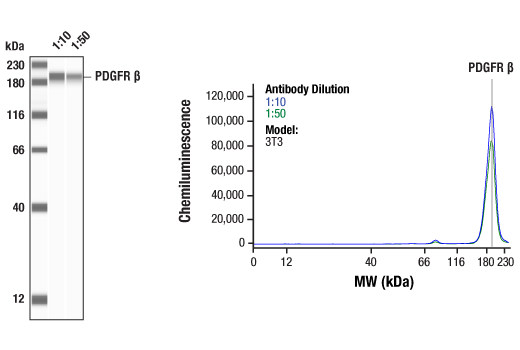
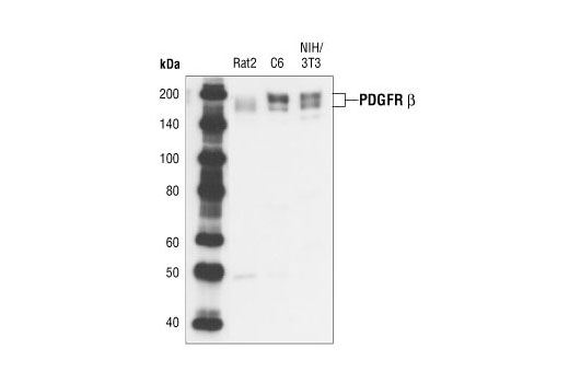
| PDGF Receptor β (28E1) Rabbit mAb | 3169 | 20 µl | 190 kDa | Rabbit IgG |
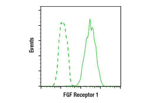
| FGF Receptor 1 (D8E4) XP® Rabbit mAb | 9740 | 20 µl | 92 , 120, 145 kDa | Rabbit IgG |
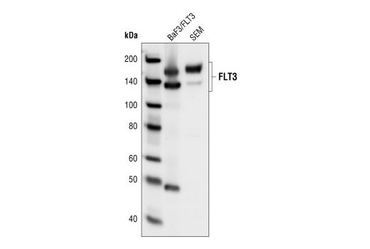
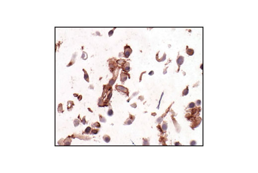
| FLT3 (8F2) Rabbit mAb | 3462 | 20 µl | 130 nonglycosylated form;160 glycosylated mature form kDa | Rabbit IgG |
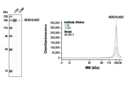
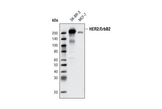
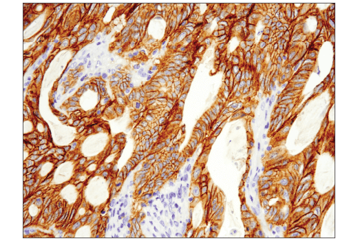
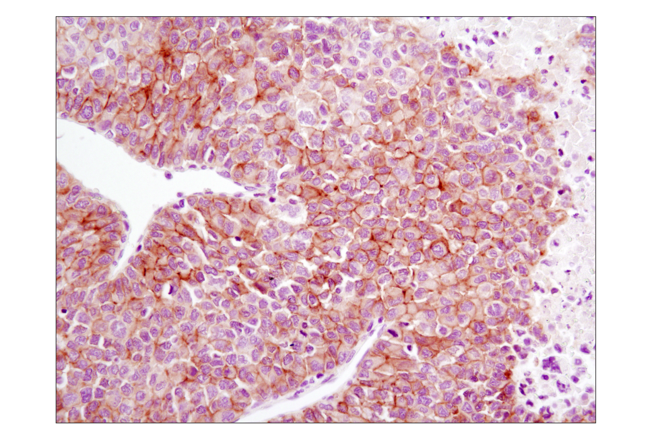
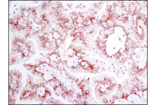
| HER2/ErbB2 (D8F12) XP® Rabbit mAb | 4290 | 20 µl | 185 kDa | Rabbit IgG |
| Anti-rabbit IgG, HRP-linked Antibody | 7074 | 100 µl | Goat |
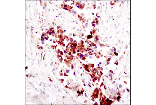
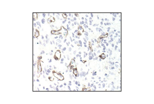
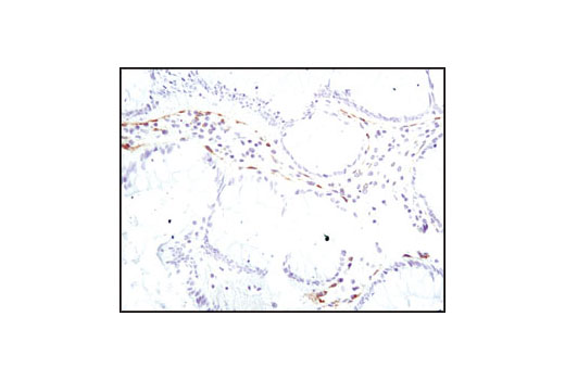
Please visit cellsignal.com for individual component applications, species cross-reactivity, dilutions, protocols, and additional product information.
Description
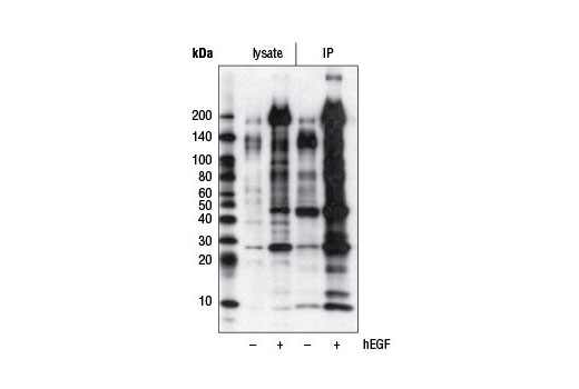
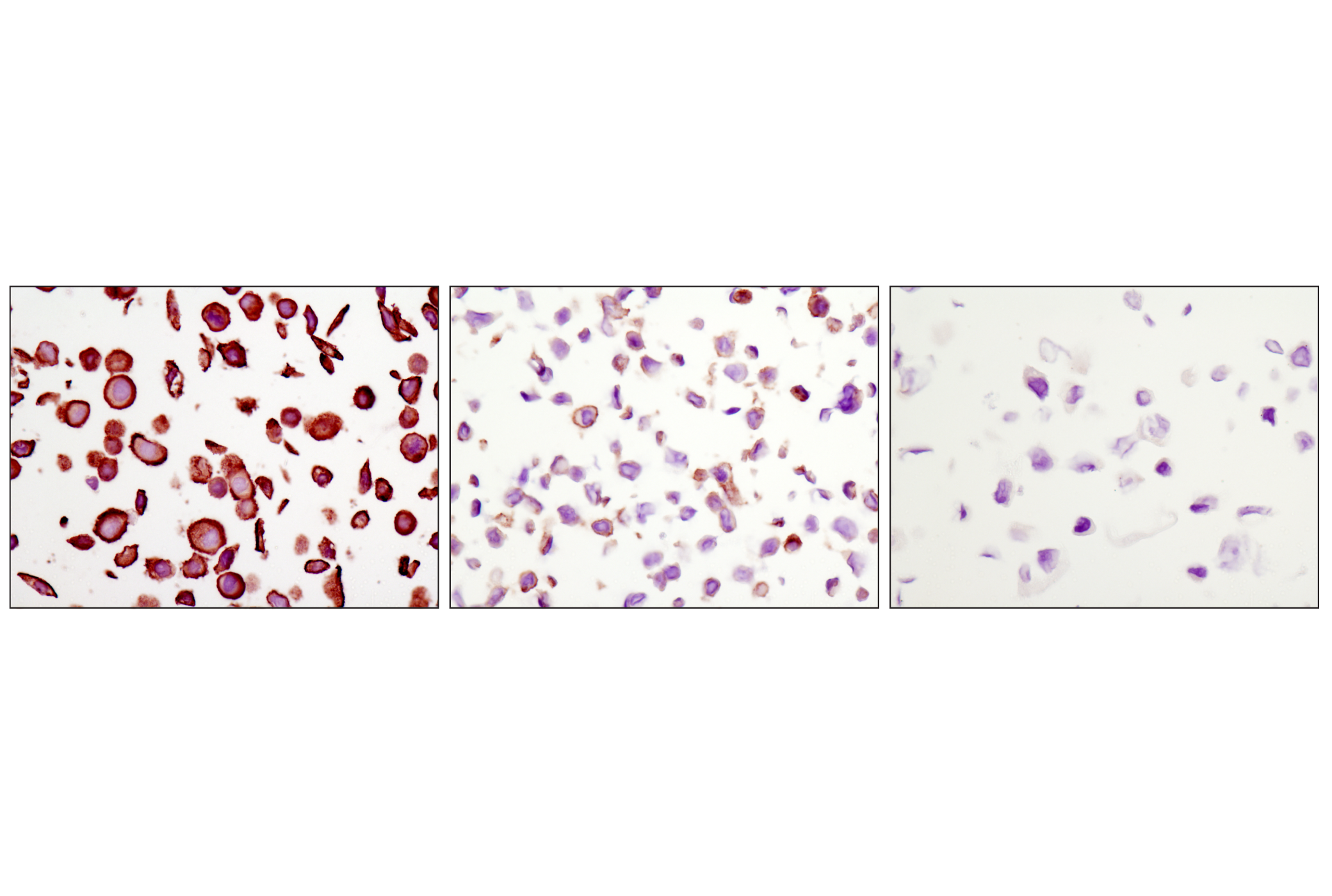
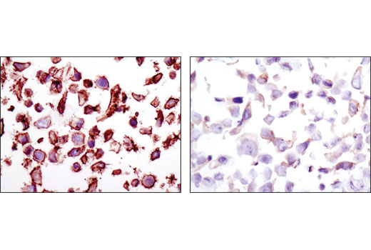
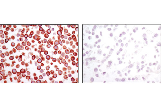
The Receptor Tyrosine Kinase Antibody Sampler Kit provides the means to detect a broad range of common receptor tyrosine kinases, as well as total phospho-tyrosine activity. The kit provides enough antibody to perform two western blot experiments with each primary antibody.
Storage
Background
Tyrosine phosphorylation plays a key role in cellular signaling (1). In cancer studies, unregulated tyrosine kinase activity can drive malignancy and tumor formation by generating inappropriate proliferation and survival signals (2). Antibodies specific for phospho-tyrosine have been invaluable reagents in these studies (3,4).
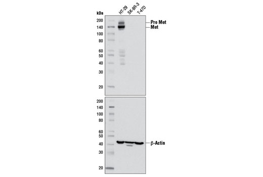
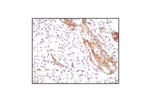
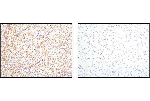
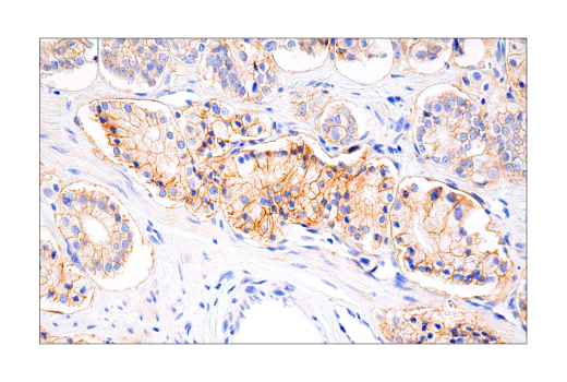
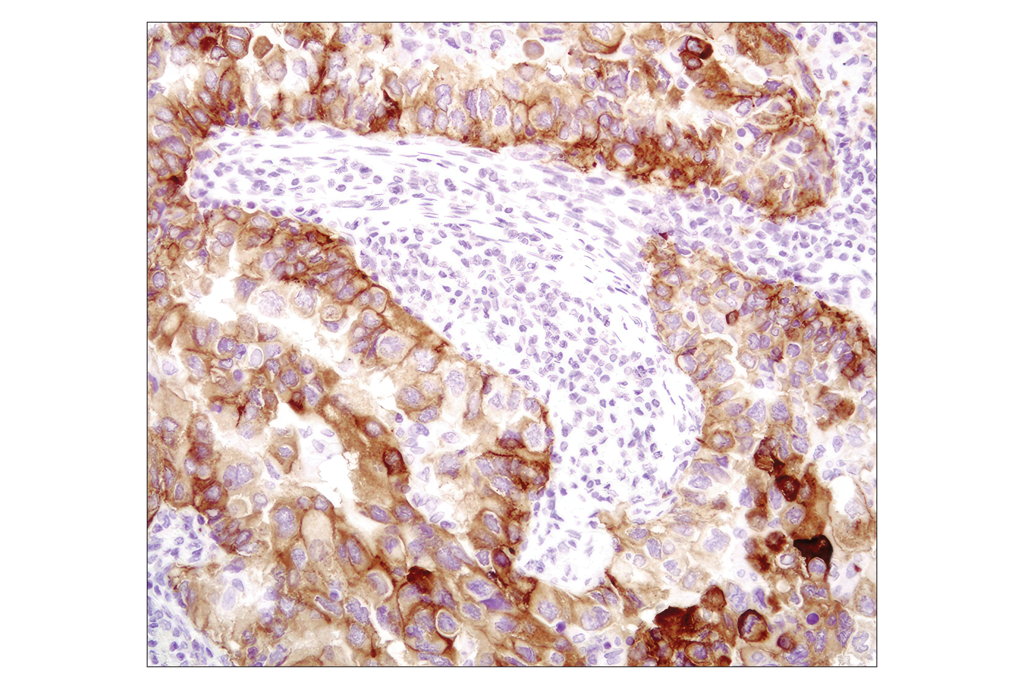
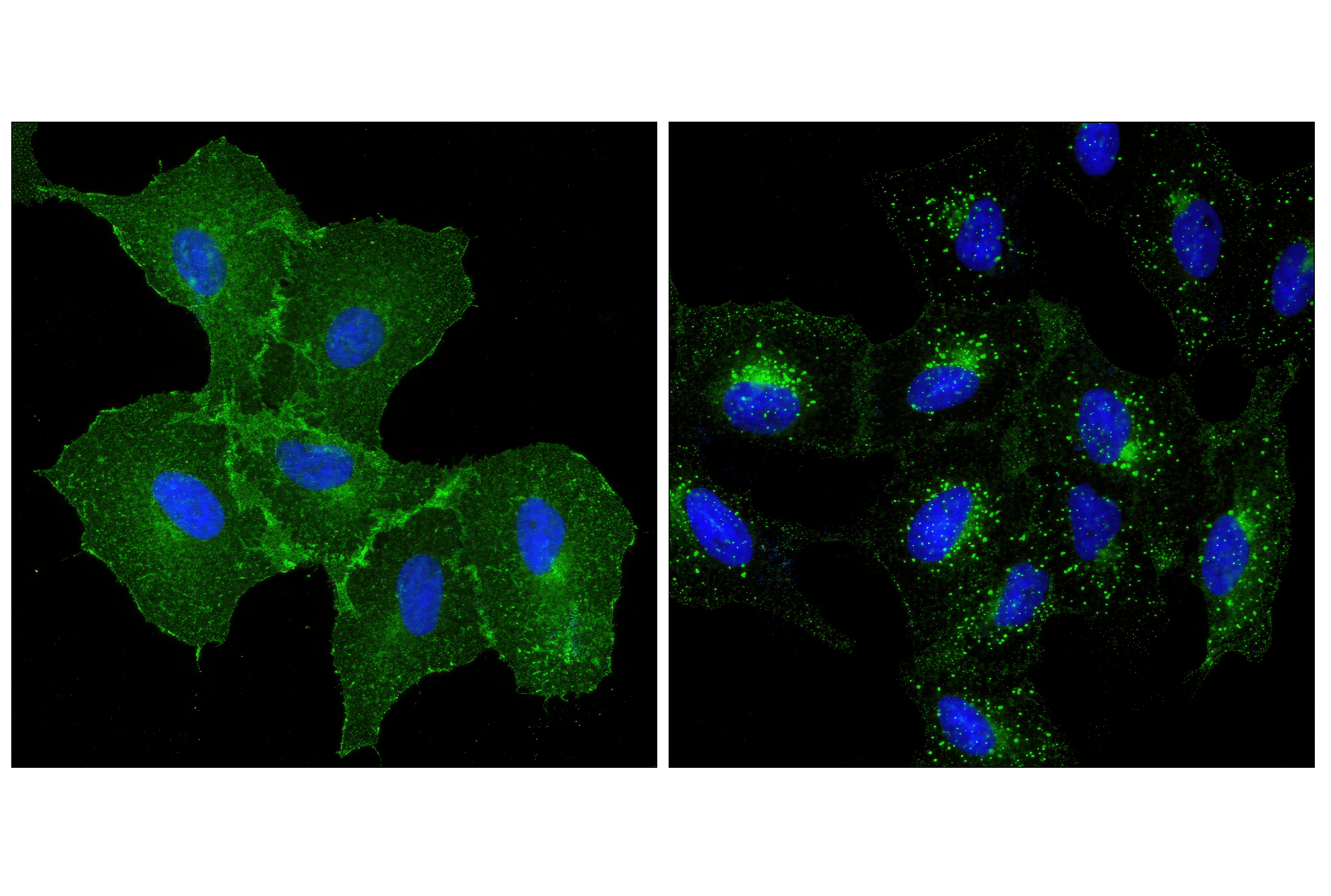
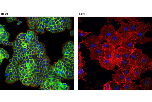
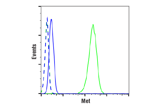
Met, a tyrosine kinase receptor for hepatocyte growth factor (HGF), is a heterodimer made of α- and β-subunits (5,6). The cytoplasmic region of the β-chain is essential for tyrosine kinase activity. Interaction of Met with HGF results in autophosphorylation at multiple tyrosines (Tyr1003, 1234/1235, 1349) which recruit downstream signaling components, including Gab1, c-Cbl, and PI3 kinase (7-9). Altered Met levels and/or tyrosine kinase activities are found in several types of tumors, including renal, colon, and breast (10,11).
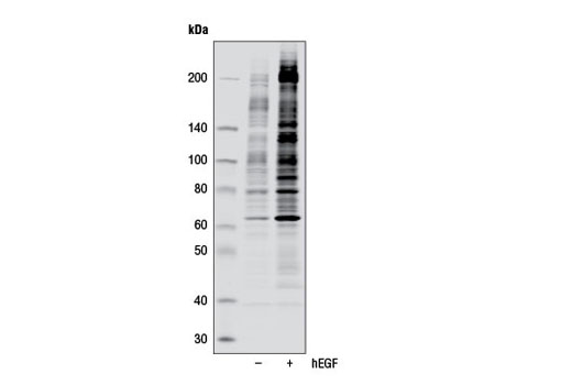
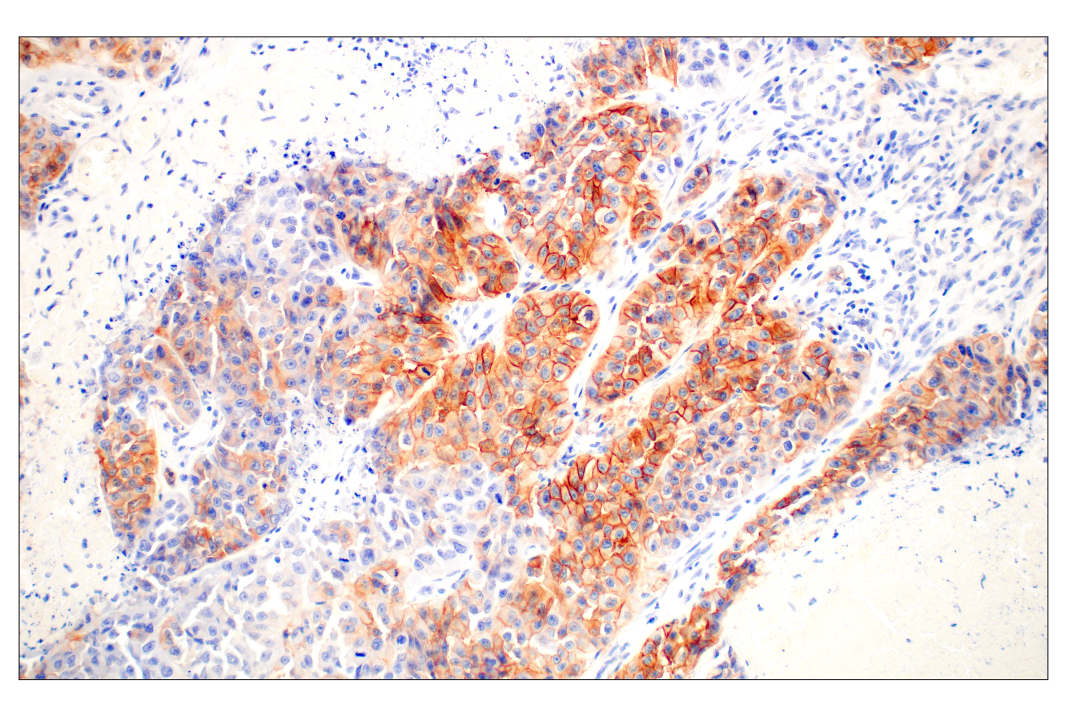
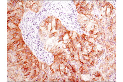
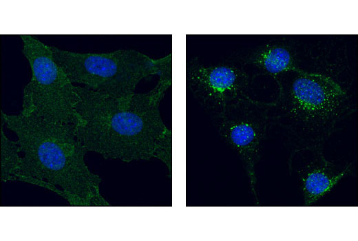
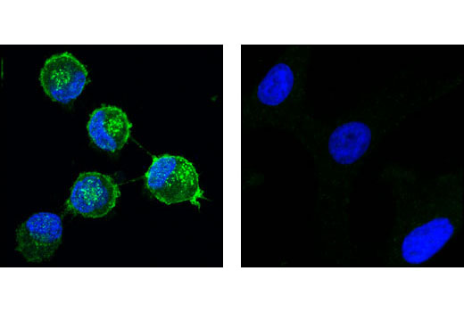
The epidermal growth factor (EGF) receptor is a transmembrane tyrosine kinase that belongs to the HER/ErbB protein family. Ligand binding results in receptor dimerization, autophosphorylation, activation of downstream signaling, internalization, and lysosomal degradation (12,13). c-Src mediated phosphorylation of EGF receptor (EGFR) at Tyr845 provides a binding surface for substrate proteins (14-16). The SH2 domain of PLCγ binds at phospho-Tyr992, activating PLCγ-mediated downstream signaling (17). Adaptor protein c-Cbl binds at phospho-Tyr1045, leading to receptor ubiquitination and degradation (18,19). The GRB2 adaptor protein binds activated EGFR at phospho-Tyr1068 (20), while phospho-Tyr1148 and -Tyr1173 provide a docking site for the Shc scaffold protein, playing a role in MAP kinase signaling (13).
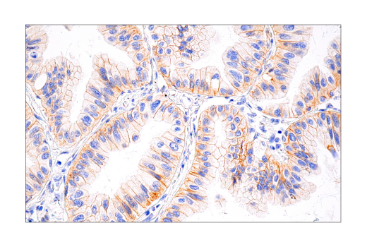
Platelet derived growth factor (PDGF) family proteins bind to two closely related receptor tyrosine kinases, PDGF receptor α (PDGFRα) and PDGF receptor β (PDGFRβ) (21). PDGFRα and PDGFRβ can each form heterodimers with EGFR, which is also activated by PDGF (22). Ligand binding induces receptor dimerization and autophosphorylation, followed by binding and activation of signal transduction molecules such as GRB2, Src, GAP, PI3 kinase, PLCγ, and NCK. Signaling pathways initiated by activated PDGF receptors lead to control of cell growth, actin reorganization, migration, and differentiation (23). Tyr751 and Tyr740 of PDGFRβ regulate binding and activation of PI3 kinase (24,25).
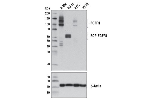
Fibroblast growth factors (FGFs) produce mitogenic and angiogenic effects in target cells by signaling through cell surface receptor tyrosine kinases, after ligand binding and dimerization (26,27). Tyr653 and Tyr654 are important for catalytic activity of activated FGFR and are essential for signaling (28). The other phosphorylated tyrosine residues (Tyr463, 583, 585, 730, and 766) may provide docking sites for downstream signaling components such as Crk and PLCγ (29,30).
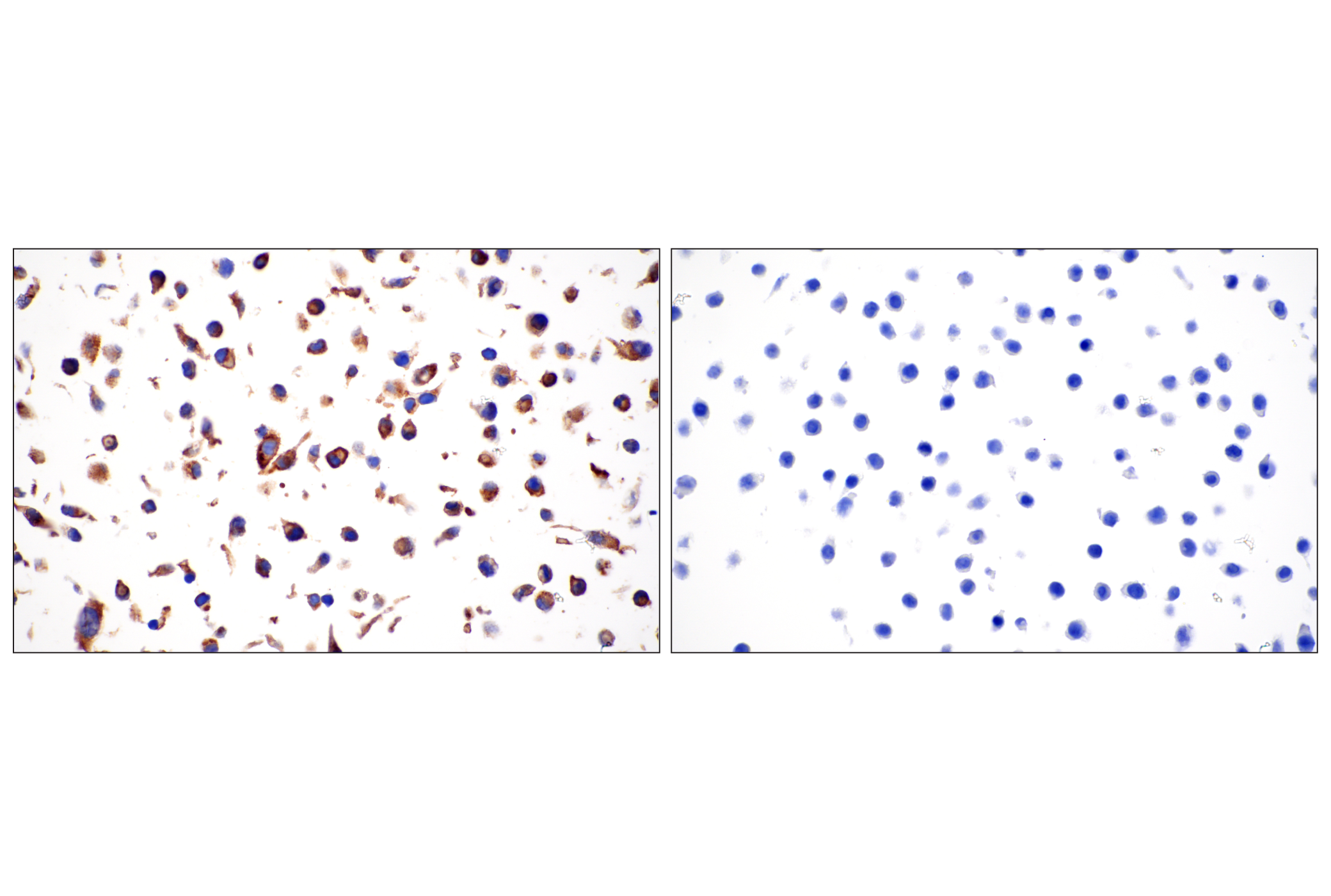
FMS-related tyrosine kinase 3 (FLT3), a member of the type III receptor tyrosine kinase family, is expressed on early hematopoietic progenitor cells and supports growth and differentiation within the hematopoietic system (31,32). FLT3 is activated after binding with its ligand FL, which results in a cascade of tyrosine autophosphorylation and tyrosine phosphorylation of downstream targets (33). The p85 subunit of PI3 kinase, SHP2, GRB2 and Shc are associated with FLT3 after FL stimulation (34-36). Tyr589/591 may play an important role in regulation of FLT3 tyrosine kinase activity (37).
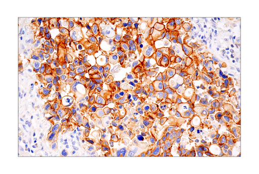
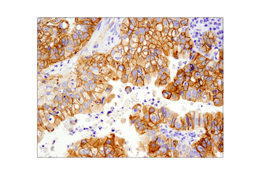
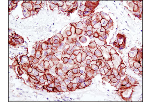
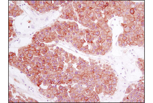
The ErbB2 (HER2) proto-oncogene encodes a transmembrane, receptor-like glycoprotein with tyrosine kinase activity (38). ErbB2 kinase activity can be activated in the absence of a ligand when overexpressed and through associations with other ErbB family members (39). Phosphorylation at Tyr877 may be involved in regulating ErbB2 activity. Autophosphorylation of ErbB2 at Tyr1248 and Tyr1221/1222 couples ErbB2 to the Ras-Raf-MAP kinase signal transduction pathway (38,40).
- Schlessinger, J. (2000) Cell 103, 211-25
- Blume-Jensen, P. and Hunter, T. (2001) Nature 411, 355-65
- Ward, S.G. et al. (1992) J Biol Chem 267, 23862-9
- Glenney, J.R. et al. (1988) J Immunol Methods 109, 277-85
- Cooper, C.S. et al. Nature 311, 29-33.
- Bottaro, D.P. et al. (1991) Science 251, 802-4.
- Bardelli, A. et al. (1997) Oncogene 15, 3103-11.
- Taher, T.E. et al. (2002) J Immunol 169, 3793-800.
- Schaeper, U. et al. (2000) J Cell Biol 149, 1419-32.
- Eder, J.P. et al. (2009) Clin Cancer Res 15, 2207-14.
- Sattler, M. and Salgia, R. (2009) Update Cancer Ther 3, 109-118.
- Hackel, P.O. et al. (1999) Curr Opin Cell Biol 11, 184-9.
- Zwick, E. et al. (1999) Trends Pharmacol Sci 20, 408-12.
- Cooper, J.A. and Howell, B. (1993) Cell 73, 1051-4.
- Hubbard, S.R. et al. Nature 372, 746-54.
- Biscardi, J.S. et al. (1999) J Biol Chem 274, 8335-43.
- Emlet, D.R. et al. (1997) J Biol Chem 272, 4079-86.
- Levkowitz, G. et al. (1999) Mol Cell 4, 1029-40.
- Ettenberg, S.A. et al. (1999) Oncogene 18, 1855-66.
- Rojas, M. et al. (1996) J Biol Chem 271, 27456-61.
- Deuel, T.F. et al. (1988) Biofactors 1, 213-7.
- Betsholtz, C. et al. (2001) Bioessays 23, 494-507.
- Ostman, A. and Heldin, C.H. (2001) Adv Cancer Res 80, 1-38.
- Panayotou, G. et al. (1992) EMBO J 11, 4261-72.
- Kashishian, A. et al. (1992) EMBO J 11, 1373-82.
- Powers, C.J. et al. (2000) Endocr Relat Cancer 7, 165-97.
- Reilly, J.F. et al. (2000) J Biol Chem 275, 7771-8.
- Mohammadi, M. et al. (1996) Mol Cell Biol 16, 977-89.
- Mohammadi, M. et al. (1991) Mol Cell Biol 11, 5068-78.
- Larsson, H. et al. (1999) J Biol Chem 274, 25726-34.
- Shurin, M.R. et al. (1998) Cytokine Growth Factor Rev 9, 37-48.
- Naoe, T. et al. (2001) Cancer Chemother Pharmacol 48 Suppl 1, S27-30.
- Namikawa, R. et al. (1996) Stem Cells 14, 388-95.
- Beslu, N. et al. (1996) J Biol Chem 271, 20075-81.
- Zhang, S. and Broxmeyer, H.E. (2000) Biochem Biophys Res Commun 277, 195-9.
- Zhang, S. et al. (1999) J Leukoc Biol 65, 372-80.
- Mizuki, M. et al. (2000) Blood 96, 3907-14.
- Muthuswamy, S.K. et al. (1999) Mol Cell Biol 19, 6845-57.
- Qian, X. et al. (1994) Proc Natl Acad Sci U S A 91, 1500-4.
- Kwon, Y.K. et al. (1997) J Neurosci 17, 8293-9.
Background References
Trademarks and Patents
使用に関する制限
法的な権限を与えられたCSTの担当者が署名した書面によって別途明示的に合意された場合を除き、 CST、その関連会社または代理店が提供する製品には以下の条件が適用されます。お客様が定める条件でここに定められた条件に含まれるものを超えるもの、 または、ここに定められた条件と異なるものは、法的な権限を与えられたCSTの担当者が別途書面にて受諾した場合を除き、拒絶され、 いかなる効力も効果も有しません。
研究専用 (For Research Use Only) またはこれに類似する表示がされた製品は、 いかなる目的についても FDA または外国もしくは国内のその他の規制機関により承認、認可または許可を受けていません。 お客様は製品を診断もしくは治療目的で使用してはならず、また、製品に表示された内容に違反する方法で使用してはなりません。 CST が販売または使用許諾する製品は、エンドユーザーであるお客様に対し、使途を研究および開発のみに限定して提供されるものです。 診断、予防もしくは治療目的で製品を使用することまたは製品を再販売 (単独であるか他の製品等の一部であるかを問いません) もしくはその他の商業的利用の目的で購入することについては、CST から別途許諾を得る必要があります。 お客様は以下の事項を遵守しなければなりません。(a) CST の製品 (単独であるか他の資材と一緒であるかを問いません) を販売、使用許諾、貸与、寄付もしくはその他の態様で第三者に譲渡したり使用させたりしてはなりません。また、商用の製品を製造するために CST の製品を使用してはなりません。(b) 複製、改変、リバースエンジニアリング、逆コンパイル、 分解または他の方法により製品の構造または技術を解明しようとしてはなりません。また、 CST の製品またはサービスと競合する製品またはサービスを開発する目的で CST の製品を使用してはなりません。(c) CST の製品の商標、商号、ロゴ、特許または著作権に関する通知または表示を除去したり改変したりしてはなりません。(d) CST の製品をCST 製品販売条件(CST’s Product Terms of Sale) および該当する書面のみに従って使用しなければなりません。(e) CST の製品に関連してお客様が使用する第三者の製品またはサービスに関する使用許諾条件、 サービス提供条件またはこれに類する合意事項を遵守しなければなりません。