| Product Includes | Product # | Quantity | Mol. Wt | Isotype/Source |
|---|---|---|---|---|
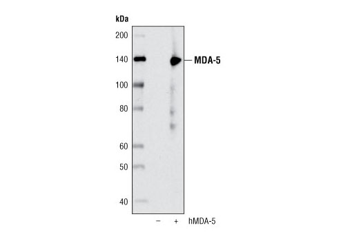
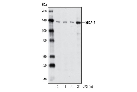
| MDA-5 (D74E4) Rabbit mAb | 5321 | 20 µl | 135 kDa | Rabbit IgG |
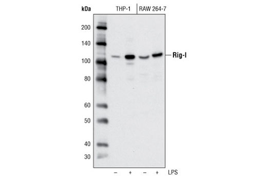
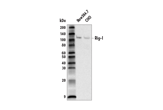
| Rig-I (D14G6) Rabbit mAb | 3743 | 20 µl | 102 kDa | Rabbit IgG |
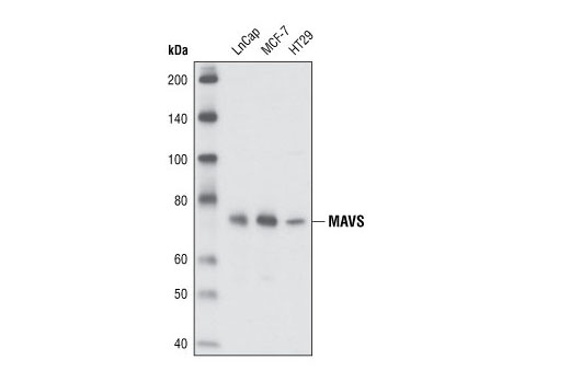
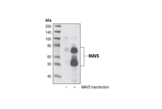
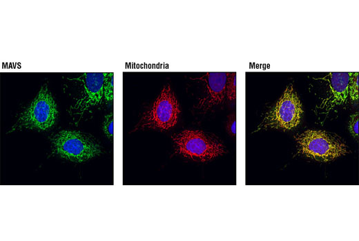
| MAVS Antibody | 3993 | 20 µl | 75, 52 kDa | Rabbit |
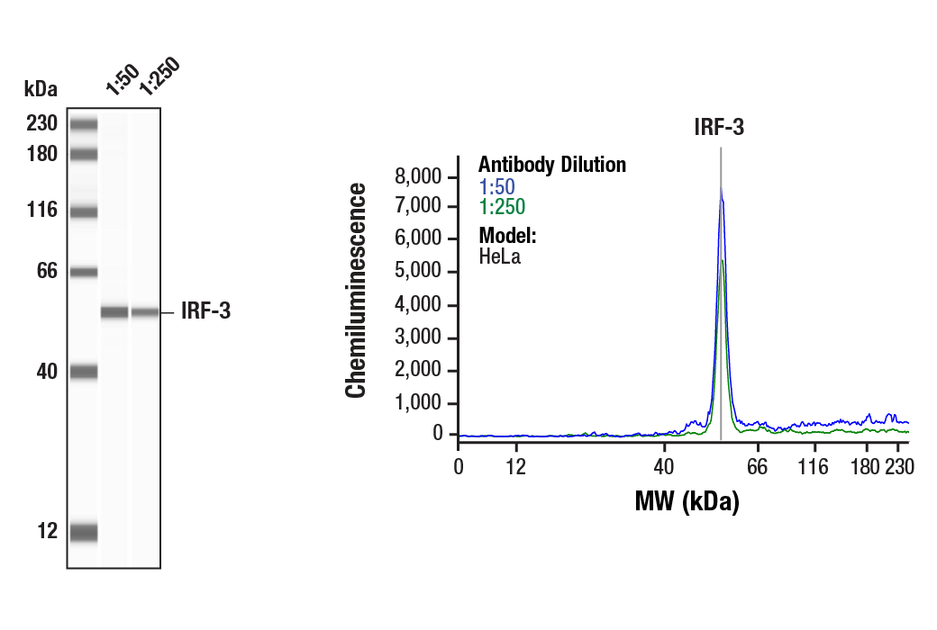
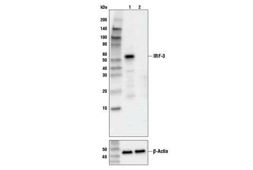
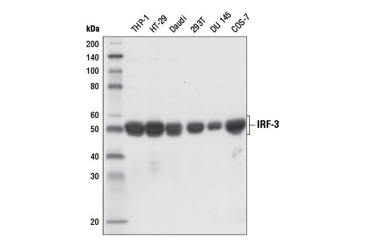
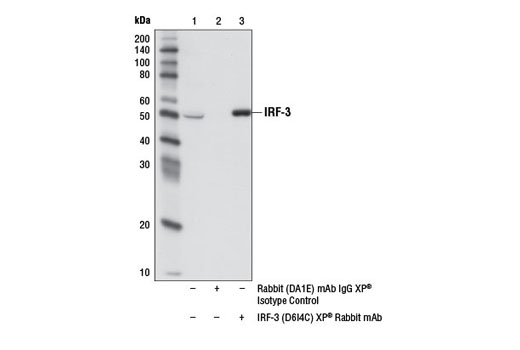
| IRF-3 (D6I4C) XP® Rabbit mAb | 11904 | 20 µl | 50-55 kDa | Rabbit IgG |
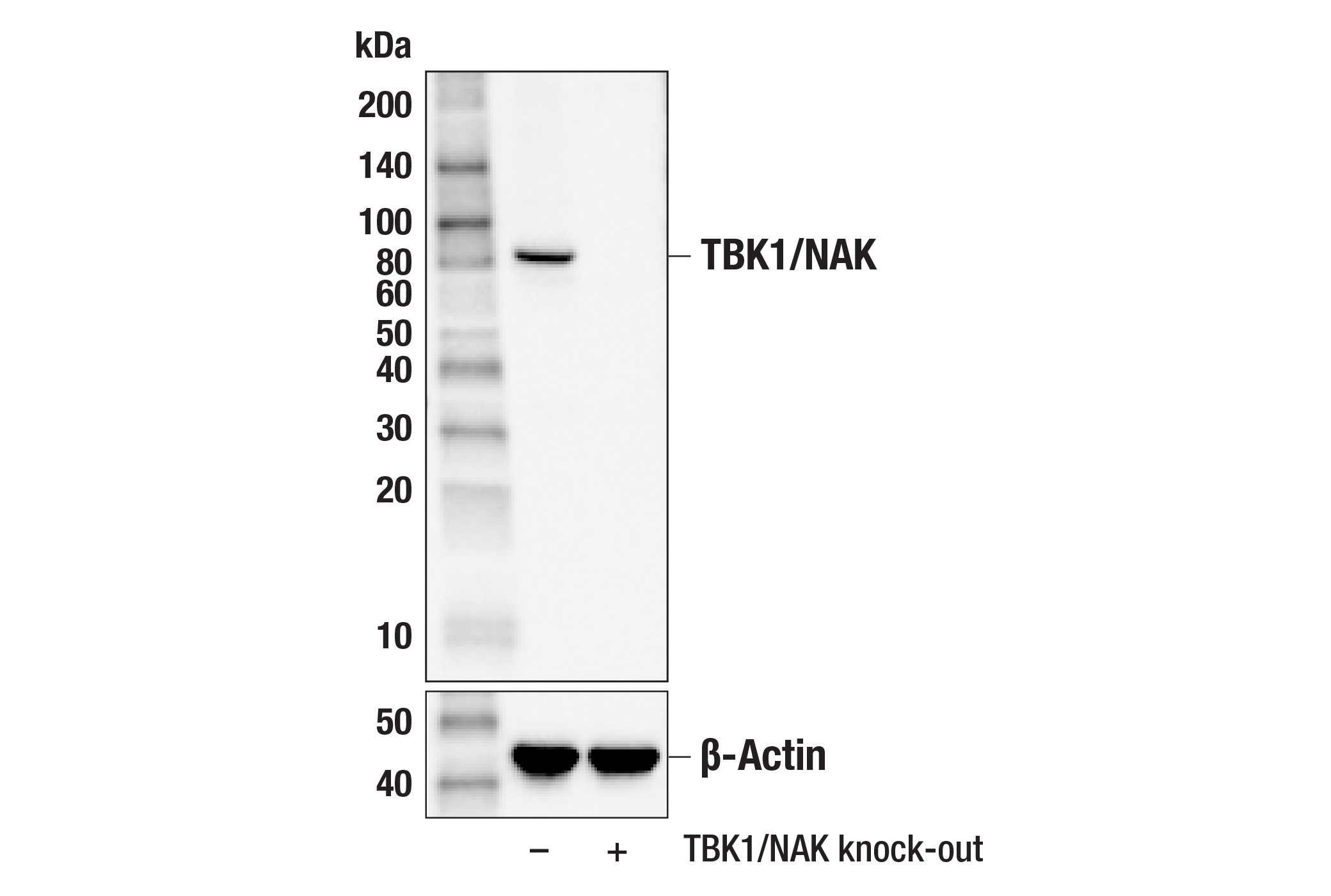
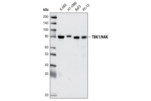
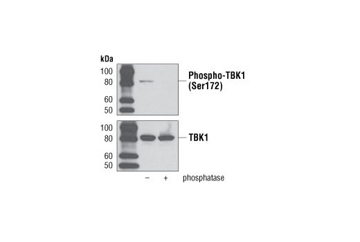
| TBK1/NAK (D1B4) Rabbit mAb | 3504 | 20 µl | 84 kDa | Rabbit IgG |
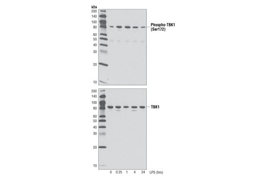
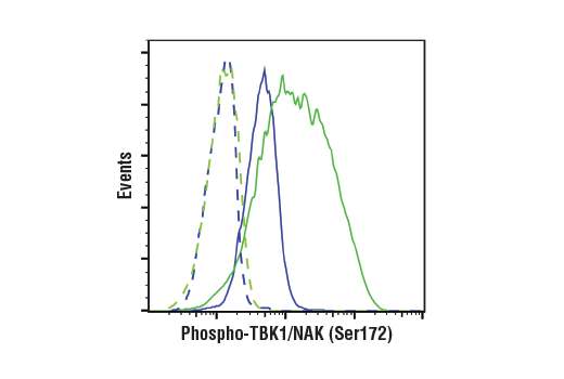
| Phospho-TBK1/NAK (Ser172) (D52C2) XP® Rabbit mAb | 5483 | 20 µl | 84 kDa | Rabbit IgG |
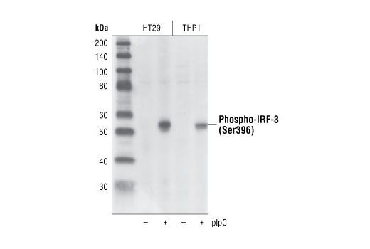
| Phospho-IRF-3 (Ser396) (4D4G) Rabbit mAb | 4947 | 20 µl | 45-55 kDa | Rabbit IgG |
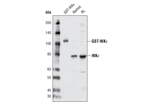
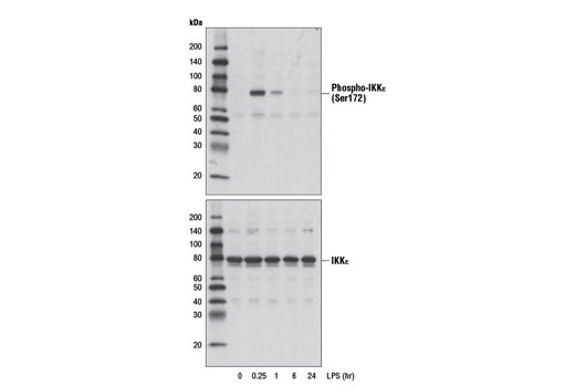
| Phospho-IKKε (Ser172) (D1B7) Rabbit mAb | 8766 | 20 µl | 80 kDa | Rabbit IgG |
| IKKε (D20G4) Rabbit mAb | 2905 | 20 µl | 80 kDa | Rabbit IgG |
| Anti-rabbit IgG, HRP-linked Antibody | 7074 | 100 µl | Goat |
Please visit cellsignal.com for individual component applications, species cross-reactivity, dilutions, protocols, and additional product information.
Description
The Rig-I Pathway Antibody Sampler Kit provides an economical means to evaluate the activation state and total protein levels of multiple members of the Rig-I pathway including Rig-I, MDA-5, MAVS, IRF-3, TBK1/NAK, and IKKε. The kit includes enough primary antibody to perform two western blot experiments per antibody.
Storage
Background
Antiviral innate immunity depends on the combination of parallel pathways triggered by virus detecting proteins in the Toll-like receptor (TLR) family and RNA helicases, such as Rig-I (retinoic acid-inducible gene I) and MDA-5 (melanoma differentiation-associated antigen 5), which promote the transcription of type I interferons (IFN) and antiviral enzymes (1-3). TLRs and helicase proteins contain sites that recognize the molecular patterns of different virus types, including DNA, single-stranded RNA (ssRNA), double-stranded RNA (dsRNA), and glycoproteins. These antiviral proteins are found in different cell compartments; TLRs (i.e. TLR3, TLR7, TLR8, and TLR9) are expressed on endosomal membranes and helicases are localized to the cytoplasm. Rig-I expression is induced by retinoic acid, LPS, IFN, and viral infection (4,5). Both Rig-I and MDA-5 share a DExD/H-box helicase domain that detects viral dsRNA and two amino-terminal caspase recruitment domains (CARD) that are required for triggering downstream signaling (4-7). Rig-I binds both dsRNA and viral ssRNA that contains a 5'-triphosphate end not seen in host RNA (8,9). Though structurally related, Rig-I and MDA-5 detect a distinct set of viruses (10,11). The CARD domain of the helicases, which is sufficient to generate signaling and IFN production, is recruited to the CARD domain of the MAVS/VISA/Cardif/IPS-1 mitochondrial protein, which triggers activation of NF-κB, TBK1/IKKε, and IRF-3/IRF-7 (12-15).
- Yoneyama, M. and Fujita, T. (2007) J Biol Chem 282, 15315-8.
- Meylan, E. and Tschopp, J. (2006) Mol Cell 22, 561-9.
- Thompson, A.J. and Locarnini, S.A. (2007) Immunol Cell Biol 85, 435-45.
- Imaizumi, T. et al. (2002) Biochem Biophys Res Commun 292, 274-9.
- Zhang, X. et al. (2000) Microb Pathog 28, 267-78.
- Yoneyama, M. et al. (2005) J Immunol 175, 2851-8.
- Yoneyama, M. et al. (2004) Nat Immunol 5, 730-7.
- Hornung, V. et al. (2006) Science 314, 994-7.
- Pichlmair, A. et al. (2006) Science 314, 997-1001.
- Kato, H. et al. (2006) Nature 441, 101-5.
- Childs, K. et al. (2007) Virology 359, 190-200.
- Meylan, E. et al. (2005) Nature 437, 1167-72.
- Xu, L.G. et al. (2005) Mol Cell 19, 727-40.
- Kawai, T. et al. (2005) Nat Immunol 6, 981-8.
- Seth, R.B. et al. (2005) Cell 122, 669-82.
Background References
Trademarks and Patents
使用に関する制限
法的な権限を与えられたCSTの担当者が署名した書面によって別途明示的に合意された場合を除き、 CST、その関連会社または代理店が提供する製品には以下の条件が適用されます。お客様が定める条件でここに定められた条件に含まれるものを超えるもの、 または、ここに定められた条件と異なるものは、法的な権限を与えられたCSTの担当者が別途書面にて受諾した場合を除き、拒絶され、 いかなる効力も効果も有しません。
研究専用 (For Research Use Only) またはこれに類似する表示がされた製品は、 いかなる目的についても FDA または外国もしくは国内のその他の規制機関により承認、認可または許可を受けていません。 お客様は製品を診断もしくは治療目的で使用してはならず、また、製品に表示された内容に違反する方法で使用してはなりません。 CST が販売または使用許諾する製品は、エンドユーザーであるお客様に対し、使途を研究および開発のみに限定して提供されるものです。 診断、予防もしくは治療目的で製品を使用することまたは製品を再販売 (単独であるか他の製品等の一部であるかを問いません) もしくはその他の商業的利用の目的で購入することについては、CST から別途許諾を得る必要があります。 お客様は以下の事項を遵守しなければなりません。(a) CST の製品 (単独であるか他の資材と一緒であるかを問いません) を販売、使用許諾、貸与、寄付もしくはその他の態様で第三者に譲渡したり使用させたりしてはなりません。また、商用の製品を製造するために CST の製品を使用してはなりません。(b) 複製、改変、リバースエンジニアリング、逆コンパイル、 分解または他の方法により製品の構造または技術を解明しようとしてはなりません。また、 CST の製品またはサービスと競合する製品またはサービスを開発する目的で CST の製品を使用してはなりません。(c) CST の製品の商標、商号、ロゴ、特許または著作権に関する通知または表示を除去したり改変したりしてはなりません。(d) CST の製品をCST 製品販売条件(CST’s Product Terms of Sale) および該当する書面のみに従って使用しなければなりません。(e) CST の製品に関連してお客様が使用する第三者の製品またはサービスに関する使用許諾条件、 サービス提供条件またはこれに類する合意事項を遵守しなければなりません。