WB, W-S, IP, IHC-P, IF-IC, FC-FP
H
Endogenous
258, 110, 50-80
Rabbit IgG
#P08922
6098
Product Information
Product Usage Information
| Application | Dilution |
|---|---|
| Western Blotting | 1:1000 |
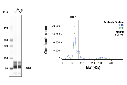
| Simple Western™ | 1:10 - 1:50 |
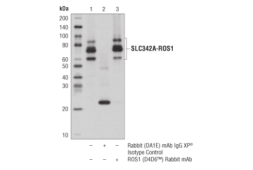
| Immunoprecipitation | 1:50 |
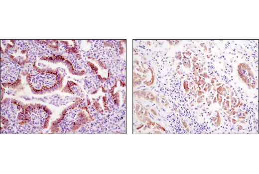
| Immunohistochemistry (Paraffin) | 1:250 |
| Immunofluorescence (Immunocytochemistry) | 1:200 - 1:800 |
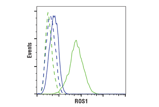
| Flow Cytometry (Fixed/Permeabilized) | 1:200 - 1:800 |
Storage
Specificity / Sensitivity
Species Reactivity:
Human
Source / Purification
Monoclonal antibody is produced by immunizing animals with a protein corresponding to residues in the carboxy terminal domain of the human ROS1 protein.
Background
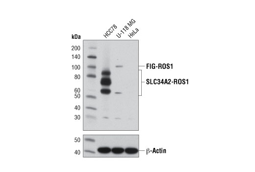
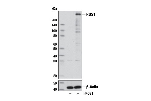
ROS1, an orphan receptor tyrosine kinase of the insulin receptor family, was initially identified as a homolog of v-ros from the UR2 sarcoma virus (1). ROS1 consists of a large extracellular domain that is composed of six fibronectin repeats, a transmembrane domain, and a C-terminal kinase domain. Being an orphan receptor, the functions of ROS1 are not well known, though it has been shown to play an important role in differentiation of epididymal epithelium (2). The first oncogenic fusion of ROS1, FIG-ROS1, was initially identified by research studies in glioblastoma (3), and subsequent studies have found this fusion in cholangiocarcinoma (4), ovarian cancer (5), and non-small cell lung cancer (NSCLC) (6). Investigators have found additional oncogenic ROS1 fusion proteins in NSCLC (at a frequency of ~1.6%), where the ROS1 kinase domain is fused to the amino-terminal region of several different proteins, including CD74 and SLC34A2 (6-8). ROS1 fusion proteins activate the SHP-2 phosphatase, PI3K/Akt/mTOR, Erk, and Stat3 pathways (3,4,9). There are two autophosphorylation sites (Tyr2274, Tyr2334) downstream of the kinase domain of ROS1, either of which may serve as biomarkers of ROS1 kinase activity, including that of ROS1 fusion proteins (10).
- Matsushime, H. et al. (1986) Mol Cell Biol 6, 3000-4.
- Yeung, C.H. et al. (1999) Biol Reprod 61, 1062-9.
- Charest, A. et al. (2003) Genes Chromosomes Cancer 37, 58-71.
- Gu, T.L. et al. (2011) PLoS One 6, e15640.
- Birch, A.H. et al. (2011) PLoS One 6, e28250.
- Rimkunas, V.M. et al. (2012) Clin Cancer Res 18, 4449-57.
- Rikova, K. et al. (2007) Cell 131, 1190-203.
- Stumpfova, M. and Jänne, P.A. (2012) Clin Cancer Res 18, 4222-4.
- Jun, H.J. et al. (2012) Cancer Res 72, 3764-74.
- Zou, H.Y. et al. (2015) Proc Natl Acad Sci U S A 112, 3493-8.
Species Reactivity
Species reactivity is determined by testing in at least one approved application (e.g., western blot).
Western Blot Buffer
IMPORTANT: For western blots, incubate membrane with diluted primary antibody in 5% w/v BSA, 1X TBS, 0.1% Tween® 20 at 4°C with gentle shaking, overnight.
Applications Key
WB: Western Blotting W-S: Simple Western™ IP: Immunoprecipitation IHC-P: Immunohistochemistry (Paraffin) IF-IC: Immunofluorescence (Immunocytochemistry) FC-FP: Flow Cytometry (Fixed/Permeabilized)
Cross-Reactivity Key
H: human M: mouse R: rat Hm: hamster Mk: monkey Vir: virus Mi: mink C: chicken Dm: D. melanogaster X: Xenopus Z: zebrafish B: bovine Dg: dog Pg: pig Sc: S. cerevisiae Ce: C. elegans Hr: horse GP: Guinea Pig Rab: rabbit All: all species expected
Trademarks and Patents
使用に関する制限
法的な権限を与えられたCSTの担当者が署名した書面によって別途明示的に合意された場合を除き、 CST、その関連会社または代理店が提供する製品には以下の条件が適用されます。お客様が定める条件でここに定められた条件に含まれるものを超えるもの、 または、ここに定められた条件と異なるものは、法的な権限を与えられたCSTの担当者が別途書面にて受諾した場合を除き、拒絶され、 いかなる効力も効果も有しません。
研究専用 (For Research Use Only) またはこれに類似する表示がされた製品は、 いかなる目的についても FDA または外国もしくは国内のその他の規制機関により承認、認可または許可を受けていません。 お客様は製品を診断もしくは治療目的で使用してはならず、また、製品に表示された内容に違反する方法で使用してはなりません。 CST が販売または使用許諾する製品は、エンドユーザーであるお客様に対し、使途を研究および開発のみに限定して提供されるものです。 診断、予防もしくは治療目的で製品を使用することまたは製品を再販売 (単独であるか他の製品等の一部であるかを問いません) もしくはその他の商業的利用の目的で購入することについては、CST から別途許諾を得る必要があります。 お客様は以下の事項を遵守しなければなりません。(a) CST の製品 (単独であるか他の資材と一緒であるかを問いません) を販売、使用許諾、貸与、寄付もしくはその他の態様で第三者に譲渡したり使用させたりしてはなりません。また、商用の製品を製造するために CST の製品を使用してはなりません。(b) 複製、改変、リバースエンジニアリング、逆コンパイル、 分解または他の方法により製品の構造または技術を解明しようとしてはなりません。また、 CST の製品またはサービスと競合する製品またはサービスを開発する目的で CST の製品を使用してはなりません。(c) CST の製品の商標、商号、ロゴ、特許または著作権に関する通知または表示を除去したり改変したりしてはなりません。(d) CST の製品をCST 製品販売条件(CST’s Product Terms of Sale) および該当する書面のみに従って使用しなければなりません。(e) CST の製品に関連してお客様が使用する第三者の製品またはサービスに関する使用許諾条件、 サービス提供条件またはこれに類する合意事項を遵守しなければなりません。