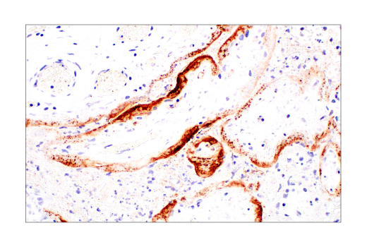
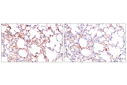
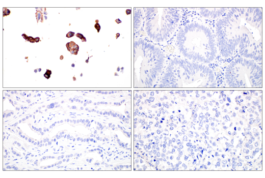
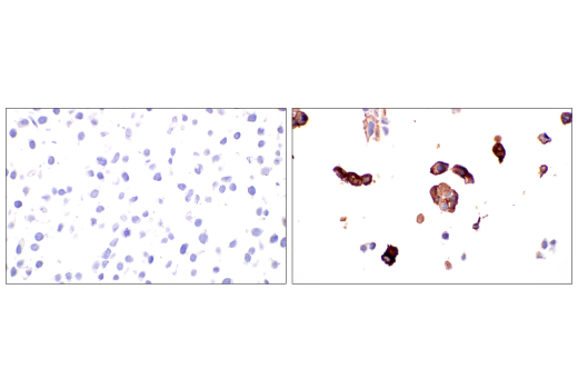
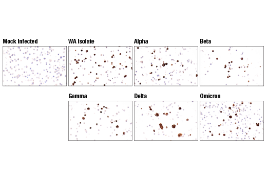
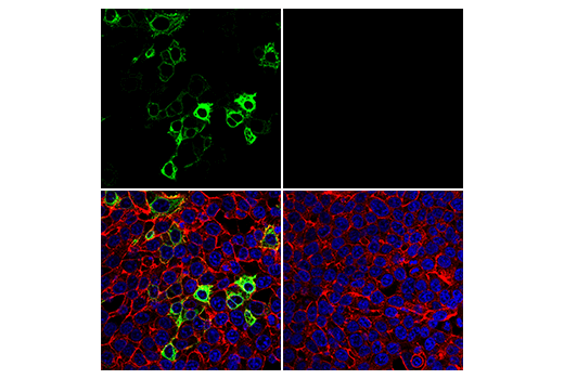
Revision 4
Cell Signaling Technology

Orders: 877-616-CELL (2355) [email protected]

Support: 877-678-TECH (8324)

Web: [email protected] cellsignal.com

3 Trask LaneDanversMassachusetts01923USA
For Research Use Only. Not for Use in Diagnostic Procedures.
Applications:

IHC-P, IF-IC

REACTIVITY:

Vir

SENSITIVITY:

Endogenous

MW (kDa):

Source/Isotype:

Mouse IgG2a

UniProt ID:

#P0DTC2

Entrez-Gene Id:

43740568

Product Information

Product Usage Information

Application Dilution
Immunohistochemistry (Paraffin) 1:200 - 1:800
Immunofluorescence (Immunocytochemistry) 1:200 - 1:800

Storage

Supplied in 10 mM sodium HEPES (pH 7.5), 150 mM NaCl, 100 µg/ml BSA, 50% glycerol and less than 0.02% sodium azide. Store at –20°C. Do not aliquot the antibody.

Specificity / Sensitivity

SARS-CoV-1/2 Spike Protein (2B3E5) Mouse mAb recognizes endogenous levels of total SARS-CoV-1 and SARS-CoV-2 spike protein. The antibody has been confirmed to detect the following SARS-CoV-2 isolates and variants: USA-WA1/2020, Alpha (B.1.1.7), Beta (B.1.351), Gamma (P.1), Delta (B.1.617.2), and Omicron (B.1.1.529).

Species Reactivity:

Virus

Source / Purification

Monoclonal antibody is produced by immunizing animals with SARS spike recombinant protein.

Background

The cause of the COVID-19 pandemic is a novel and highly pathogenic coronavirus, termed SARS-CoV-2 (severe acute respiratory syndrome coronavirus-2). SARS-CoV-2 is a member of the Coronaviridae family of viruses (1). The genome of SARS-CoV-2 is similar to other coronaviruses, and is comprised of four key structural proteins: S, the spike protein, E, the envelope protein, M, the membrane protein, and N, the nucleocapsid protein (2). Coronavirus spike proteins are class I fusion proteins and harbor an ectodomain, a transmembrane domain, and an intracellular tail (3,4). The highly glycosylated ectodomain projects from the viral envelope surface and facilitates attachment and fusion with the host cell plasma membrane. The ectodomain can be further subdivided into host receptor-binding domain (RBD) (S1) and membrane-fusion (S2) subunits, which are produced upon proteolysis by host proteases at S1/S2 and S2’ sites. S1 and S2 subunits remain associated after cleavage and assemble into crown-like homotrimers (2,4). In humans, both SARS-CoV and SARS-CoV-2 spike proteins utilize the angiotensin-converting enzyme 2 (ACE2) protein as a receptor for cellular entry (5-7). Spike protein subunits represent a key antigenic feature of coronavirus virions, and therefore represent an important target of vaccines, novel therapeutic antibodies, and small-molecule inhibitors (8,9).

  1. Zhou, P. et al. (2020) Nature 579, 270-3.
  2. Tortorici, M.A. and Veesler, D. (2019) Adv Virus Res 105, 93-116.
  3. Li, F. et al. (2006) J Virol 80, 6794-800.
  4. Li, F. (2016) Annu Rev Virol 3, 237-61.
  5. Shang, J. et al. (2020) Nature 581, 221-4.
  6. Wrapp, D. et al. (2020) Science 367, 1260-3.
  7. Yan, R. et al. (2020) Science 367, 1444-8.
  8. Yuan, Y. et al. (2017) Nat Commun 8, 15092.
  9. Amanat, F. and Krammer, F. (2020) Immunity 52, 583-9.

Species Reactivity

Species reactivity is determined by testing in at least one approved application (e.g., western blot).

Applications Key

IHC-P: Immunohistochemistry (Paraffin) IF-IC: Immunofluorescence (Immunocytochemistry)

Cross-Reactivity Key

H: human M: mouse R: rat Hm: hamster Mk: monkey Vir: virus Mi: mink C: chicken Dm: D. melanogaster X: Xenopus Z: zebrafish B: bovine Dg: dog Pg: pig Sc: S. cerevisiae Ce: C. elegans Hr: horse GP: Guinea Pig Rab: rabbit All: all species expected

Trademarks and Patents

Cell Signaling Technology is a trademark of Cell Signaling Technology, Inc.
SignalStain is a registered trademark of Cell Signaling Technology, Inc.
All other trademarks are the property of their respective owners. Visit cellsignal.com/trademarks for more information.

使用に関する制限

法的な権限を与えられたCSTの担当者が署名した書面によって別途明示的に合意された場合を除き、 CST、その関連会社または代理店が提供する製品には以下の条件が適用されます。お客様が定める条件でここに定められた条件に含まれるものを超えるもの、 または、ここに定められた条件と異なるものは、法的な権限を与えられたCSTの担当者が別途書面にて受諾した場合を除き、拒絶され、 いかなる効力も効果も有しません。

研究専用 (For Research Use Only) またはこれに類似する表示がされた製品は、 いかなる目的についても FDA または外国もしくは国内のその他の規制機関により承認、認可または許可を受けていません。 お客様は製品を診断もしくは治療目的で使用してはならず、また、製品に表示された内容に違反する方法で使用してはなりません。 CST が販売または使用許諾する製品は、エンドユーザーであるお客様に対し、使途を研究および開発のみに限定して提供されるものです。 診断、予防もしくは治療目的で製品を使用することまたは製品を再販売 (単独であるか他の製品等の一部であるかを問いません) もしくはその他の商業的利用の目的で購入することについては、CST から別途許諾を得る必要があります。 お客様は以下の事項を遵守しなければなりません。(a) CST の製品 (単独であるか他の資材と一緒であるかを問いません) を販売、使用許諾、貸与、寄付もしくはその他の態様で第三者に譲渡したり使用させたりしてはなりません。また、商用の製品を製造するために CST の製品を使用してはなりません。(b) 複製、改変、リバースエンジニアリング、逆コンパイル、 分解または他の方法により製品の構造または技術を解明しようとしてはなりません。また、 CST の製品またはサービスと競合する製品またはサービスを開発する目的で CST の製品を使用してはなりません。(c) CST の製品の商標、商号、ロゴ、特許または著作権に関する通知または表示を除去したり改変したりしてはなりません。(d) CST の製品をCST 製品販売条件(CST’s Product Terms of Sale) および該当する書面のみに従って使用しなければなりません。(e) CST の製品に関連してお客様が使用する第三者の製品またはサービスに関する使用許諾条件、 サービス提供条件またはこれに類する合意事項を遵守しなければなりません。

Revision 4
#52342

SARS-CoV-1/2 Spike Protein (2B3E5) Mouse mAb

Immunohistochemistry Image 1: SARS-CoV-1/2 Spike Protein (2B3E5) Mouse mAb Expand Image
Immunohistochemical analysis of paraffin-embedded SARS-CoV-2 positive human placenta using SARS-CoV-1/2 Spike Protein (2B3E5) Mouse mAb.
Immunohistochemistry Image 2: SARS-CoV-1/2 Spike Protein (2B3E5) Mouse mAb Expand Image
Immunohistochemical analysis of paraffin-embedded SARS-CoV-2 positive human placenta (top) or SARS-CoV-2 positive human lung (bottom) using SARS-CoV-1/2 Spike Protein (2B3E5) Mouse mAb (left) compared to SARS-CoV-2 Nucleocapsid Protein (E8R1L) Mouse mAb #33717 (right). The differential signal observed between SARS-CoV-1/2 Spike Protein (2B3E5) Mouse mAb and SARS-CoV-2 Nucleocapsid Protein (E8R1L) Mouse mAb #33717 antibodies is likely related to differences in antigen abundance of spike and nucleocapsid proteins.
Immunohistochemistry Image 3: SARS-CoV-1/2 Spike Protein (2B3E5) Mouse mAb Expand Image
Immunohistochemical analysis of paraffin-embedded SARS-CoV-2-infected hACE2 K18 mouse lung using SARS-CoV-1/2 Spike Protein (2B3E5) Mouse mAb (left) compared to secondary-only negative control (right). Note the mouse on mouse background staining in the absence of primary antibody. Human ACE2 transgenic mouse lung tissue was generously provided by Dr. Nicholas Crossland and Dr. Florian Douam, National Emerging Infectious Diseases Laboratories (NEIDL), Boston University.
Immunohistochemistry Image 4: SARS-CoV-1/2 Spike Protein (2B3E5) Mouse mAb Expand Image
Immunohistochemical analysis of paraffin-embedded 293T cell pellet transfected with SARS-CoV-2 spike protein (left-top) and various paraffin-embedded human tissues: colon adenocarcinoma (right-top), squamous cell lung carcinoma (right-bottom), and ductal breast carcinoma (left-bottom) using SARS-CoV-1/2 Spike Protein (2B3E5) Mouse mAb. Note the lack of staining in the tissues, all of which were procured prior to 2019, and therefore serve as reliable negative controls.
Immunohistochemistry Image 5: SARS-CoV-1/2 Spike Protein (2B3E5) Mouse mAb Expand Image
Immunohistochemical analysis of paraffin-embedded 293T cell pellet, untransfected (left) or SARS-CoV-2 spike protein transfected (right), using SARS-CoV-1/2 Spike Protein (2B3E5) Mouse mAb.
Immunohistochemistry Image 6: SARS-CoV-1/2 Spike Protein (2B3E5) Mouse mAb Expand Image
Immunohistochemical analysis of paraffin-embedded A549-hACE2-hTMPRSS2 cell pellet, mock infected or infected with SARS-CoV-2 2019-nCoV/USA-WA1/2020 variant, SARS-CoV-2 Alpha (B.1.1.7) variant, SARS-CoV-2 Beta (B.1.351) variant (from top left to right as indicated), SARS-CoV-2 Gamma (P.1) variant, SARS-CoV-2 Delta (B.1.617.2) variant, or SARS-CoV-2 Omicron (B.1.1.529) variant (from bottom left to right as indicated), using SARS-CoV-1/2 Spike Protein (2B3E5) Mouse mAb. A549-hACE2-hTMPRSS2 cell pellets were generously provided by Dr. Nicholas Crossland and Dr. Florian Douam, National Emerging Infectious Diseases Laboratories, Boston University.
Immunofluorescence Image 1: SARS-CoV-1/2 Spike Protein (2B3E5) Mouse mAb Expand Image
Confocal immunofluorescent analysis of HCT 116 cells transiently transfected with SARS-CoV-2 spike protein (left, positive) or mock transfected (right, negative), using SARS-CoV-1/2 Spike Protein (2B3E5) Mouse mAb (green), β-Actin (13E5) Rabbit mAb #4970 (red), and DAPI #4083 (blue).