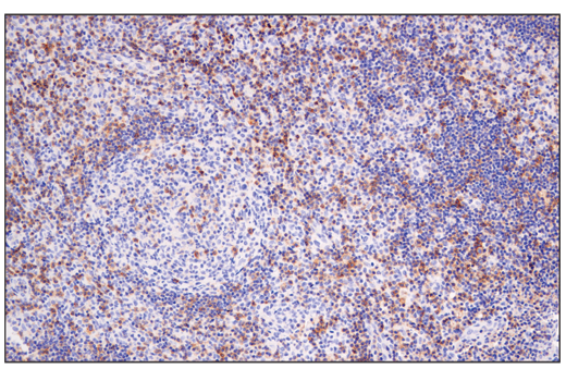
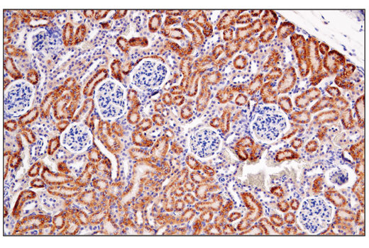
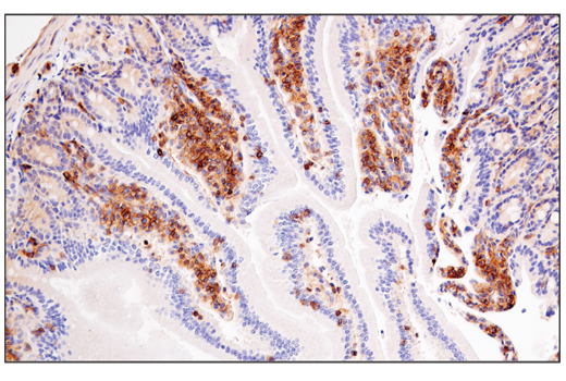
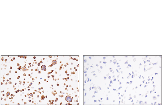
WB, IP, IHC-Bond, IHC-P
H M
Endogenous
140
Rabbit IgG
#Q92854
10507
Product Information
Product Usage Information
| Application | Dilution |
|---|---|
| Western Blotting | 1:1000 |
| Immunoprecipitation | 1:50 |
| IHC Leica Bond | 1:200 |
| Immunohistochemistry (Paraffin) | 1:400 |
Storage
For a carrier free (BSA and azide free) version of this product see product #61515.
Specificity / Sensitivity
Species Reactivity:
Human, Mouse
Source / Purification
Monoclonal antibody is produced by immunizing animals with a synthetic peptide corresponding to residues surrounding Gly814 of human Semaphorin-4D/CD100 protein.
Background
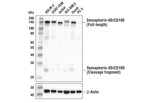
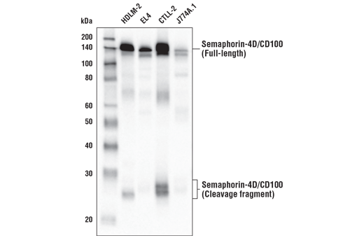
Semaphorin-4D/CD100 (Sema4D) is a disulfide-linked homodimeric type 1 transmembrane glycoprotein belonging to the class IV family of membrane bound semaphorins. The extracellular domain of Sema4D contains a cysteine-rich semaphorin-like domain, an Ig-like domain, and a PSI domain (1). Research studies have suggested that the cytoplasmic domain has a signaling function as it is phosphorylated on serine residues (2). Initial studies involving Sema4D revealed that it was implicated in axon guidance within the central nervous system through binding its high affinity receptor, plexin-B1 (3). Sema4D function has also been extensively characterized in the immune system and is the first semaphorin found to be expressed on the surface of many types of immune cells (4-6). In the immune system, CD72 serves as a low-affinity receptor for Sema4D (7) and research studies have shown that Sema4D not only regulates T-cell activation (8,9) but is also involved in the regulation of B-cell survival and differentiation (5). Many of the physiologic effects of Sema4D in the immune system are regulated by a soluble extracellular domain-containing fragment generated through proteolytic cleavage (10).
Sema4D has also been implicated in oncogenesis as research studies have demonstrated overexpression in multiple types of solid tumors (11,12). The role of Sema4D in oncogenesis, in part, has been linked to its ability to promote tumor angiogenesis (13), cell invasion (14), and immunosuppression through enhancement of myeloid derived suppressor cell function (15).
- Love, C.A. et al. (2003) Nat Struct Biol 10, 843-8.
- Elhabazi, A. et al. (1997) J Biol Chem 272, 23515-20.
- Kolodkin, A.L. et al. (1993) Cell 75, 1389-99.
- Bougeret, C. et al. (1992) J Immunol 148, 318-23.
- Hall, K.T. et al. (1996) Proc Natl Acad Sci U S A 93, 11780-5.
- Furuyama, T. et al. (1996) J Biol Chem 271, 33376-81.
- Kumanogoh, A. et al. (2000) Immunity 13, 621-31.
- Hérold, C. et al. (1995) Int Immunol 7, 1-8.
- Jiang, X. et al. (2017) Front Immunol 8, 765.
- Elhabazi, A. et al. (2001) J Immunol 166, 4341-7.
- Chen, Y. et al. (2012) Int J Mol Sci 13, 13264-74.
- Liu, H. et al. (2014) Microvasc Res 93, 1-8.
- Basile, J.R. et al. (2006) Proc Natl Acad Sci U S A 103, 9017-22.
- Kato, S. et al. (2011) Cancer Sci 102, 2029-37.
- Younis, R.H. et al. (2016) J Immunol 196, 1419-29.
Species Reactivity
Species reactivity is determined by testing in at least one approved application (e.g., western blot).
Western Blot Buffer
IMPORTANT: For western blots, incubate membrane with diluted primary antibody in 5% w/v BSA, 1X TBS, 0.1% Tween® 20 at 4°C with gentle shaking, overnight.
Applications Key
WB: Western Blotting IP: Immunoprecipitation IHC-Bond: IHC Leica Bond IHC-P: Immunohistochemistry (Paraffin)
Cross-Reactivity Key
H: human M: mouse R: rat Hm: hamster Mk: monkey Vir: virus Mi: mink C: chicken Dm: D. melanogaster X: Xenopus Z: zebrafish B: bovine Dg: dog Pg: pig Sc: S. cerevisiae Ce: C. elegans Hr: horse GP: Guinea Pig Rab: rabbit All: all species expected
Trademarks and Patents
使用に関する制限
法的な権限を与えられたCSTの担当者が署名した書面によって別途明示的に合意された場合を除き、 CST、その関連会社または代理店が提供する製品には以下の条件が適用されます。お客様が定める条件でここに定められた条件に含まれるものを超えるもの、 または、ここに定められた条件と異なるものは、法的な権限を与えられたCSTの担当者が別途書面にて受諾した場合を除き、拒絶され、 いかなる効力も効果も有しません。
研究専用 (For Research Use Only) またはこれに類似する表示がされた製品は、 いかなる目的についても FDA または外国もしくは国内のその他の規制機関により承認、認可または許可を受けていません。 お客様は製品を診断もしくは治療目的で使用してはならず、また、製品に表示された内容に違反する方法で使用してはなりません。 CST が販売または使用許諾する製品は、エンドユーザーであるお客様に対し、使途を研究および開発のみに限定して提供されるものです。 診断、予防もしくは治療目的で製品を使用することまたは製品を再販売 (単独であるか他の製品等の一部であるかを問いません) もしくはその他の商業的利用の目的で購入することについては、CST から別途許諾を得る必要があります。 お客様は以下の事項を遵守しなければなりません。(a) CST の製品 (単独であるか他の資材と一緒であるかを問いません) を販売、使用許諾、貸与、寄付もしくはその他の態様で第三者に譲渡したり使用させたりしてはなりません。また、商用の製品を製造するために CST の製品を使用してはなりません。(b) 複製、改変、リバースエンジニアリング、逆コンパイル、 分解または他の方法により製品の構造または技術を解明しようとしてはなりません。また、 CST の製品またはサービスと競合する製品またはサービスを開発する目的で CST の製品を使用してはなりません。(c) CST の製品の商標、商号、ロゴ、特許または著作権に関する通知または表示を除去したり改変したりしてはなりません。(d) CST の製品をCST 製品販売条件(CST’s Product Terms of Sale) および該当する書面のみに従って使用しなければなりません。(e) CST の製品に関連してお客様が使用する第三者の製品またはサービスに関する使用許諾条件、 サービス提供条件またはこれに類する合意事項を遵守しなければなりません。