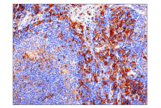
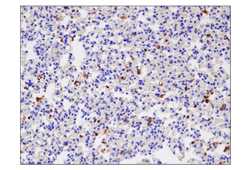
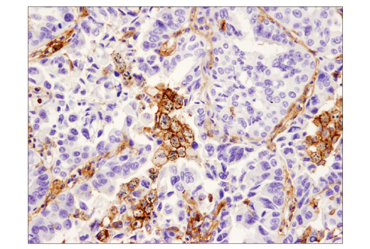
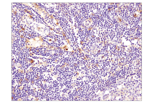
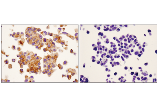
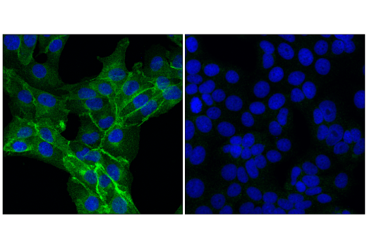
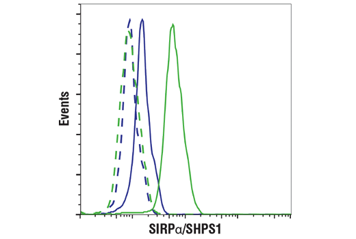
WB, IHC-Bond, IHC-P, IF-IC, FC-FP
H M R Mk
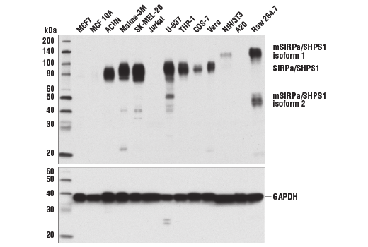
Endogenous
85 (human, monkey), 100 (rat), 120 (murine isoform 1), 55 (murine isoform 2)
Rabbit IgG
#P78324
140885
Product Information
Product Usage Information
This formulation is ideal for use with technologies requiring specialized or custom antibody labeling, including fluorophores, metals, lanthanides, and oligonucleotides. It is not recommended for ChIP, ChIP-seq, CUT&RUN, or CUT&Tag assays. If you require a carrier-free formulation for chromatin profiling, please contact us. Optimal dilutions/concentrations should be determined by the end user.
Formulation
Storage
Specificity / Sensitivity
Species Reactivity:
Human, Mouse, Rat, Monkey
Source / Purification
Monoclonal antibody is produced by immunizing animals with a synthetic peptide corresponding to residues surrounding Pro413 of human SIRPα/SHPS1 protein.
Background
SHP-substrate 1 (SHPS1, SIRPα) is a single-pass membrane protein and member of both the immunoglobulin superfamily and the signal regulatory protein (SIRP) family. Following growth hormone stimulation or integrin binding, SHPS1 is phosphorylated at several tyrosine residues within its cytoplasmic tail. These phosphorylation events promote association between SHPS1 and multiple signaling proteins, including SHP-1, SHP-2, Grb2 and Shc via their SH2 domains (1-4). Recruitment of SHP-1 and SHP-2 results in SHPS1 dephosphorylation and suppression of tyrosine kinase signaling (1-3,5). The tyrosine kinase JAK2 associates with SHPS1 via its carboxy terminus and phosphorylates SHPS1 in response to extracellular stimuli (5). Research studies show that Src associates with and may phosphorylate SHPS1 in response to insulin (4). In macrophages, SHPS1 can form a complex with the Src pathway adaptor protein SKAP2, Fyn-binding protein FYB, and the tyrosine kinase PYK2 (6). The SHPS1 extracellular domain contains at least three IgG-like domains that interact with CD47, a ubiquitously expressed, integrin-associated protein that acts as a repressive cue in both immune and neuronal cells (7,8). The interaction between CD47 and SHPS1 on opposing cells can inhibit cellular migration (9), promote "tethering" between macrophages and target cells during engulfment (10), facilitate self versus non-self recognition (11), and maintain immune homeostasis (12). SHPS1 plays a critical role in modulating the immune response and inflammation, and may play a role in neuronal development (13,14). The interaction between SHPS1 and CD47 may be an exploitable target in cancer therapy (15-17).
- Kharitonenkov, A. et al. (1997) Nature 386, 181-6.
- Ochi, F. et al. (1997) Biochem Biophys Res Commun 239, 483-7.
- Takada, T. et al. (1998) J Biol Chem 273, 9234-42.
- Shen, X. et al. (2009) Mol Cell Proteomics 8, 1539-51.
- Stofega, M.R. et al. (2000) J Biol Chem 275, 28222-9.
- Timms, J.F. et al. (1999) Curr Biol 9, 927-30.
- Seiffert, M. et al. (1999) Blood 94, 3633-43.
- Vernon-Wilson, E.F. et al. (2000) Eur J Immunol 30, 2130-7.
- Motegi, S. et al. (2003) EMBO J 22, 2634-44.
- Tada, K. et al. (2003) J Immunol 171, 5718-26.
- van Beek, E.M. et al. (2005) J Immunol 175, 7781-7.
- Legrand, N. et al. (2011) Proc Natl Acad Sci U S A 108, 13224-9.
- Sarfati, M. et al. (2008) Curr Drug Targets 9, 842-50.
- Matozaki, T. et al. (2009) Trends Cell Biol 19, 72-80.
- Hara, K. et al. (2011) Cancer Res 71, 1229-34.
- Willingham, S.B. et al. (2012) Proc Natl Acad Sci U S A 109, 6662-7.
- Weiskopf, K. et al. (2013) Science 341, 88-91.
Species Reactivity
Species reactivity is determined by testing in at least one approved application (e.g., western blot).
Applications Key
WB: Western Blotting IHC-Bond: IHC Leica Bond IHC-P: Immunohistochemistry (Paraffin) IF-IC: Immunofluorescence (Immunocytochemistry) FC-FP: Flow Cytometry (Fixed/Permeabilized)
Cross-Reactivity Key
H: human M: mouse R: rat Hm: hamster Mk: monkey Vir: virus Mi: mink C: chicken Dm: D. melanogaster X: Xenopus Z: zebrafish B: bovine Dg: dog Pg: pig Sc: S. cerevisiae Ce: C. elegans Hr: horse GP: Guinea Pig Rab: rabbit All: all species expected
Trademarks and Patents
使用に関する制限
法的な権限を与えられたCSTの担当者が署名した書面によって別途明示的に合意された場合を除き、 CST、その関連会社または代理店が提供する製品には以下の条件が適用されます。お客様が定める条件でここに定められた条件に含まれるものを超えるもの、 または、ここに定められた条件と異なるものは、法的な権限を与えられたCSTの担当者が別途書面にて受諾した場合を除き、拒絶され、 いかなる効力も効果も有しません。
研究専用 (For Research Use Only) またはこれに類似する表示がされた製品は、 いかなる目的についても FDA または外国もしくは国内のその他の規制機関により承認、認可または許可を受けていません。 お客様は製品を診断もしくは治療目的で使用してはならず、また、製品に表示された内容に違反する方法で使用してはなりません。 CST が販売または使用許諾する製品は、エンドユーザーであるお客様に対し、使途を研究および開発のみに限定して提供されるものです。 診断、予防もしくは治療目的で製品を使用することまたは製品を再販売 (単独であるか他の製品等の一部であるかを問いません) もしくはその他の商業的利用の目的で購入することについては、CST から別途許諾を得る必要があります。 お客様は以下の事項を遵守しなければなりません。(a) CST の製品 (単独であるか他の資材と一緒であるかを問いません) を販売、使用許諾、貸与、寄付もしくはその他の態様で第三者に譲渡したり使用させたりしてはなりません。また、商用の製品を製造するために CST の製品を使用してはなりません。(b) 複製、改変、リバースエンジニアリング、逆コンパイル、 分解または他の方法により製品の構造または技術を解明しようとしてはなりません。また、 CST の製品またはサービスと競合する製品またはサービスを開発する目的で CST の製品を使用してはなりません。(c) CST の製品の商標、商号、ロゴ、特許または著作権に関する通知または表示を除去したり改変したりしてはなりません。(d) CST の製品をCST 製品販売条件(CST’s Product Terms of Sale) および該当する書面のみに従って使用しなければなりません。(e) CST の製品に関連してお客様が使用する第三者の製品またはサービスに関する使用許諾条件、 サービス提供条件またはこれに類する合意事項を遵守しなければなりません。