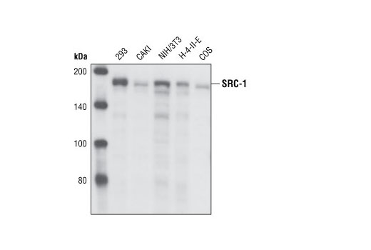
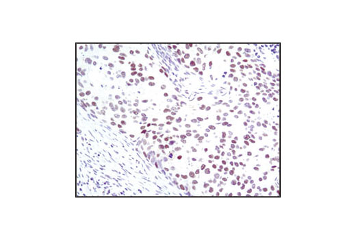
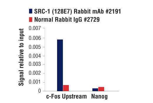
WB, IP, IHC-P, ChIP
H M R Mk
Endogenous
180
Rabbit IgG
#Q15788
8648
Product Information
Product Usage Information
For optimal ChIP results, use 5 μl of antibody and 10 μg of chromatin (approximately 4 x 10⁶ cells) per IP. This antibody has been validated using SimpleChIP® Enzymatic Chromatin IP Kits.
| Application | Dilution |
|---|---|
| Western Blotting | 1:1000 |
| Immunoprecipitation | 1:100 |
| Immunohistochemistry (Paraffin) | 1:200 |
| Chromatin IP | 1:100 |
Storage
Specificity / Sensitivity
Species Reactivity:
Human, Mouse, Rat, Monkey
Source / Purification
Monoclonal antibody is produced by immunizing animals with a synthetic peptide corresponding to the sequence of human SRC-1 protein.
Background
There are three members of the steroid receptor co-activator (SRC) family of proteins: SRC-1 (NCoA-1), SRC-2 (TIF2/GRIP1/NCoA-2), and SRC-3 (ACTR/pCIP/RAC3/TRAM-1/AIB1). All SRC family members share significant structural homology and function to stimulate transcription mediated by nuclear hormone receptors and other transcriptional activators such as Stat3, NF-κB, E2F1, and p53 (1-4). Two SRC proteins, SRC-1 and SRC-3, function as histone acetyltransferases (5,6). In addition, all three family members can recruit other histone acetyltransferases (CBP/p300, PCAF) and histone methyltransferases (PRMT1, CARM1) to target promoters and cooperate to enhance expression of many genes (5-8). The SRC proteins play important roles in multiple physiological processes including cell proliferation, cell survival, somatic cell growth, mammary gland development, female reproductive function, and vasoprotection (9). SRC-1 and SRC-3 are conduits for kinase-mediated growth factor signaling to the estrogen receptor and other transcriptional activators. Seven SRC-1 phosphorylation sites and six SRC-3 phosphorylation sites have been identified, which are induced by steroids, cytokines, and growth factors and involve multiple kinase signaling pathways (9-11). Research has shown that all three SRC family members are associated with increased activity of nuclear receptors in breast, prostate, and ovarian carcinomas. According to the literature, SRC-3 is frequently amplified or overexpressed in a number of cancers (12), and SRC-1/PAX3 and SRC-2/MYST3 translocations are found associated with rhabdomyosarcoma and acute myeloid leukemia, respectively (13,14).
- Giraud, S. et al. (2002) J. Biol. Chem. 277, 8004-8011.
- Na, S.Y. et al. (1998) J. Biol. Chem. 273, 10831-10834.
- Louie, M.C. et al. (2004) Mol. Cell Biol. 24, 5157-5171.
- Lee, S.K. et al. (1999) Mol. Endocrinol. 13, 1924-1933.
- Spencer, T.E. et al. (1997) Nature 389, 194-198.
- Chen, H. et al. (1997) Cell 90, 569-580.
- Koh, S.S. et al. (2001) J. Biol. Chem. 276, 1089-1098.
- Chen, D. et al. (1999) Science 284, 2174-2177.
- Wu, R.C. et al. (2004) Mol. Cell 15, 937-949.
- Rowan, B.G. et al. (2000) J. Biol. Chem. 275, 4475-4483.
- Zhou, H.J. et al. (2005) Cancer Res. 65, 7976-7983.
- Torres-Arzayus, M.I. et al. (2004) Cancer Cell 6, 263-274.
- Wachtel, M. et al. (2004) Cancer Res. 64, 5539-5545.
- Deguchi, K. et al. (2003) Cancer Cell 3, 259-271.
Species Reactivity
Species reactivity is determined by testing in at least one approved application (e.g., western blot).
Western Blot Buffer
IMPORTANT: For western blots, incubate membrane with diluted primary antibody in 5% w/v BSA, 1X TBS, 0.1% Tween® 20 at 4°C with gentle shaking, overnight.
Applications Key
WB: Western Blotting IP: Immunoprecipitation IHC-P: Immunohistochemistry (Paraffin) ChIP: Chromatin IP
Cross-Reactivity Key
H: human M: mouse R: rat Hm: hamster Mk: monkey Vir: virus Mi: mink C: chicken Dm: D. melanogaster X: Xenopus Z: zebrafish B: bovine Dg: dog Pg: pig Sc: S. cerevisiae Ce: C. elegans Hr: horse GP: Guinea Pig Rab: rabbit All: all species expected
Trademarks and Patents
使用に関する制限
法的な権限を与えられたCSTの担当者が署名した書面によって別途明示的に合意された場合を除き、 CST、その関連会社または代理店が提供する製品には以下の条件が適用されます。お客様が定める条件でここに定められた条件に含まれるものを超えるもの、 または、ここに定められた条件と異なるものは、法的な権限を与えられたCSTの担当者が別途書面にて受諾した場合を除き、拒絶され、 いかなる効力も効果も有しません。
研究専用 (For Research Use Only) またはこれに類似する表示がされた製品は、 いかなる目的についても FDA または外国もしくは国内のその他の規制機関により承認、認可または許可を受けていません。 お客様は製品を診断もしくは治療目的で使用してはならず、また、製品に表示された内容に違反する方法で使用してはなりません。 CST が販売または使用許諾する製品は、エンドユーザーであるお客様に対し、使途を研究および開発のみに限定して提供されるものです。 診断、予防もしくは治療目的で製品を使用することまたは製品を再販売 (単独であるか他の製品等の一部であるかを問いません) もしくはその他の商業的利用の目的で購入することについては、CST から別途許諾を得る必要があります。 お客様は以下の事項を遵守しなければなりません。(a) CST の製品 (単独であるか他の資材と一緒であるかを問いません) を販売、使用許諾、貸与、寄付もしくはその他の態様で第三者に譲渡したり使用させたりしてはなりません。また、商用の製品を製造するために CST の製品を使用してはなりません。(b) 複製、改変、リバースエンジニアリング、逆コンパイル、 分解または他の方法により製品の構造または技術を解明しようとしてはなりません。また、 CST の製品またはサービスと競合する製品またはサービスを開発する目的で CST の製品を使用してはなりません。(c) CST の製品の商標、商号、ロゴ、特許または著作権に関する通知または表示を除去したり改変したりしてはなりません。(d) CST の製品をCST 製品販売条件(CST’s Product Terms of Sale) および該当する書面のみに従って使用しなければなりません。(e) CST の製品に関連してお客様が使用する第三者の製品またはサービスに関する使用許諾条件、 サービス提供条件またはこれに類する合意事項を遵守しなければなりません。