Revision 1
Cell Signaling Technology

Orders: 877-616-CELL (2355) [email protected]

Support: 877-678-TECH (8324)

Web: [email protected] cellsignal.com

3 Trask LaneDanversMassachusetts01923USA
For Research Use Only. Not for Use in Diagnostic Procedures.
:

:

:

:

UniProt ID:

#P42224

Entrez-Gene Id:

6772

Product Information

Storage

Supplied in 10 mM sodium HEPES (pH 7.5), 150 mM NaCl, 100 µg/ml BSA, 50% glycerol and less than 0.02% sodium azide. Store at –20°C. Do not aliquot the antibody.

Specificity / Sensitivity

Phospho-Stat1 (Tyr701) (D4A7) Rabbit mAb recognizes endogenous levels of Stat1 protein only when phosphorylated at Tyr701. Stat1 antibody detects endogenous levels of total Stat1 α (91kDa) and Stat1 β (84kDa) protein.

Source / Purification

Phospho-Stat1 (Tyr701) (D4A7) monoclonal antibody is produced by immunizing animals with a synthetic peptide corresponding to residues surrounding Tyr701 of human Stat1 protein. Stat1 polyclonal antibody is produced using a synthetic peptide corresponding to a sequence of human Stat1 (Stat1 antibody). Polyclonal antibodies are purified by protein A and peptide affinity chromatography.

Background

The Stat1 transcription factor is activated in response to a large number of ligands (1) and is essential for responsiveness to IFN-α and IFN-γ (2,3). Phosphorylation of Stat1 at Tyr701 induces Stat1 dimerization, nuclear translocation, and DNA binding (4). Stat1 protein exists as a pair of isoforms, Stat1α (91 kDa) and the splice variant Stat1β (84 kDa). In most cells, both isoforms are activated by IFN-α, but only Stat1α is activated by IFN-γ. The inappropriate activation of Stat1 occurs in many tumors (5). In addition to tyrosine phosphorylation, Stat1 is also phosphorylated at Ser727 through a p38 mitogen-activated protein kinase (MAPK)-dependent pathway in response to IFN-α and other cellular stresses (6). Serine phosphorylation may be required for the maximal induction of Stat1-mediated gene activation.

  1. Heim, M.H. (1999) J Recept Signal Transduct Res 19, 75-120.
  2. Durbin, J.E. et al. (1996) Cell 84, 443-50.
  3. Meraz, M.A. et al. (1996) Cell 84, 431-42.
  4. Ihle, J.N. et al. (1994) Trends Biochem Sci 19, 222-7.
  5. Frank, D.A. (1999) Mol Med 5, 432-56.
  6. Wen, Z. et al. (1995) Cell 82, 241-50.

Species Reactivity

Species reactivity is determined by testing in at least one approved application (e.g., western blot).

Cross-Reactivity Key

H: human M: mouse R: rat Hm: hamster Mk: monkey Vir: virus Mi: mink C: chicken Dm: D. melanogaster X: Xenopus Z: zebrafish B: bovine Dg: dog Pg: pig Sc: S. cerevisiae Ce: C. elegans Hr: horse GP: Guinea Pig Rab: rabbit All: all species expected

Trademarks and Patents

Cell Signaling Technology is a trademark of Cell Signaling Technology, Inc.
PhosphoPlus is a registered trademark of Cell Signaling Technology, Inc.
All other trademarks are the property of their respective owners. Visit cellsignal.com/trademarks for more information.

使用に関する制限

法的な権限を与えられたCSTの担当者が署名した書面によって別途明示的に合意された場合を除き、 CST、その関連会社または代理店が提供する製品には以下の条件が適用されます。お客様が定める条件でここに定められた条件に含まれるものを超えるもの、 または、ここに定められた条件と異なるものは、法的な権限を与えられたCSTの担当者が別途書面にて受諾した場合を除き、拒絶され、 いかなる効力も効果も有しません。

研究専用 (For Research Use Only) またはこれに類似する表示がされた製品は、 いかなる目的についても FDA または外国もしくは国内のその他の規制機関により承認、認可または許可を受けていません。 お客様は製品を診断もしくは治療目的で使用してはならず、また、製品に表示された内容に違反する方法で使用してはなりません。 CST が販売または使用許諾する製品は、エンドユーザーであるお客様に対し、使途を研究および開発のみに限定して提供されるものです。 診断、予防もしくは治療目的で製品を使用することまたは製品を再販売 (単独であるか他の製品等の一部であるかを問いません) もしくはその他の商業的利用の目的で購入することについては、CST から別途許諾を得る必要があります。 お客様は以下の事項を遵守しなければなりません。(a) CST の製品 (単独であるか他の資材と一緒であるかを問いません) を販売、使用許諾、貸与、寄付もしくはその他の態様で第三者に譲渡したり使用させたりしてはなりません。また、商用の製品を製造するために CST の製品を使用してはなりません。(b) 複製、改変、リバースエンジニアリング、逆コンパイル、 分解または他の方法により製品の構造または技術を解明しようとしてはなりません。また、 CST の製品またはサービスと競合する製品またはサービスを開発する目的で CST の製品を使用してはなりません。(c) CST の製品の商標、商号、ロゴ、特許または著作権に関する通知または表示を除去したり改変したりしてはなりません。(d) CST の製品をCST 製品販売条件(CST’s Product Terms of Sale) および該当する書面のみに従って使用しなければなりません。(e) CST の製品に関連してお客様が使用する第三者の製品またはサービスに関する使用許諾条件、 サービス提供条件またはこれに類する合意事項を遵守しなければなりません。
Revision 1
#9170

PhosphoPlus® Stat1 (Tyr701) Antibody Kit

PhosphoPlus® Stat1 (Tyr701) Antibody Kit: Image 1 Expand Image
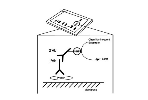
After the primary antibody is bound to the target protein, a complex with HRP-linked secondary antibody is formed. The LumiGLO® is added and emits light during enzyme catalyzed decomposition.
PhosphoPlus® Stat1 (Tyr701) Antibody Kit: Image 2 Expand Image
After the primary antibody is bound to the target protein, a complex with HRP-linked secondary antibody is formed. The LumiGLO® is added and emits light during enzyme catalyzed decomposition.
PhosphoPlus® Stat1 (Tyr701) Antibody Kit: Image 3 Expand Image
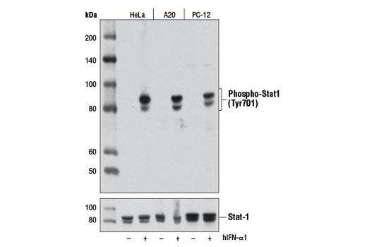
Western blot analysis of extracts from HeLa, A20, and PC-12 cells, untreated or treated with Human Interferon-α1 (hIFN-α1) #8927 (10 ng/ml, 30 min), using Phospho-Stat1 (Tyr701) (D4A7) Rabbit mAb (upper) or Stat1 Antibody #9172 (lower).
PhosphoPlus® Stat1 (Tyr701) Antibody Kit: Image 4 Expand Image
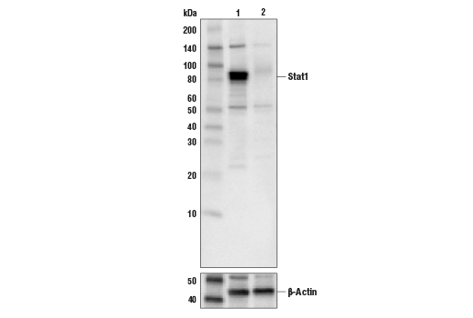
Western blot analysis of extracts from A549 cells (lane 1) or STAT1 knock-out cells (lane 2) using Stat1 Antibody #9172 (upper), and β-actin (D6A8) Rabbit mAb #8457 (lower). The absence of signal in the STAT1 knock-out A549 cells confirms specificity of the antibody for STAT1.
PhosphoPlus® Stat1 (Tyr701) Antibody Kit: Image 5 Expand Image
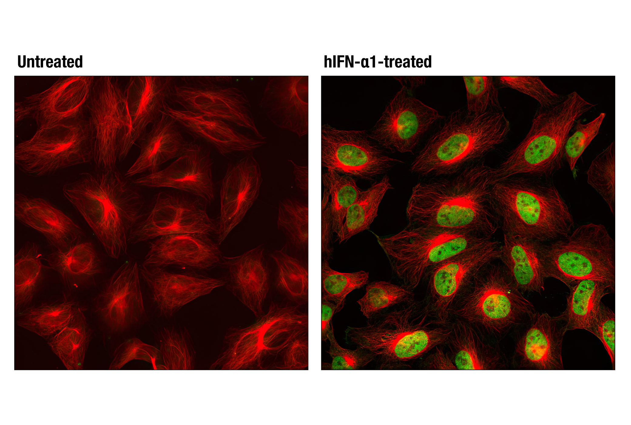
Confocal immunofluorescent analysis of HeLa cells, untreated (left) or treated with hIFN-α1 #8927 (100 ng/mL, 30 min; right), using Phospho-Stat1 (Tyr701) (D4A7) Rabbit mAb (green) and β-Tubulin (9F3) Rabbit mAb (Alexa Fluor® 555 Conjugate) #2116 (red).
PhosphoPlus® Stat1 (Tyr701) Antibody Kit: Image 6 Expand Image
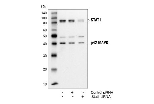
Western blot analysis of extracts from HeLa cells 48 hours following mock transfection, transfection with non-targeted (control) siRNA or transfection with Stat1 siRNA. Stat1 was detected using Stat1 Antibody #9172, and p42 was detected using p42 MAPK Antibody #9108. The Stat1 Antibody confirms silencing of Stat1 expression, and the p42 MAPK Antibody is used to control for protein loading and siRNA specificity.
PhosphoPlus® Stat1 (Tyr701) Antibody Kit: Image 7 Expand Image
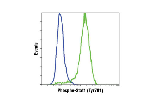
Flow cytometric analysis of Jurkat cells, untreated (blue) or treated with hIFN-α1 #8927 (green), using Phospho-Stat1 (Tyr701) (D4A7) Rabbit mAb.
PhosphoPlus® Stat1 (Tyr701) Antibody Kit: Image 8 Expand Image
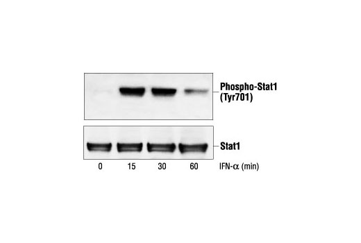
Western blot analysis of extracts from SK-MEL-28 cells, untreated or IFN-alpha-treated (100 ng/ml), using Phospho-Stat1 (Tyr701) Antibody #9171 (upper) or Stat1 Antibody (lower).
PhosphoPlus® Stat1 (Tyr701) Antibody Kit: Image 9 Expand Image
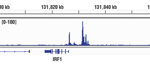
Chromatin immunoprecipitations were performed with cross-linked chromatin from HT-1080 cells treated with IFN-γ (50 ng/ml) for 30 minutes and Phospho-Stat1 (Tyr701) (D4A7) Rabbit mAb, using SimpleChIP® Enzymatic Chromatin IP Kit (Magnetic Beads) #9003. DNA Libraries were prepared using DNA Library Prep Kit for Illumina® (ChIP-seq, CUT&RUN) #56795. The figure shows binding across IRF-1, a known target gene of Phospho-Stat1 (see additional figure containing ChIP-qPCR data). For additional ChIP-seq tracks, please download the product datasheet.
PhosphoPlus® Stat1 (Tyr701) Antibody Kit: Image 10 Expand Image
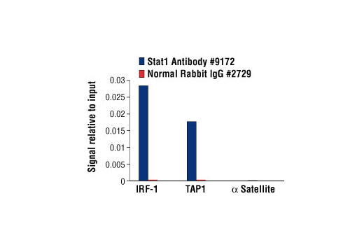
Chromatin immunoprecipitations were performed with cross-linked chromatin from HT-1080 cells treated with IFN-γ (50 ng/ml) for 30 minutes and either Stat1 Antibody or Normal Rabbit IgG #2729 using SimpleChIP® Enzymatic Chromatin IP Kit (Magnetic Beads) #9003. The enriched DNA was quantified by real-time PCR using human IRF-1 promoter primers, SimpleChIP® Human TAP1 Promoter Primers #5148, and SimpleChIP® Human α Satellite Repeat Primers #4486. The amount of immunoprecipitated DNA in each sample is represented as signal relative to the total amount of input chromatin, which is equivalent to one.
PhosphoPlus® Stat1 (Tyr701) Antibody Kit: Image 11 Expand Image
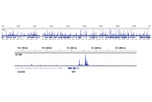
Chromatin immunoprecipitations were performed with cross-linked chromatin from 4 x 10⁶ HT-1080 cells treated with IFN-γ (50 ng/ml) for 30 minutes and 5 μl of Phospho-Stat1 (Tyr701) (D4A7) Rabbit mAb, using SimpleChIP® Enzymatic Chromatin IP Kit (Magnetic Beads) #9003. DNA Libraries were prepared using DNA Library Prep Kit for Illumina® (ChIP-seq, CUT&RUN) #56795. The figure shows binding across chromosome 5 (upper), including IRF1 (lower), a known target gene of Phospho-Stat1 (see additional figure containing ChIP-qPCR data).
PhosphoPlus® Stat1 (Tyr701) Antibody Kit: Image 12 Expand Image
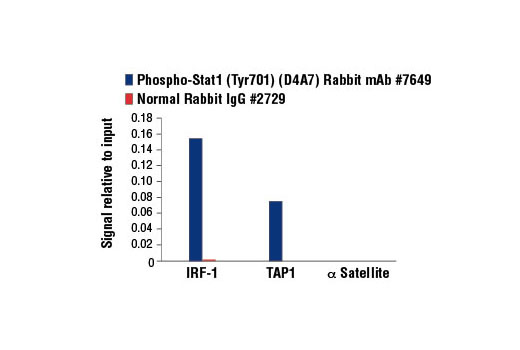
Chromatin immunoprecipitations were performed with cross-linked chromatin from HT-1080 cells treated with IFN-γ (50 ng/ml) for 30 minutes and either Phospho-Stat1 (Tyr701) (D4A7) Rabbit mAb or Normal Rabbit IgG #2729 using SimpleChIP® Enzymatic Chromatin IP Kit (Magnetic Beads) #9003. The enriched DNA was quantified by real-time PCR using human IRF-1 promoter primers, SimpleChIP® Human TAP1 Promoter Primers #5148, and SimpleChIP® Human α Satellite Repeat Primers #4486. The amount of immunoprecipitated DNA in each sample is represented as signal relative to the total amount of input chromatin, which is equivalent to one.