| Product Includes | Product # | Quantity | Mol. Wt | Isotype/Source |
|---|---|---|---|---|
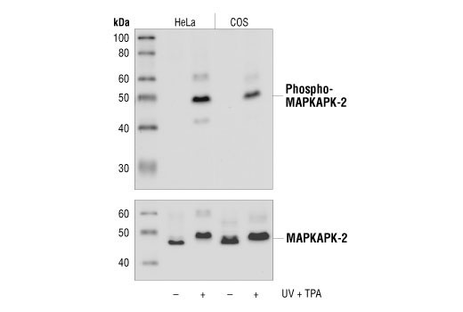
| Phospho-MAPKAPK-2 (Thr334) (27B7) Rabbit mAb | 3007 | 20 µl | 49 kDa | Rabbit IgG |
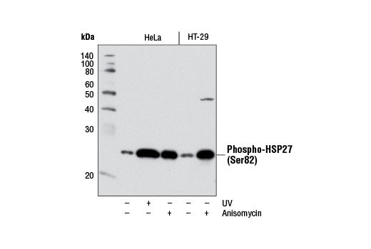
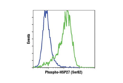
| Phospho-HSP27 (Ser82) (D1H2F6) XP® Rabbit mAb | 9709 | 20 µl | 27 kDa | Rabbit IgG |
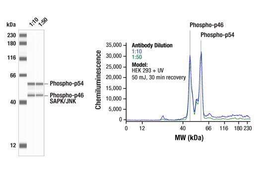
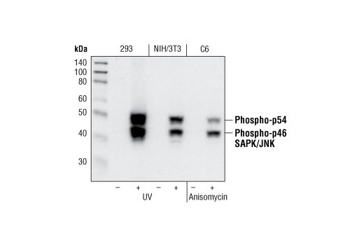
| Phospho-SAPK/JNK (Thr183/Tyr185) (81E11) Rabbit mAb | 4668 | 20 µl | 46, 54 kDa | Rabbit IgG |
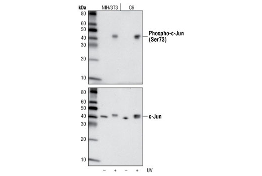
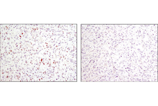
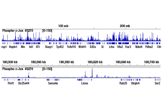
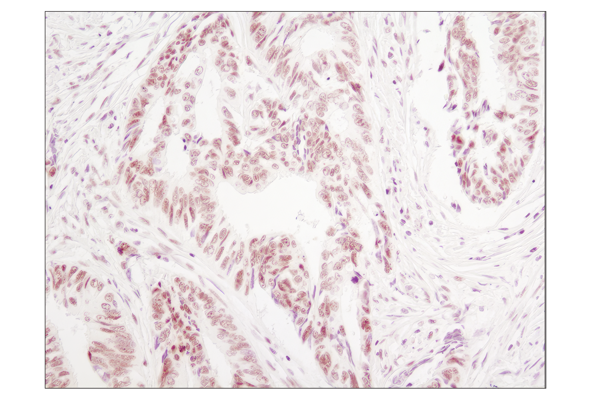
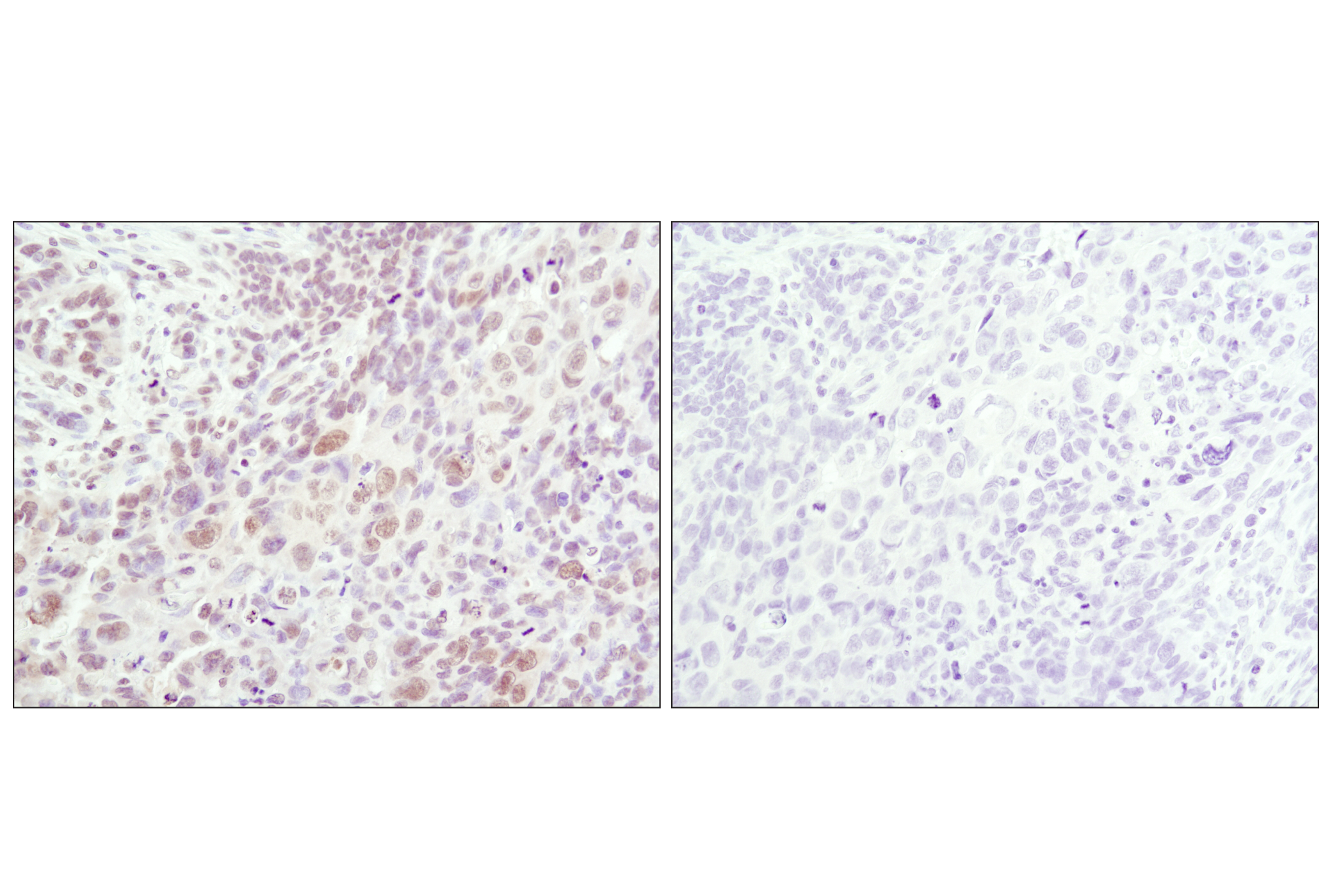
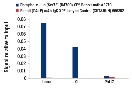
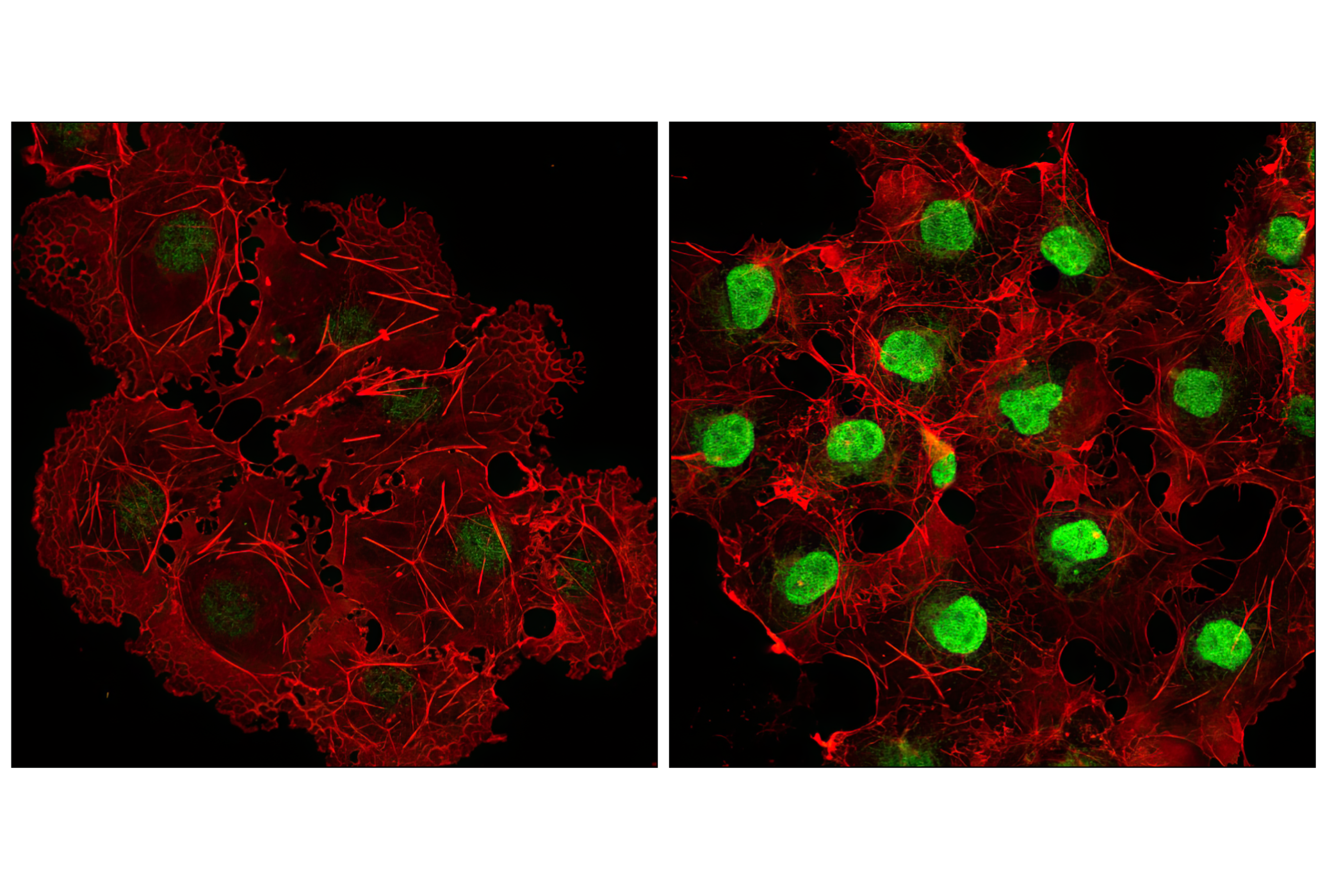
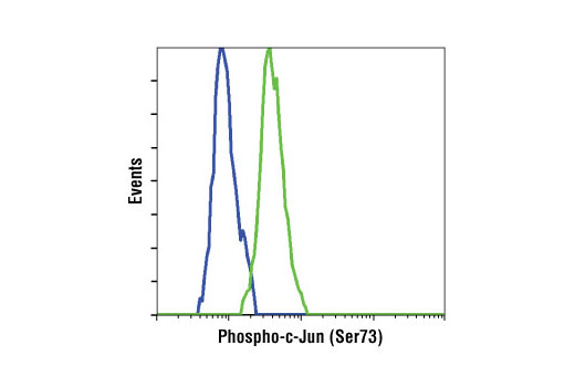
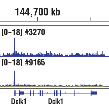
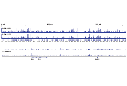
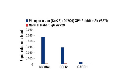
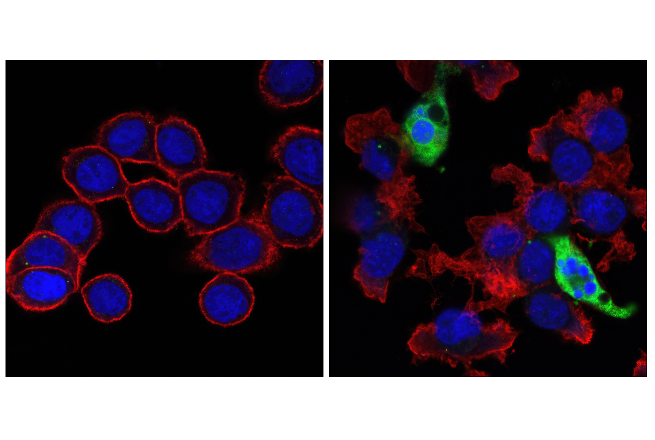
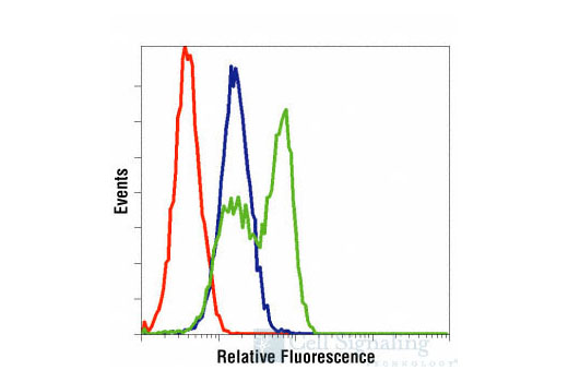
| Phospho-c-Jun (Ser73) (D47G9) XP® Rabbit mAb | 3270 | 20 µl | 48 kDa | Rabbit IgG |
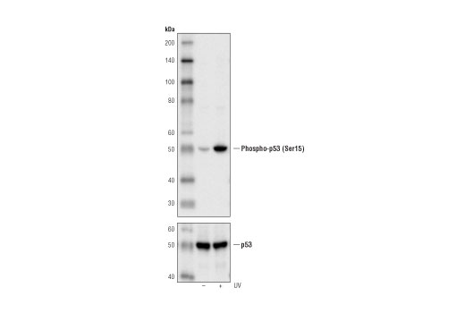
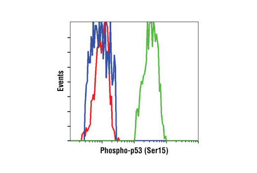
| Phospho-p53 (Ser15) (16G8) Mouse mAb | 9286 | 20 µl | 53 kDa | Mouse IgG1 |
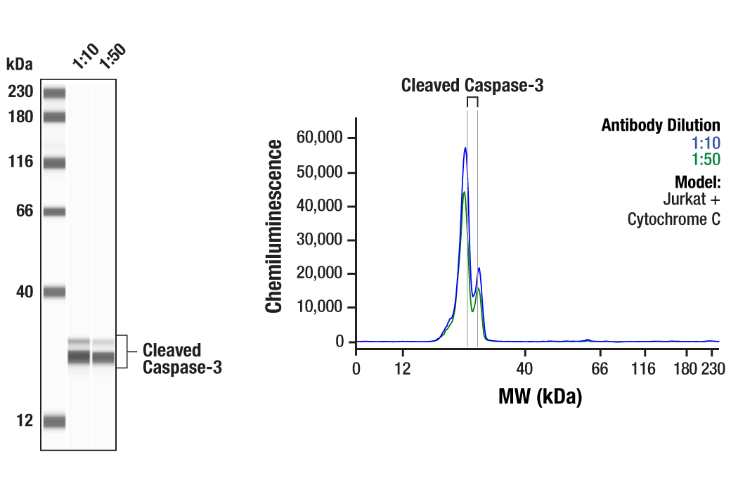
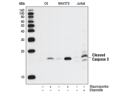
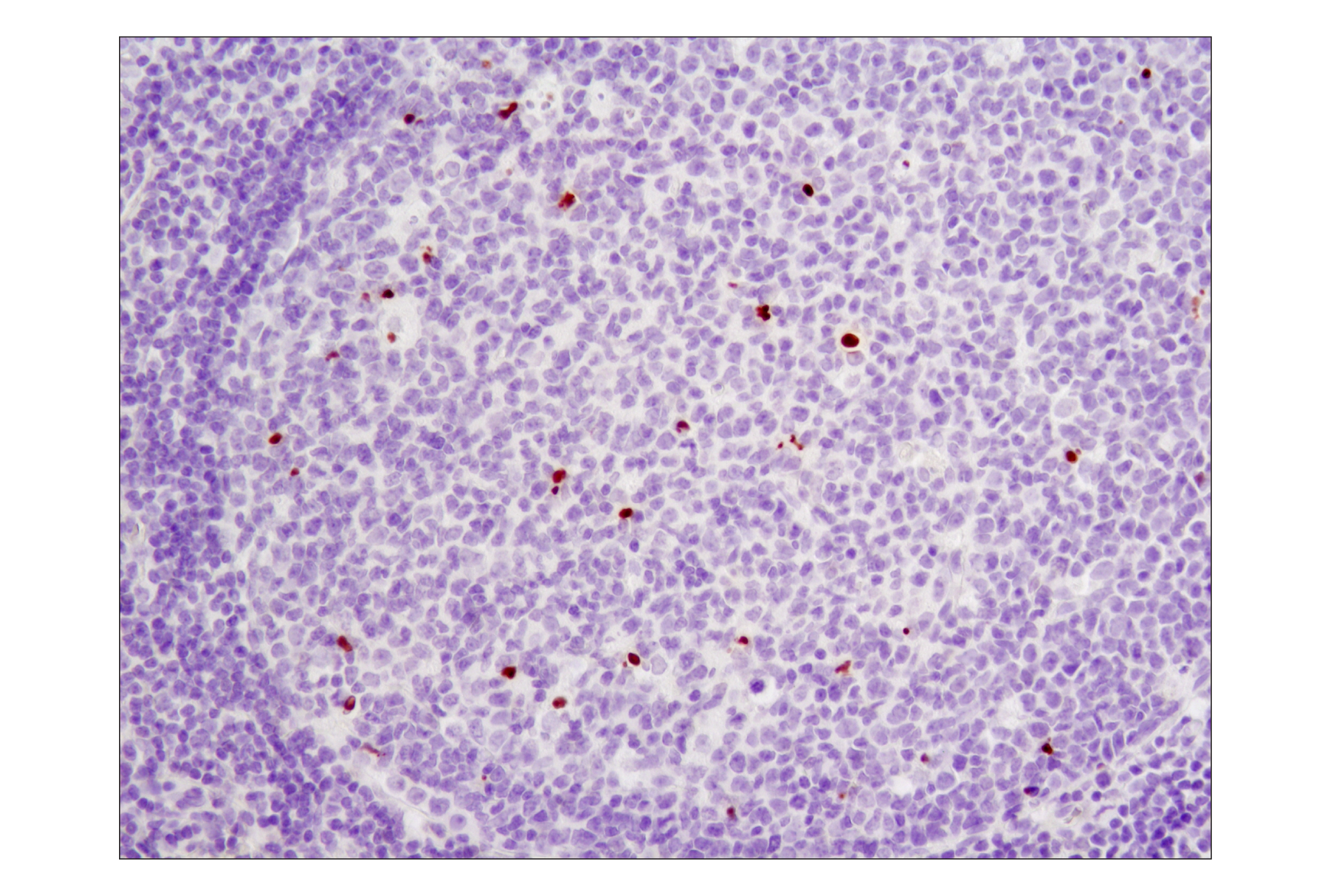
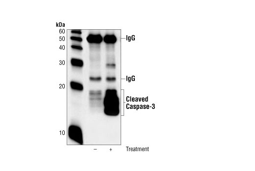
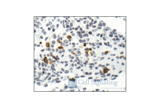
| Cleaved Caspase-3 (Asp175) (5A1E) Rabbit mAb | 9664 | 20 µl | 17, 19 kDa | Rabbit IgG |
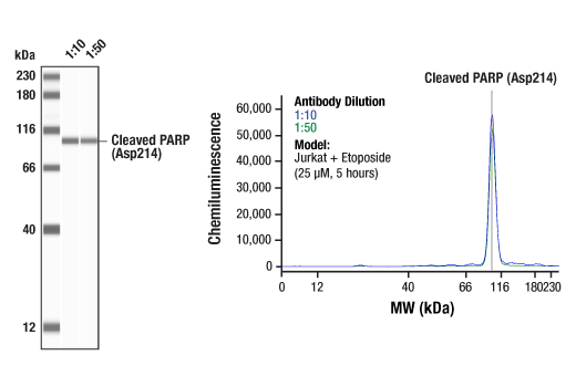
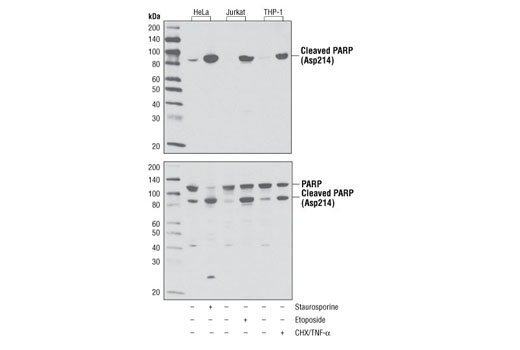
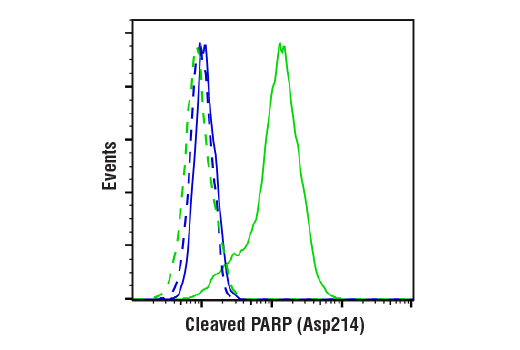
| Cleaved PARP (Asp214) (D64E10) XP® Rabbit mAb | 5625 | 20 µl | 89 kDa | Rabbit IgG |
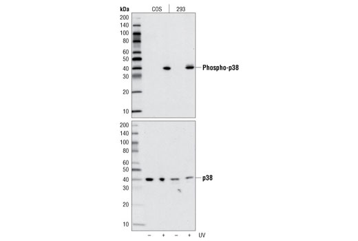
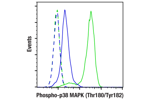
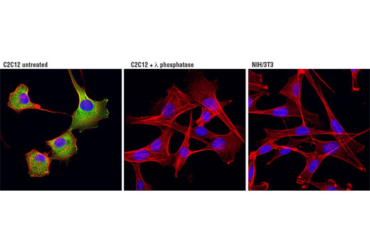
| Phospho-p38 MAPK (Thr180/Tyr182) (D3F9) XP® Rabbit mAb | 4511 | 20 µl | 43 kDa | Rabbit IgG |
| Anti-rabbit IgG, HRP-linked Antibody | 7074 | 100 µl | Goat | |
| Anti-mouse IgG, HRP-linked Antibody | 7076 | 100 µl | Horse |
Please visit cellsignal.com for individual component applications, species cross-reactivity, dilutions, protocols, and additional product information.
Description
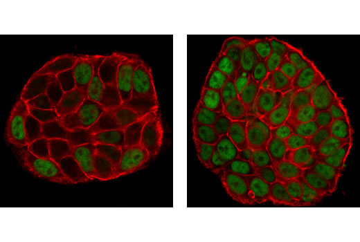
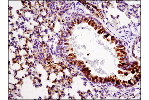
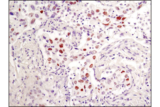
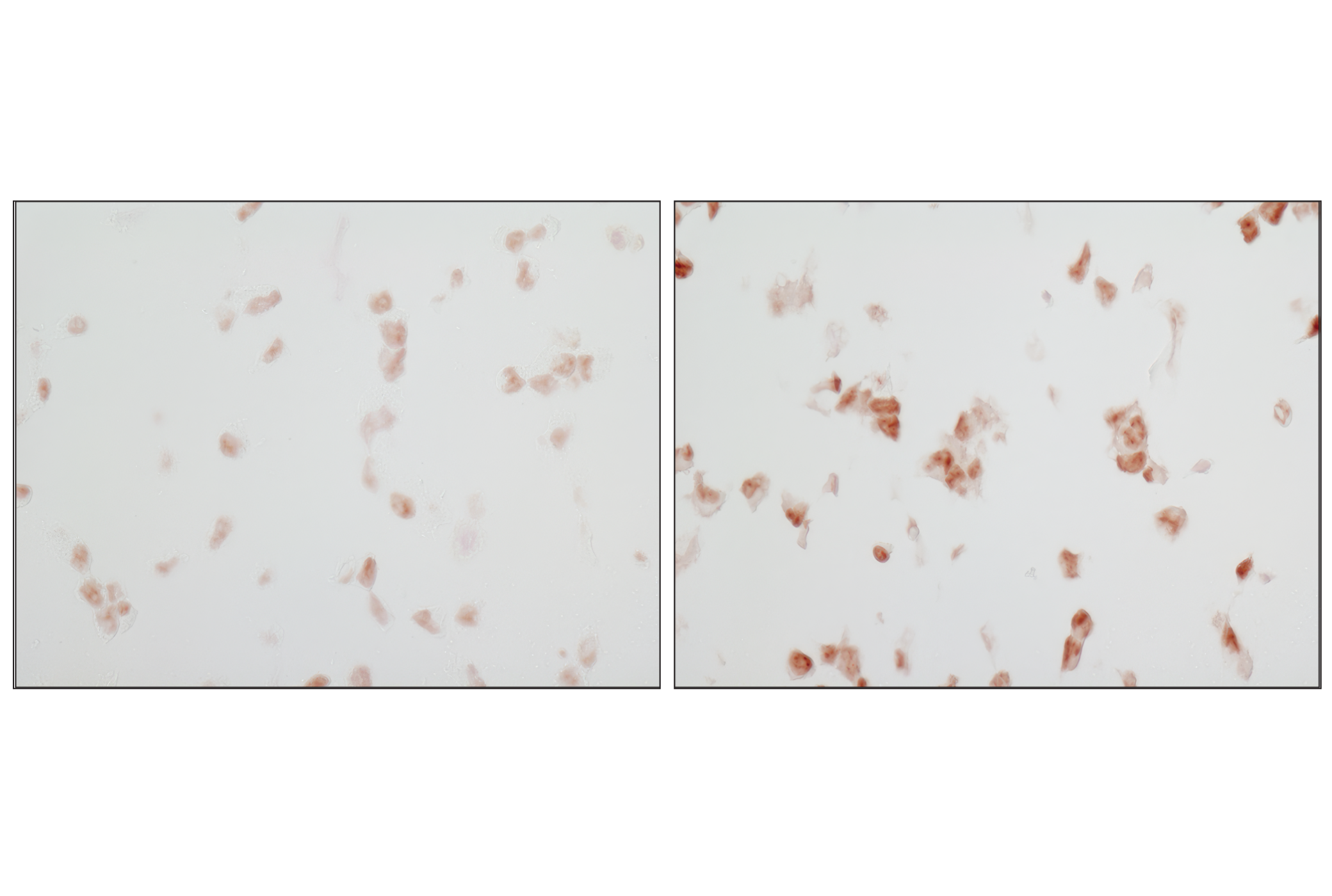
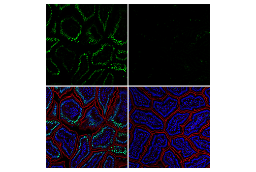
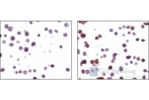
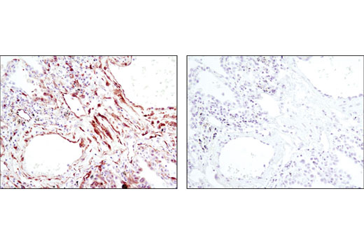
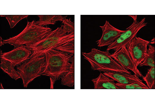
The Stress and Apoptosis Antibody Sampler Kit provides an economical means of evaluating stress and apoptotic responses of each protein. The kit contains enough primary and secondary antibody to perform two western blot experiments per primary antibody.
Storage
Background
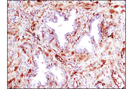
Cells respond to environmental or intracellular stresses through various mechanisms ranging from initiation of prosurvival strategies to activation of cell death pathways that remove damaged cells from the organism. Many of the proteins and cellular processes involved in normal signaling and survival pathways also play dual roles in cell death-promoting mechanisms. Apoptosis is a regulated cellular suicide mechanism characterized by nuclear condensation, cell shrinkage, membrane blebbing, and DNA fragmentation. Caspase-3 (CPP-32, Apoptain, Yama, SCA-1) is a critical executioner of apoptosis, as it is either partially or totally responsible for the proteolytic cleavage of many key proteins such as the nuclear enzyme poly (ADP-ribose) polymerase (PARP) (1). PARP appears to be involved in DNA repair in response to environmental stress (2). This protein can be cleaved by many ICE-like caspases in vitro (3,4) and is one of the main cleavage targets of caspase-3 in vivo (5,6). PARP helps cells to maintain their viability; cleavage of PARP facilitates cellular disassembly and serves as a marker of cells undergoing apoptosis (7). The p53 tumor suppressor protein plays a major role in cellular response to DNA damage and other genomic aberrations. Activation of p53 can lead to either cell cycle arrest and DNA repair or apoptosis (8). DNA damage induces phosphorylation of p53 at Ser15 and Ser20 and leads to a reduced interaction between p53 and its negative regulator, the oncoprotein MDM2 (9). MDM2 inhibits p53 accumulation by targeting it for ubiquitination and proteasomal degradation (10,11). Stress-activated protein kinases (SAPK)/Jun amino-terminal kinases (JNK) are members of the MAPK family that are activated by a variety of environmental stresses, inflammatory cytokines, growth factors, and GPCR agonists. Stress signals are delivered to this cascade by small GTPases of the Rho family (Rac, Rho, cdc42) (12). SAPK/JNK, when active as a dimer, can translocate to the nucleus and regulate transcription through its effects on c-Jun, ATF-2, and other transcription factors (12,13). c-Jun is a member of the Jun Family, containing c-Jun, JunB, and JunD, and is a component of the transcription factor AP-1 (activator protein-1). Extracellular signals from growth factors, chemokines, and stress activate AP-1-dependent transcription. The transcriptional activity of c-Jun is regulated by phosphorylation at Ser63 and Ser73 through SAPK/JNK (reviewed in 14). AP-1 regulated genes exert diverse biological functions including cell proliferation, differentiation, and apoptosis, as well as transformation, invasion and metastasis, depending on cell type and context (13, 15-17). p38 MAP kinase (MAPK), also called RK (18) or CSBP (19), is the mammalian orthologue of the yeast HOG kinase that participates in a signaling cascade controlling cellular responses to cytokines and stress (17-20). MKK3, MKK6, and SEK activate p38 MAP kinase by phosphorylation at Thr180 and Tyr182. MAPKAPK-2 is a direct target of p38 MAPK (17). Multiple residues of MAPKAPK-2 are phosphorylated in vivo in response to stress. However, only four residues (Thr25, Thr222, Ser272 and Thr334) are phosphorylated by p38 MAPK in an in vitro kinase assay (21). Phosphorylation at Thr222, Ser272, and Thr334 appears to be essential for the activity of MAPKAPK-2 (6). Heat shock protein (HSP) 27 is one of the small HSPs that are constitutively expressed at different levels in various cell types and tissues. In response to stress, the expression level of HSP27 increases several-fold to confer cellular resistance to the adverse environmental change. HSP27 is phosphorylated at Ser15, Ser78, and Ser82 by MAPKAPK-2 as a result of the activation of the p38 MAP kinase pathway (19,22).
- Fernandes-Alnemri, T. et al. (1994) J Biol Chem 269, 30761-4.
- Satoh, M.S. and Lindahl, T. (1992) Nature 356, 356-8.
- Lazebnik, Y.A. et al. (1994) Nature 371, 346-7.
- Cohen, G.M. (1997) Biochem J 326 ( Pt 1), 1-16.
- Nicholson, D.W. et al. (1995) Nature 376, 37-43.
- Tewari, M. et al. (1995) Cell 81, 801-9.
- Oliver, F.J. et al. (1998) J Biol Chem 273, 33533-9.
- Levine, A.J. (1997) Cell 88, 323-31.
- Shieh, S.Y. et al. (1997) Cell 91, 325-34.
- Chehab, N.H. et al. (1999) Proc Natl Acad Sci U S A 96, 13777-82.
- Honda, R. et al. (1997) FEBS Lett 420, 25-7.
- Kyriakis, J.M. and Avruch, J. (2001) Physiol Rev 81, 807-69.
- Leppä, S. and Bohmann, D. (1999) Oncogene 18, 6158-62.
- Davis, R.J. (2000) Cell 103, 239-52.
- Shaulian, E. and Karin, M. (2002) Nat Cell Biol 4, E131-6.
- Weiss, C. and Bohmann, D. (2004) Cell Cycle 3, 111-3.
- Rouse, J. et al. (1994) Cell 78, 1027-37.
- Han, J. et al. (1994) Science 265, 808-11.
- Lee, J.C. et al. Nature 372, 739-46.
- Freshney, N.W. et al. (1994) Cell 78, 1039-49.
- Ben-Levy, R. et al. (1995) EMBO J 14, 5920-30.
- Landry, J. et al. (1992) J Biol Chem 267, 794-803.
Background References
Trademarks and Patents
使用に関する制限
法的な権限を与えられたCSTの担当者が署名した書面によって別途明示的に合意された場合を除き、 CST、その関連会社または代理店が提供する製品には以下の条件が適用されます。お客様が定める条件でここに定められた条件に含まれるものを超えるもの、 または、ここに定められた条件と異なるものは、法的な権限を与えられたCSTの担当者が別途書面にて受諾した場合を除き、拒絶され、 いかなる効力も効果も有しません。
研究専用 (For Research Use Only) またはこれに類似する表示がされた製品は、 いかなる目的についても FDA または外国もしくは国内のその他の規制機関により承認、認可または許可を受けていません。 お客様は製品を診断もしくは治療目的で使用してはならず、また、製品に表示された内容に違反する方法で使用してはなりません。 CST が販売または使用許諾する製品は、エンドユーザーであるお客様に対し、使途を研究および開発のみに限定して提供されるものです。 診断、予防もしくは治療目的で製品を使用することまたは製品を再販売 (単独であるか他の製品等の一部であるかを問いません) もしくはその他の商業的利用の目的で購入することについては、CST から別途許諾を得る必要があります。 お客様は以下の事項を遵守しなければなりません。(a) CST の製品 (単独であるか他の資材と一緒であるかを問いません) を販売、使用許諾、貸与、寄付もしくはその他の態様で第三者に譲渡したり使用させたりしてはなりません。また、商用の製品を製造するために CST の製品を使用してはなりません。(b) 複製、改変、リバースエンジニアリング、逆コンパイル、 分解または他の方法により製品の構造または技術を解明しようとしてはなりません。また、 CST の製品またはサービスと競合する製品またはサービスを開発する目的で CST の製品を使用してはなりません。(c) CST の製品の商標、商号、ロゴ、特許または著作権に関する通知または表示を除去したり改変したりしてはなりません。(d) CST の製品をCST 製品販売条件(CST’s Product Terms of Sale) および該当する書面のみに従って使用しなければなりません。(e) CST の製品に関連してお客様が使用する第三者の製品またはサービスに関する使用許諾条件、 サービス提供条件またはこれに類する合意事項を遵守しなければなりません。