Revision 3
Cell Signaling Technology

Orders: 877-616-CELL (2355) [email protected]

Support: 877-678-TECH (8324)

Web: [email protected] cellsignal.com

3 Trask LaneDanversMassachusetts01923USA
For Research Use Only. Not for Use in Diagnostic Procedures.
:

:

:

:

UniProt ID:

#Q9GZV5

Entrez-Gene Id:

25937

Please visit cellsignal.com for individual component applications, species cross-reactivity, dilutions, protocols, and additional product information.

Description

PhosphoPlus® Duets from Cell Signaling Technology (CST) provide a means to assess protein activation status. Each Duet contains an activation-state and total protein antibody to your target of interest. These antibodies have been selected from CST's product offering based upon superior performance in specified applications.

Storage

Supplied in 10 mM sodium HEPES (pH 7.5), 150 mM NaCl, 100 µg/ml BSA, 50% glycerol and less than 0.02% sodium azide. Store at –20°C. Do not aliquot the antibodies.

Background

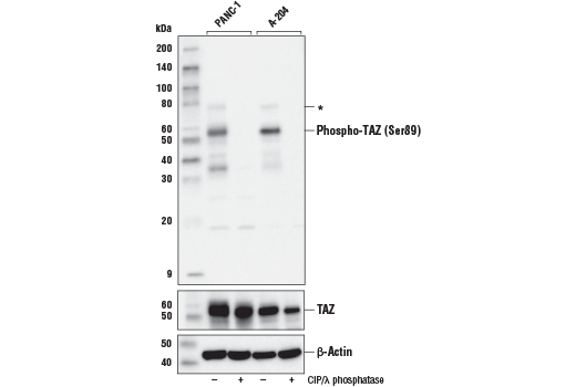
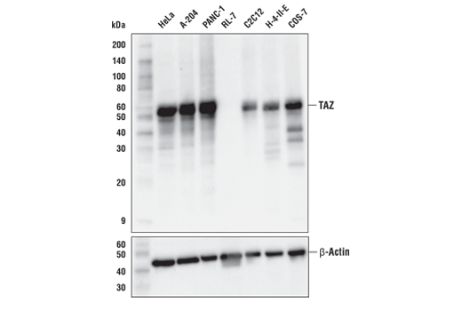
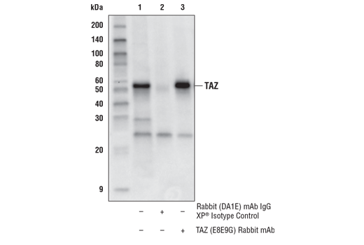
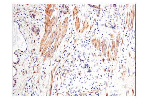
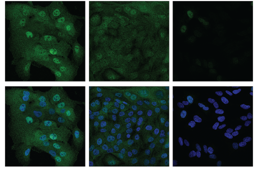
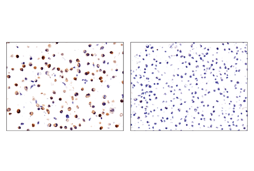
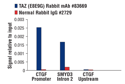
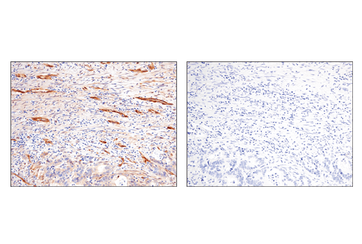
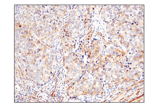
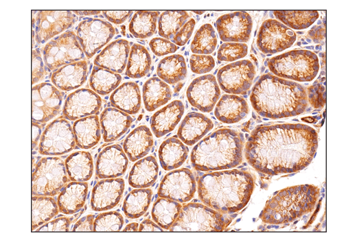
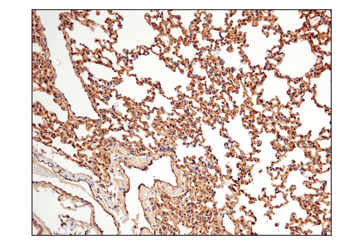
TAZ is a transcriptional co-activator with a PDZ-binding motif that is regulated by its interaction with 14-3-3 proteins (1). TAZ shares homology with the WW domain of Yes-associated protein (YAP) (1). TAZ is proposed to modulate the switch between proliferation and differentiation of mesenchymal stem cells (MSC) via interaction with transcription factors Runx2 and PPARγ. This process is critical to normal tissue development and the prevention of tumor formation. Due to its role in determination of MSC fate, TAZ may have clinical relevance to several human diseases caused by an imbalance of MSC differentiation (2,3). TAZ is negatively regulated via phosphorylation by LATS1/2, core kinases in the Hippo signaling pathway that controls stem cell development, tissue growth and tumor development (4).
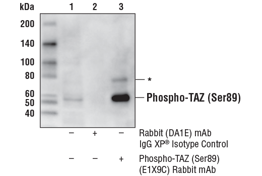
Phosphorylation of TAZ at Ser89 functions to destabilize TAZ protein by promoting 14-3-3 binding, cytoplasmic sequestration, and proteasomal degradation, thereby reducing the ability of TAZ to co-activate transcription of downstream target genes. Mutation of Ser89 to alanine (S89A) yields a constitutively active form of TAZ; expression of TAZ (S89A) in breast cancer cells was shown to promote a cancer stem cell phenotype (5).

  1. Kanai, F. et al. (2000) EMBO J 19, 6778-91.
  2. Hong, J.H. et al. (2005) Science 309, 1074-8.
  3. Hong, J.H. and Yaffe, M.B. (2006) Cell Cycle 5, 176-9.
  4. Lei, Q.Y. et al. (2008) Mol Cell Biol 28, 2426-36.
  5. Cordenonsi, M. et al. (2011) Cell 147, 759-72.

Background References

    Trademarks and Patents

    Cell Signaling Technology is a trademark of Cell Signaling Technology, Inc.
    PhosphoPlus is a trademark of Cell Signaling Technology, Inc.
    All other trademarks are the property of their respective owners. Visit cellsignal.com/trademarks for more information.

    使用に関する制限

    法的な権限を与えられたCSTの担当者が署名した書面によって別途明示的に合意された場合を除き、 CST、その関連会社または代理店が提供する製品には以下の条件が適用されます。お客様が定める条件でここに定められた条件に含まれるものを超えるもの、 または、ここに定められた条件と異なるものは、法的な権限を与えられたCSTの担当者が別途書面にて受諾した場合を除き、拒絶され、 いかなる効力も効果も有しません。

    研究専用 (For Research Use Only) またはこれに類似する表示がされた製品は、 いかなる目的についても FDA または外国もしくは国内のその他の規制機関により承認、認可または許可を受けていません。 お客様は製品を診断もしくは治療目的で使用してはならず、また、製品に表示された内容に違反する方法で使用してはなりません。 CST が販売または使用許諾する製品は、エンドユーザーであるお客様に対し、使途を研究および開発のみに限定して提供されるものです。 診断、予防もしくは治療目的で製品を使用することまたは製品を再販売 (単独であるか他の製品等の一部であるかを問いません) もしくはその他の商業的利用の目的で購入することについては、CST から別途許諾を得る必要があります。 お客様は以下の事項を遵守しなければなりません。(a) CST の製品 (単独であるか他の資材と一緒であるかを問いません) を販売、使用許諾、貸与、寄付もしくはその他の態様で第三者に譲渡したり使用させたりしてはなりません。また、商用の製品を製造するために CST の製品を使用してはなりません。(b) 複製、改変、リバースエンジニアリング、逆コンパイル、 分解または他の方法により製品の構造または技術を解明しようとしてはなりません。また、 CST の製品またはサービスと競合する製品またはサービスを開発する目的で CST の製品を使用してはなりません。(c) CST の製品の商標、商号、ロゴ、特許または著作権に関する通知または表示を除去したり改変したりしてはなりません。(d) CST の製品をCST 製品販売条件(CST’s Product Terms of Sale) および該当する書面のみに従って使用しなければなりません。(e) CST の製品に関連してお客様が使用する第三者の製品またはサービスに関する使用許諾条件、 サービス提供条件またはこれに類する合意事項を遵守しなければなりません。

    Revision 3
    #80074

    PhosphoPlus® TAZ (Ser89) Antibody Duet

    PhosphoPlus® TAZ (Ser89) Antibody Duet: Image 1 Expand Image
    Western blot analysis of extracts from PANC-1 and A-204 cells, untreated (-) or treated with calf intestinal phosphatase (CIP) and λ phosphatase (+) using Phospho-TAZ (Ser89) (E1X9C) Rabbit mAb (upper), TAZ (D3I6D) Rabbit MAb #70148 (middle) and β-Actin (D6A8) Rabbit mAb #8457 (lower). Asterisk (*) indicates weak detection of phosphorylated YAP, due to sequence similarity in the regions surrounding TAZ (Ser89) and YAP (Ser127).
    PhosphoPlus® TAZ (Ser89) Antibody Duet: Image 2 Expand Image
    Western blot analysis of extracts from various cell lines using TAZ (E8E9G) Rabbit mAb (upper) and β-Actin (D6A8) Rabbit mAb #8457 (lower). Expression levels of TAZ among cell lines are consistent with expectations based on publicly available bioinformatic databases.
    PhosphoPlus® TAZ (Ser89) Antibody Duet: Image 3 Expand Image
    Immunoprecipitation of Phospho-TAZ (Ser89) from A-204 cell extracts. Lane 1 is 10% input, lane 2 is Rabbit (DA1E) mAb IgG XP® Isotype Control #3900, and lane 3 is Phospho-TAZ (Ser89) (E1X9C) Rabbit mAb. Western blot analysis was performed using Phospho-TAZ (Ser89) (E1X9C) Rabbit mAb. Mouse Anti-Rabbit IgG (Conformation Specific) (L27A9) mAb (HRP Conjugate) #5127 was used as the secondary antibody. Asterisk (*) indicates weak detection of phosphorylated YAP, due to sequence similarity in the regions surrouding TAZ (Ser89) and YAP (Ser127).
    PhosphoPlus® TAZ (Ser89) Antibody Duet: Image 4 Expand Image
    Immunoprecipitation of TAZ protein from HeLa cell extracts. Lane 1 is 10% input, lane 2 is Rabbit (DA1E) mAb IgG XP® Isotype Control #3900, and lane 3 is TAZ (E8E9G) Rabbit mAb. Western blot analysis was performed using TAZ (D3I6D) Rabbit mAb #70148. Mouse anti-rabbit IgG (Conformation Specific) (L27A9) mAb (HRP Conjugate) #5127 was used for detection to avoid cross-reactivity with IgG.
    PhosphoPlus® TAZ (Ser89) Antibody Duet: Image 5 Expand Image
    Immunohistochemical analysis of paraffin-embedded human urothelial carcinoma using Phospho-TAZ (Ser89) (E1X9C) Rabbit mAb.
    PhosphoPlus® TAZ (Ser89) Antibody Duet: Image 6 Expand Image
    Confocal immunofluorescent analysis of MCF 10A cells (high-expressing) at low (left) and high-confluence (middle) versus SH-SY5Y cells (right, low-expressing) using TAZ (E8E9G) Rabbit mAb (green). Nuclei are labeled with DAPI #4083 (blue). Note that decreased nuclear localization of TAZ protein is seen in high-confluence MCF 10A cells.
    PhosphoPlus® TAZ (Ser89) Antibody Duet: Image 7 Expand Image
    Immunohistochemical analysis of paraffin-embedded SK-MEL-28 cell pellet (left, positive) or RL cell pellet (right, negative) using Phospho-TAZ (Ser89) (E1X9C) Rabbit mAb.
    PhosphoPlus® TAZ (Ser89) Antibody Duet: Image 8 Expand Image
    Chromatin immunoprecipitations were performed with cross-linked chromatin from NCI-H2052 cells and either TAZ (E8E9G) Rabbit mAb #83669 or Normal Rabbit IgG #2729 using SimpleChIP® Plus Enzymatic Chromatin IP Kit (Magnetic Beads) #9005. The enriched DNA was quantified by real-time PCR using SimpleChIP® Human CTGF Promoter Primers #14927, human SMYD3 intron 2 primers, and SimpleChIP® Human CTGF Upstream Primers #14928. The amount of immunoprecipitated DNA in each sample is represented as signal relative to the total amount of input chromatin, which is equivalent to one.
    PhosphoPlus® TAZ (Ser89) Antibody Duet: Image 9 Expand Image
    Immunohistochemical analysis of paraffin-embedded human colon adenocarcinoma, untreated (left) or lambda phosphatase treated (right), using Phospho-TAZ (Ser89) (E1X9C) Rabbit mAb.
    PhosphoPlus® TAZ (Ser89) Antibody Duet: Image 10 Expand Image
    Immunohistochemical analysis of paraffin-embedded human lung adenocarcinoma using Phospho-TAZ (Ser89) (E1X9C) Rabbit mAb.
    PhosphoPlus® TAZ (Ser89) Antibody Duet: Image 11 Expand Image
    Immunohistochemical analysis of paraffin-embedded mouse colon using Phospho-TAZ (Ser89) (E1X9C) Rabbit mAb.
    PhosphoPlus® TAZ (Ser89) Antibody Duet: Image 12 Expand Image
    Immunohistochemical analysis of paraffin-embedded mouse lung using Phospho-TAZ (Ser89) (E1X9C) Rabbit mAb.
    PhosphoPlus® TAZ (Ser89) Antibody Duet: Image 13 Expand Image
    Immunohistochemical analysis of paraffin-embedded human ovarian clear cell carcinoma using Phospho-TAZ (Ser89) (E1X9C) Rabbit mAb.