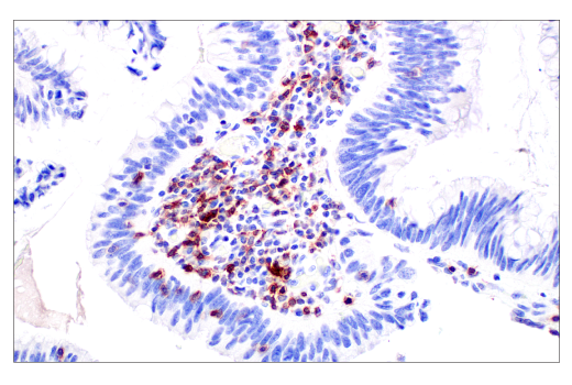
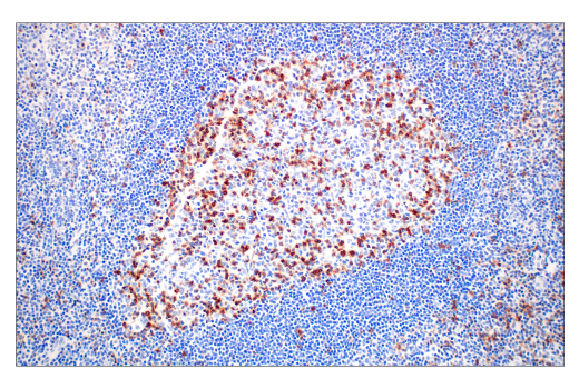
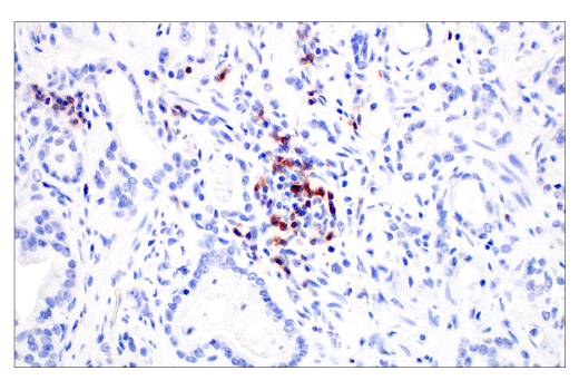
WB, IHC-P, IF-IC
H
Endogenous
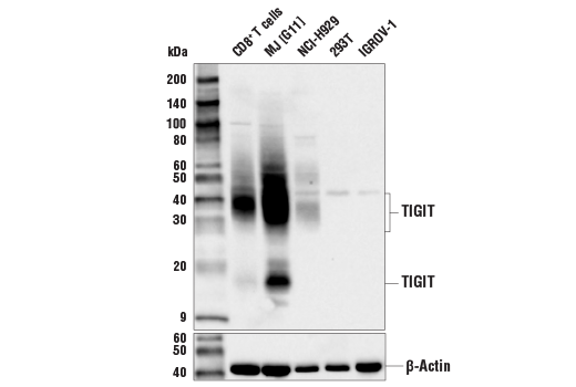
18, 30-40
Rabbit IgG
#Q495A1
201633
Product Information
Product Usage Information
This product is the carrier free version of product #99567. All data were generated using the same antibody clone in the standard formulation which contains BSA and glycerol.
This formulation is ideal for use with technologies requiring specialized or custom antibody labeling, including fluorophores, metals, lanthanides, and oligonucleotides. It is not recommended for ChIP, ChIP-seq, CUT&RUN or CUT&Tag assays. If you require a carrier free formulation for chromatin profiling, please contact us. Optimal dilutions/concentrations should be determined by the end user.
Formulation
Storage
Specificity / Sensitivity
Species Reactivity:
Human
Source / Purification
Monoclonal antibody is produced by immunizing animals with recombinant protein specific to the carboxy terminus of human TIGIT protein. This antibody cross-reacts with an unidentified protein of 42 kDa in some cell extracts.
Background
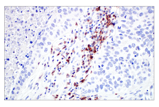
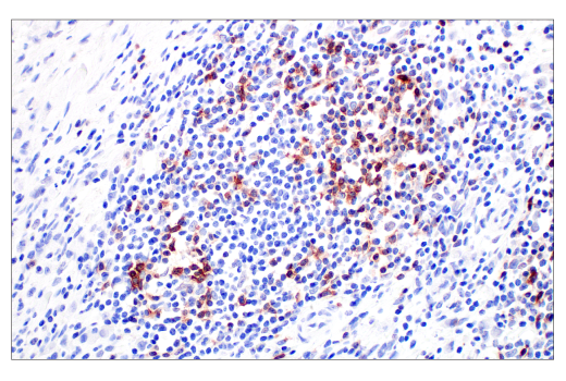
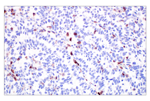
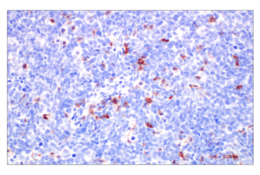
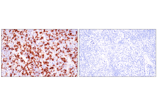
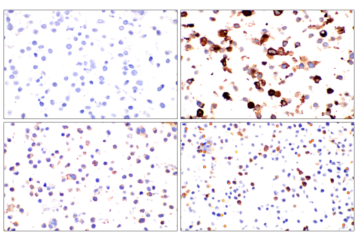
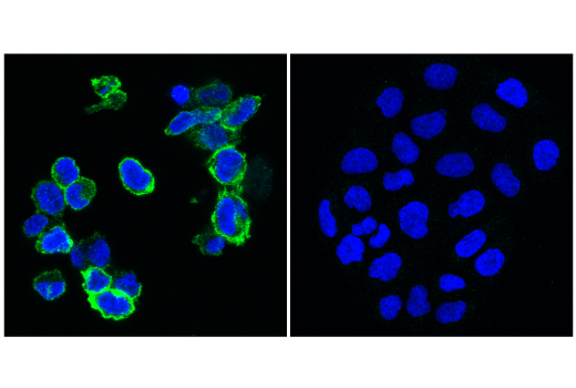
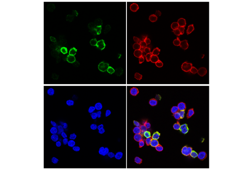
T-cell immunoreceptor with Ig and ITIM domains (TIGIT), also known as VSIG9, VSTM3, and WUCAM, is a member of the poliovirus receptor family of immunoglobulin proteins (1-3). TIGIT is expressed at low levels on subsets of T cells and NK cells, and is upregulated at the protein level following activation of these cells (1-4). TIGIT marks exhausted T cells in the tumor microenvironment (5) and during human immunodeficiency virus (HIV) infection (6). Research has shown TIGIT interacts with several receptors expressed on antigen presenting cells, such as dendritic cells and macrophages, as well as tumor cells and cells of the microenvironment. TIGIT binds with high affinity to PVR/CD155, and with low affinity to Nectin-2/CD112 and Nectin-3/CD113 (2,4,7). Upon binding to its ligands, TIGIT suppresses T cell activation, and inhibits T and NK cell cytotoxicity. This inhibition can be blocked using monoclonal antibodies directed at the extracellular domain of TIGIT, resulting in rejuvenated antigen-specific CD8+ T cell responses in tumors and during HIV infection (5,6,8). Three potential isoforms of TIGIT have been computationally mapped (9).
- Yu, X. et al. (2009) Nat Immunol 10, 48-57.
- Levin, S.D. et al. (2011) Eur J Immunol 41, 902-15.
- Boles, K.S. et al. (2009) Eur J Immunol 39, 695-703.
- Stanietsky, N. et al. (2009) Proc Natl Acad Sci U S A 106, 17858-63.
- Chauvin, J.M. et al. (2015) J Clin Invest 125, 2046-58.
- Chew, G.M. et al. (2016) PLoS Pathog 12, e1005349.
- Stengel, K.F. et al. (2012) Proc Natl Acad Sci U S A 109, 5399-404.
- Johnston, R.J. et al. (2014) Cancer Cell 26, 923-37.
- Bechtel, S. et al. (2007) BMC Genomics 8, 399.
Species Reactivity
Species reactivity is determined by testing in at least one approved application (e.g., western blot).
Applications Key
WB: Western Blotting IHC-P: Immunohistochemistry (Paraffin) IF-IC: Immunofluorescence (Immunocytochemistry)
Cross-Reactivity Key
H: human M: mouse R: rat Hm: hamster Mk: monkey Vir: virus Mi: mink C: chicken Dm: D. melanogaster X: Xenopus Z: zebrafish B: bovine Dg: dog Pg: pig Sc: S. cerevisiae Ce: C. elegans Hr: horse GP: Guinea Pig Rab: rabbit All: all species expected
Trademarks and Patents
使用に関する制限
法的な権限を与えられたCSTの担当者が署名した書面によって別途明示的に合意された場合を除き、 CST、その関連会社または代理店が提供する製品には以下の条件が適用されます。お客様が定める条件でここに定められた条件に含まれるものを超えるもの、 または、ここに定められた条件と異なるものは、法的な権限を与えられたCSTの担当者が別途書面にて受諾した場合を除き、拒絶され、 いかなる効力も効果も有しません。
研究専用 (For Research Use Only) またはこれに類似する表示がされた製品は、 いかなる目的についても FDA または外国もしくは国内のその他の規制機関により承認、認可または許可を受けていません。 お客様は製品を診断もしくは治療目的で使用してはならず、また、製品に表示された内容に違反する方法で使用してはなりません。 CST が販売または使用許諾する製品は、エンドユーザーであるお客様に対し、使途を研究および開発のみに限定して提供されるものです。 診断、予防もしくは治療目的で製品を使用することまたは製品を再販売 (単独であるか他の製品等の一部であるかを問いません) もしくはその他の商業的利用の目的で購入することについては、CST から別途許諾を得る必要があります。 お客様は以下の事項を遵守しなければなりません。(a) CST の製品 (単独であるか他の資材と一緒であるかを問いません) を販売、使用許諾、貸与、寄付もしくはその他の態様で第三者に譲渡したり使用させたりしてはなりません。また、商用の製品を製造するために CST の製品を使用してはなりません。(b) 複製、改変、リバースエンジニアリング、逆コンパイル、 分解または他の方法により製品の構造または技術を解明しようとしてはなりません。また、 CST の製品またはサービスと競合する製品またはサービスを開発する目的で CST の製品を使用してはなりません。(c) CST の製品の商標、商号、ロゴ、特許または著作権に関する通知または表示を除去したり改変したりしてはなりません。(d) CST の製品をCST 製品販売条件(CST’s Product Terms of Sale) および該当する書面のみに従って使用しなければなりません。(e) CST の製品に関連してお客様が使用する第三者の製品またはサービスに関する使用許諾条件、 サービス提供条件またはこれに類する合意事項を遵守しなければなりません。