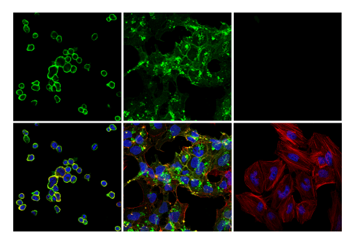
WB, IHC-Bond, IHC-P, IF-IC, FC-L
H Mk
Endogenous
90, 120
Rabbit IgG
#P28908
943
Product Information
Product Usage Information
| Application | Dilution |
|---|---|
| Western Blotting | 1:1000 |
| IHC Leica Bond | 1:50 |
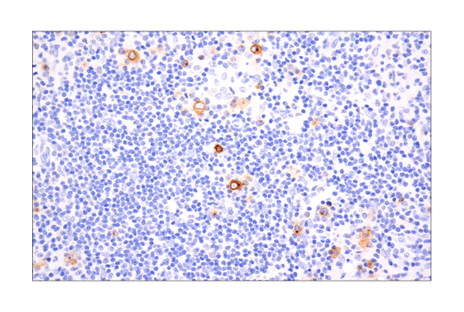
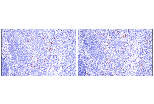
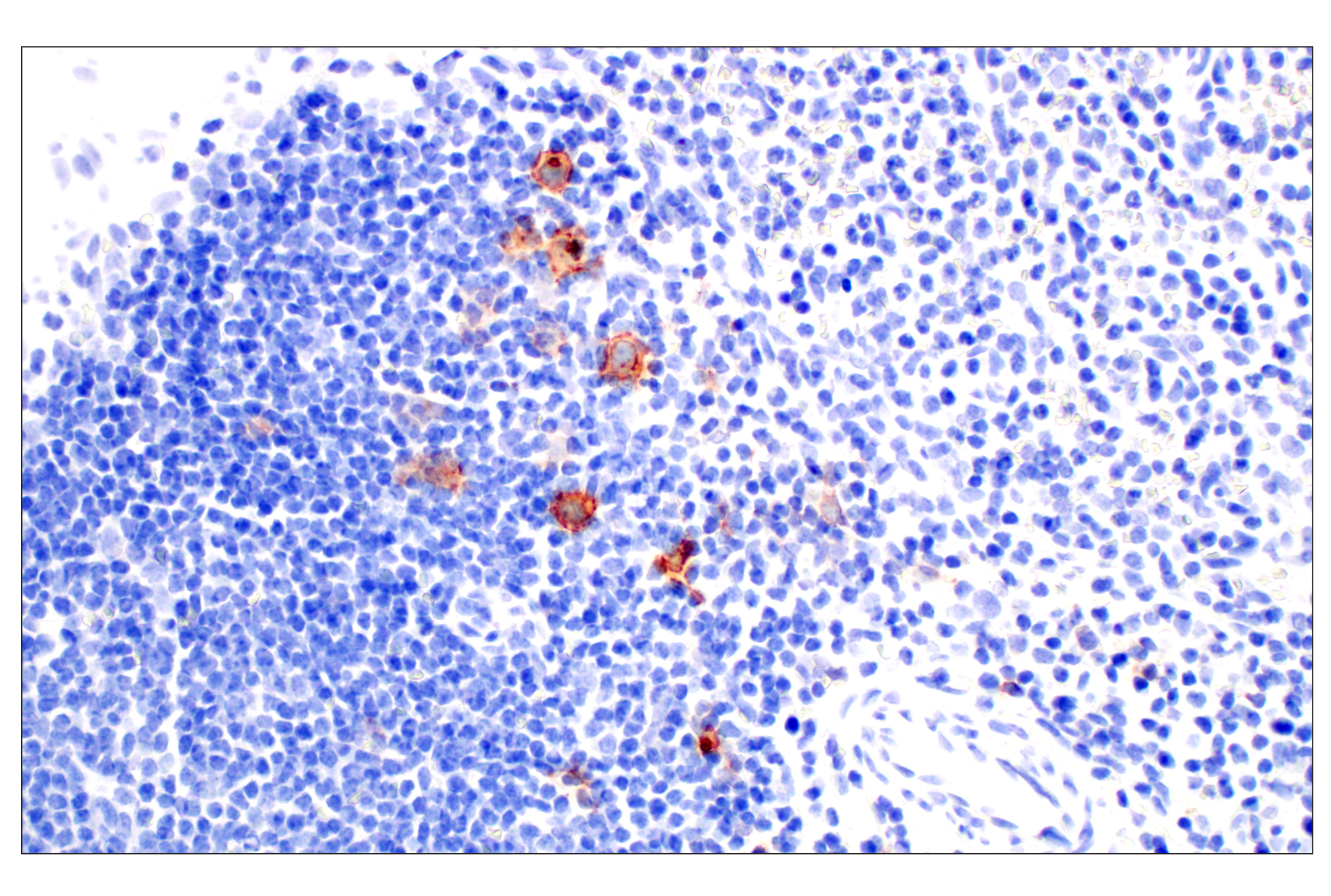
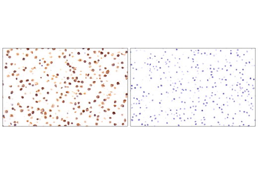
| Immunohistochemistry (Paraffin) | 1:50 |
| Immunofluorescence (Immunocytochemistry) | 1:400 |
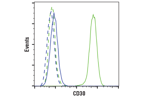
| Flow Cytometry (Live) | 1:200 - 1:800 |
Storage
For a carrier free (BSA and azide free) version of this product see product #18445.
Specificity / Sensitivity
Species Reactivity:
Human, Monkey
Source / Purification
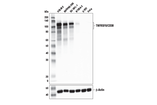
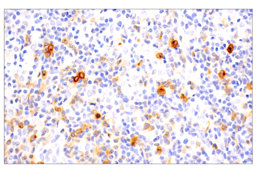
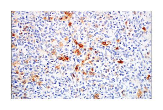
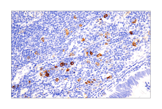
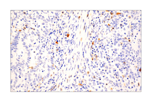
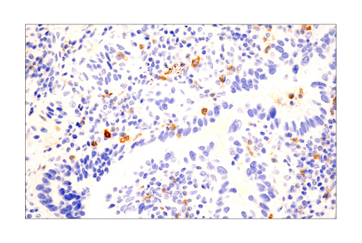
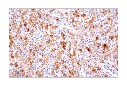
Monoclonal antibody is produced by immunizing animals with recombinant protein specific to the extracellular domain of human TNFRSF8/CD30 protein.
Background
TNFRSF8/CD30 is a type-I transmembrane glycoprotein that is a member of the TNFR superfamily. CD30 is synthesized as a precursor protein that undergoes extensive post-translational modification before becoming embedded in the plasma membrane as a 120-kDa transmembrane protein (1,2). The expression of CD30 is upregulated in activated T cells and may trigger costimulatory signaling pathways upon its engagement (3,4). While its expression is normally restricted to subsets of activated T cells and B cells, CD30 expression is robustly upregulated in hematologic malignancies, such as Hodgkin lymphoma (HL), anaplastic large cell lymphoma (ALCL), and adult T-cell leukemia, thus making it an attractive target for therapeutic intervention (5,6). Research studies have suggested that in certain disease contexts, CD30 recruits TRAF2 and TRAF5 adaptor proteins to drive NF-kappa B activation, aberrant cell growth, and cytokine production (7-9). CD30 signaling is also regulated by TACE-dependent proteolytic cleavage of its ectodomain, which results in reduced CD30L-dependent activation of CD30+ cells (10,11).
- Froese, P. et al. (1987) J Immunol 139, 2081-7.
- Nawrocki, J.F. et al. (1988) J Immunol 141, 672-80.
- Del Prete, G. et al. (1995) J Exp Med 182, 1655-61.
- Gilfillan, M.C. et al. (1998) J Immunol 160, 2180-7.
- Stein, H. et al. (1985) Blood 66, 848-58.
- Chiarle, R. et al. (1999) Clin Immunol 90, 157-64.
- Horie, R. et al. (2002) Am J Pathol 160, 1647-54.
- Horie, R. et al. (2002) Oncogene 21, 2493-503.
- Horie, R. et al. (2004) Cancer Cell 5, 353-64.
- Hansen, H.P. et al. (2000) J Immunol 165, 6703-9.
- Gruss, H.J. et al. (1997) Immunol Today 18, 156-63.
Species Reactivity
Species reactivity is determined by testing in at least one approved application (e.g., western blot).
Western Blot Buffer
IMPORTANT: For western blots, incubate membrane with diluted primary antibody in 5% w/v BSA, 1X TBS, 0.1% Tween® 20 at 4°C with gentle shaking, overnight.
Applications Key
WB: Western Blotting IHC-Bond: IHC Leica Bond IHC-P: Immunohistochemistry (Paraffin) IF-IC: Immunofluorescence (Immunocytochemistry) FC-L: Flow Cytometry (Live)
Cross-Reactivity Key
H: human M: mouse R: rat Hm: hamster Mk: monkey Vir: virus Mi: mink C: chicken Dm: D. melanogaster X: Xenopus Z: zebrafish B: bovine Dg: dog Pg: pig Sc: S. cerevisiae Ce: C. elegans Hr: horse GP: Guinea Pig Rab: rabbit All: all species expected
Trademarks and Patents
使用に関する制限
法的な権限を与えられたCSTの担当者が署名した書面によって別途明示的に合意された場合を除き、 CST、その関連会社または代理店が提供する製品には以下の条件が適用されます。お客様が定める条件でここに定められた条件に含まれるものを超えるもの、 または、ここに定められた条件と異なるものは、法的な権限を与えられたCSTの担当者が別途書面にて受諾した場合を除き、拒絶され、 いかなる効力も効果も有しません。
研究専用 (For Research Use Only) またはこれに類似する表示がされた製品は、 いかなる目的についても FDA または外国もしくは国内のその他の規制機関により承認、認可または許可を受けていません。 お客様は製品を診断もしくは治療目的で使用してはならず、また、製品に表示された内容に違反する方法で使用してはなりません。 CST が販売または使用許諾する製品は、エンドユーザーであるお客様に対し、使途を研究および開発のみに限定して提供されるものです。 診断、予防もしくは治療目的で製品を使用することまたは製品を再販売 (単独であるか他の製品等の一部であるかを問いません) もしくはその他の商業的利用の目的で購入することについては、CST から別途許諾を得る必要があります。 お客様は以下の事項を遵守しなければなりません。(a) CST の製品 (単独であるか他の資材と一緒であるかを問いません) を販売、使用許諾、貸与、寄付もしくはその他の態様で第三者に譲渡したり使用させたりしてはなりません。また、商用の製品を製造するために CST の製品を使用してはなりません。(b) 複製、改変、リバースエンジニアリング、逆コンパイル、 分解または他の方法により製品の構造または技術を解明しようとしてはなりません。また、 CST の製品またはサービスと競合する製品またはサービスを開発する目的で CST の製品を使用してはなりません。(c) CST の製品の商標、商号、ロゴ、特許または著作権に関する通知または表示を除去したり改変したりしてはなりません。(d) CST の製品をCST 製品販売条件(CST’s Product Terms of Sale) および該当する書面のみに従って使用しなければなりません。(e) CST の製品に関連してお客様が使用する第三者の製品またはサービスに関する使用許諾条件、 サービス提供条件またはこれに類する合意事項を遵守しなければなりません。