| Product Includes | Product # | Quantity | Mol. Wt | Isotype/Source |
|---|---|---|---|---|
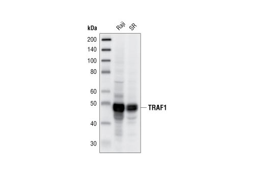
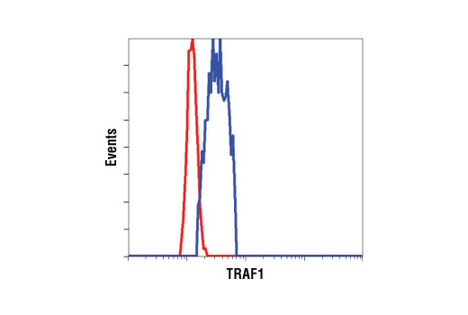
| TRAF1 (45D3) Rabbit mAb | 4715 | 20 µl | 50 kDa | Rabbit |
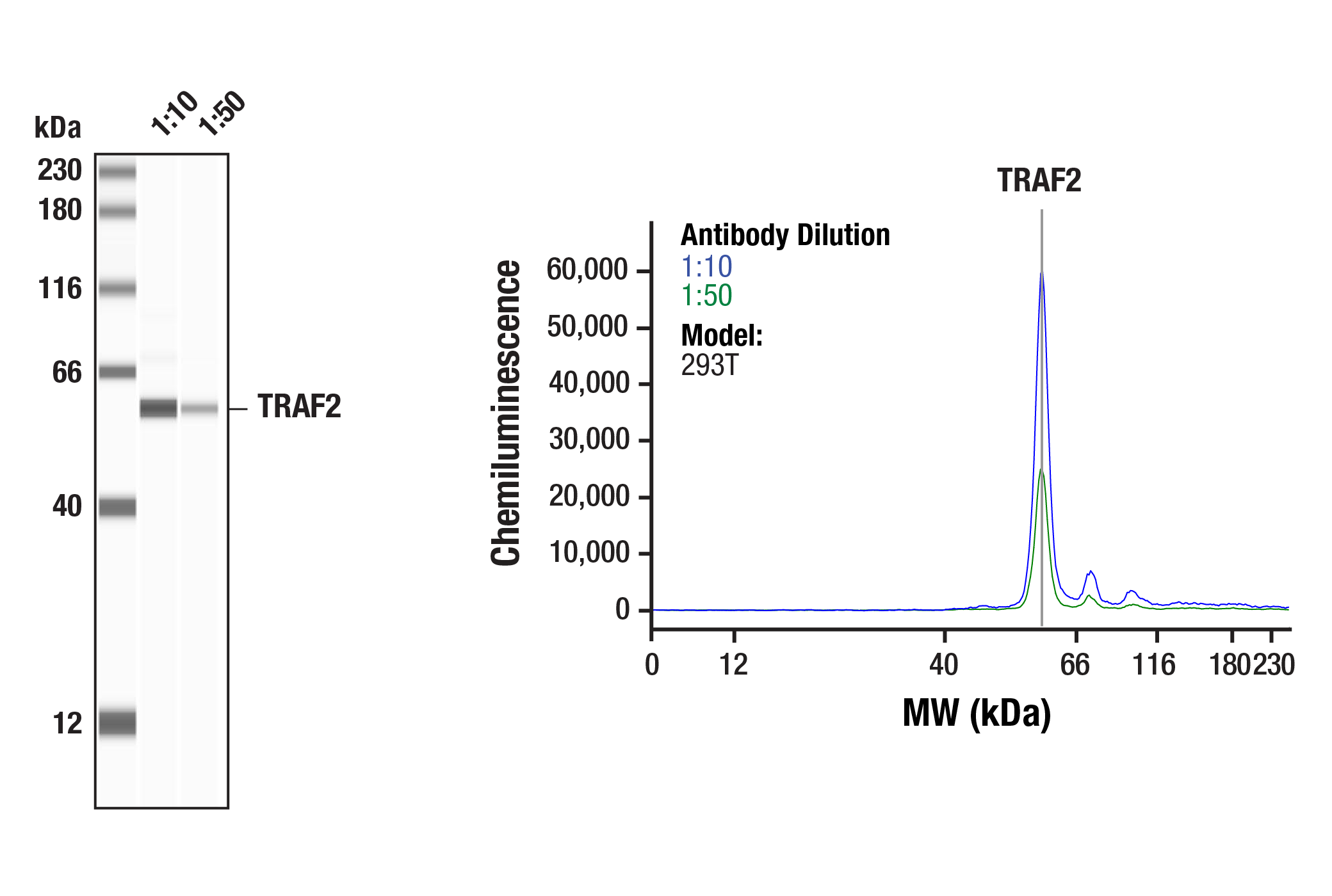
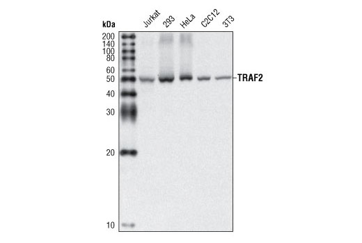
| TRAF2 (C192) Antibody | 4724 | 20 µl | 53 kDa | Rabbit |
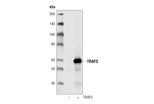
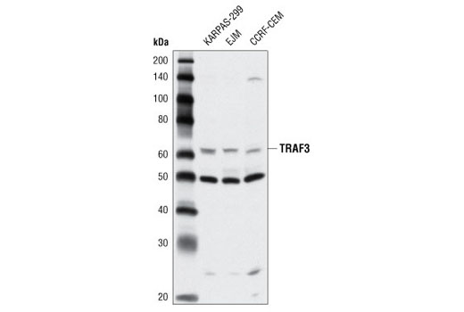
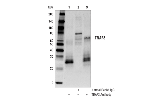
| TRAF3 Antibody | 4729 | 20 µl | 62 kDa | Rabbit |
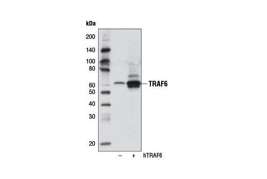
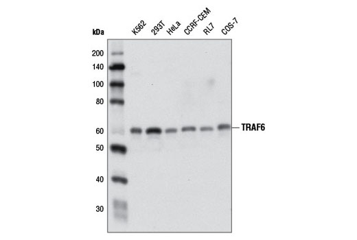
| TRAF6 (D21G3) Rabbit mAb | 8028 | 20 µl | 60 kDa | Rabbit IgG |
| Anti-rabbit IgG, HRP-linked Antibody | 7074 | 100 µl | Goat |
Please visit cellsignal.com for individual component applications, species cross-reactivity, dilutions, protocols, and additional product information.
Description
The TRAF Antibody Sampler Kit provides an economical means to evaluate endogenous levels of TRAF1, 2, 3, and 6. The kit contains enough primary and secondary antibodies to perform two western mini-blot experiments.
Storage
Background
TRAFs (TNF receptor-associated factors) are a family of multifunctional adaptor proteins that bind to surface receptors and recruit additional proteins to form multiprotein signaling complexes capable of promoting cellular responses (1-3). Members of the TRAF family share a common carboxy-terminal "TRAF domain", which mediates interactions with associated proteins; many also contain amino-terminal Zinc/RING finger motifs. The first TRAFs identified, TRAF1 and TRAF2, were found by virtue of their interactions with the cytoplasmic domain of TNF-receptor 2 (TNFRII) (4). The six known TRAFs (TRAF1-6) act as adaptor proteins for a wide range of cell surface receptors and participate in the regulation of cell survival, proliferation, differentiation, and stress responses.
While TRAF2 was originally described through its interaction with TNFRII, it has since been shown to interact with other surface receptors including CD27, CD30, CD40, 4-1BB, Ox40, HVEM/ATAR, and LMP-1 (1-3). TRAF2 also associates with a large number of intracellular proteins, including TRADD, FADD, I-TRAF/TANK, TRIP, A20, c-IAP1 and 2, Casper, RIP, and NIK, which help to regulate cell survival. Dominant negative and knockout studies have shown that TRAF2 plays an important role in TNF-mediated activation of NF-κB and the MAPK/JNK kinase pathway (5-7).
TRAF6 plays a critical role in innate and adaptive immunity, bone metabolism, and development of certain tissues including the nervous system (8). TRAF6 deficiency results in osteopetrosis and defective IL-1, CD40, and LPS signaling (9) as well as defects in neuronal development (10). Unlike other TRAF family members that mediate signaling through TNF, TRAF6 has unique binding activities (11) that result in signaling responses from the interleukin-1 receptor (IL-1R) (12), toll-like receptor (13,14), CD (15), RANK (16,17), and p75 neurotrophin receptor (18). TRAF6 associates directly with CD40 and RANK, and indirectly with IL-1R/TLR through IRAK (13). This leads to activation of NF-κB and MAP kinase signaling pathways through downstream association with the TAB/TAK-1 complex (19). TRAF6 also activates Src family nonreceptor tyrosine kinases leading to Akt activation (20).
- Arch, R.H. et al. (1998) Genes Dev 12, 2821-30.
- Chung, J.Y. et al. (2002) J Cell Sci 115, 679-88.
- Bradley, J.R. and Pober, J.S. (2001) Oncogene 20, 6482-91.
- Rothe, M. et al. (1994) Cell 78, 681-92.
- Yeh, W.C. et al. (1997) Immunity 7, 715-25.
- Reinhard, C. et al. (1997) EMBO J 16, 1080-92.
- Rothe, M. et al. (1995) Science 269, 1424-7.
- Wu, H. and Arron, J.R. (2003) Bioessays 25, 1096-105.
- Lomaga, M.A. et al. (1999) Genes Dev 13, 1015-24.
- Lomaga, M.A. et al. (2000) J Neurosci 20, 7384-93.
- Ye, H. et al. (2002) Nature 418, 443-7.
- Cao, Z. et al. (1996) Nature 383, 443-6.
- Muzio, M. et al. (1997) Science 278, 1612-5.
- Medzhitov, R. et al. (1998) Mol Cell 2, 253-8.
- Ishida, T. et al. (1996) J Biol Chem 271, 28745-8.
- Darnay, B.G. et al. (1998) J Biol Chem 273, 20551-5.
- Wong, B.R. et al. (1998) J Biol Chem 273, 28355-9.
- Khursigara, G. et al. (1999) J Biol Chem 274, 2597-600.
- Ninomiya-Tsuji, J. et al. (1999) Nature 398, 252-6.
- Wong, B.R. et al. (1999) Mol Cell 4, 1041-9.
Background References
Trademarks and Patents
使用に関する制限
法的な権限を与えられたCSTの担当者が署名した書面によって別途明示的に合意された場合を除き、 CST、その関連会社または代理店が提供する製品には以下の条件が適用されます。お客様が定める条件でここに定められた条件に含まれるものを超えるもの、 または、ここに定められた条件と異なるものは、法的な権限を与えられたCSTの担当者が別途書面にて受諾した場合を除き、拒絶され、 いかなる効力も効果も有しません。
研究専用 (For Research Use Only) またはこれに類似する表示がされた製品は、 いかなる目的についても FDA または外国もしくは国内のその他の規制機関により承認、認可または許可を受けていません。 お客様は製品を診断もしくは治療目的で使用してはならず、また、製品に表示された内容に違反する方法で使用してはなりません。 CST が販売または使用許諾する製品は、エンドユーザーであるお客様に対し、使途を研究および開発のみに限定して提供されるものです。 診断、予防もしくは治療目的で製品を使用することまたは製品を再販売 (単独であるか他の製品等の一部であるかを問いません) もしくはその他の商業的利用の目的で購入することについては、CST から別途許諾を得る必要があります。 お客様は以下の事項を遵守しなければなりません。(a) CST の製品 (単独であるか他の資材と一緒であるかを問いません) を販売、使用許諾、貸与、寄付もしくはその他の態様で第三者に譲渡したり使用させたりしてはなりません。また、商用の製品を製造するために CST の製品を使用してはなりません。(b) 複製、改変、リバースエンジニアリング、逆コンパイル、 分解または他の方法により製品の構造または技術を解明しようとしてはなりません。また、 CST の製品またはサービスと競合する製品またはサービスを開発する目的で CST の製品を使用してはなりません。(c) CST の製品の商標、商号、ロゴ、特許または著作権に関する通知または表示を除去したり改変したりしてはなりません。(d) CST の製品をCST 製品販売条件(CST’s Product Terms of Sale) および該当する書面のみに従って使用しなければなりません。(e) CST の製品に関連してお客様が使用する第三者の製品またはサービスに関する使用許諾条件、 サービス提供条件またはこれに類する合意事項を遵守しなければなりません。