WB, W-S, IP, IF-IC, FC-FP
H
Endogenous
28
Rabbit IgG
#Q9NZC2
54209
Product Information
Product Usage Information
| Application | Dilution |
|---|---|
| Western Blotting | 1:1000 |
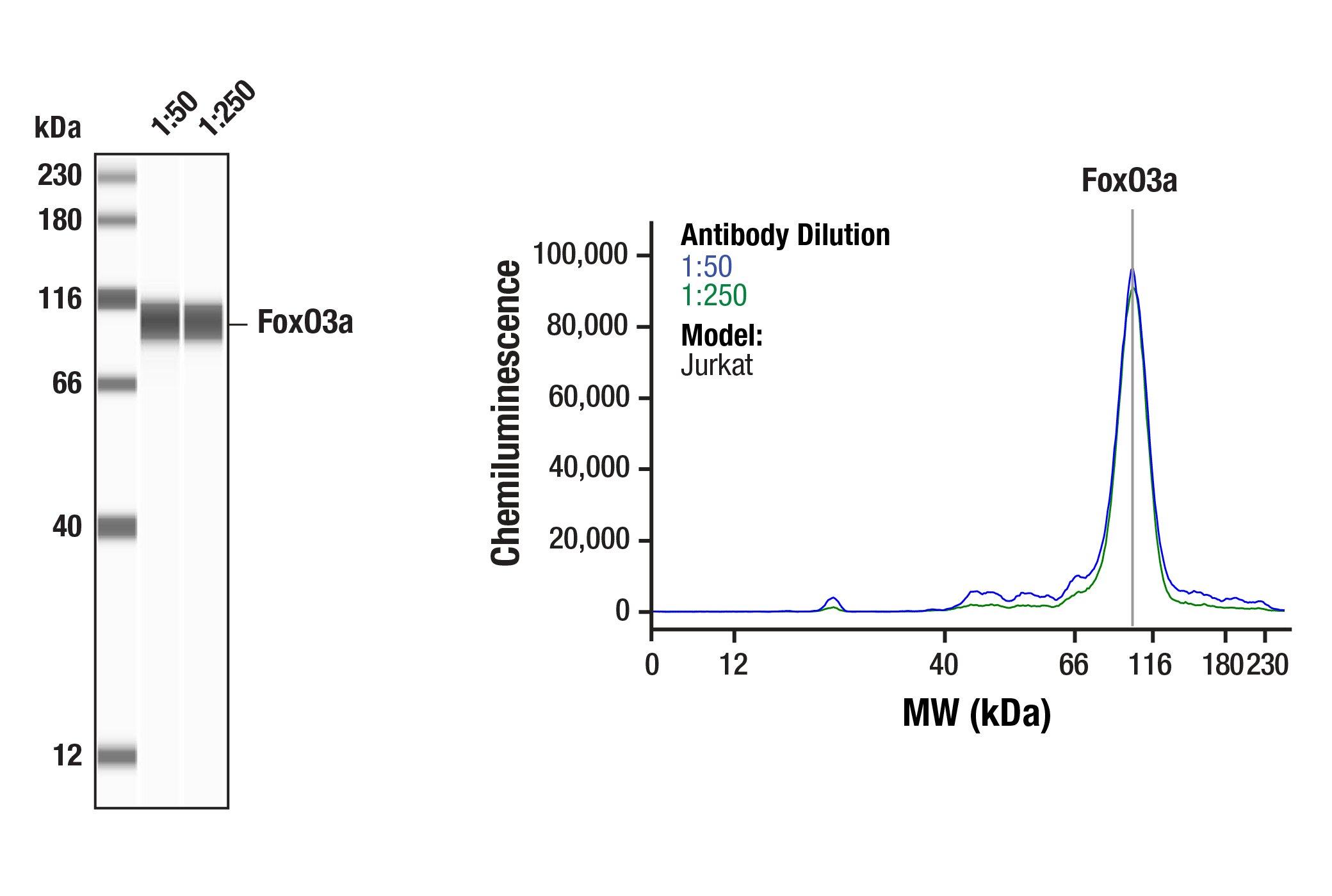
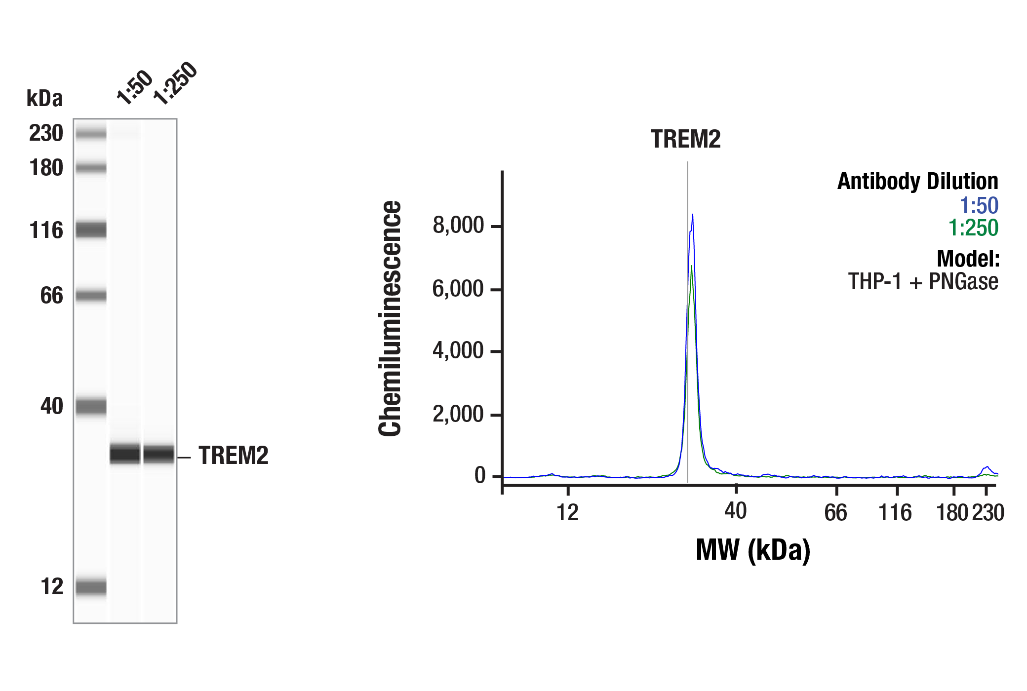
| Simple Western™ | 1:50 - 1:250 |
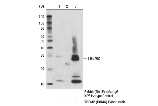
| Immunoprecipitation | 1:50 |
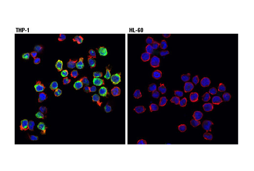
| Immunofluorescence (Immunocytochemistry) | 1:200 - 1:800 |
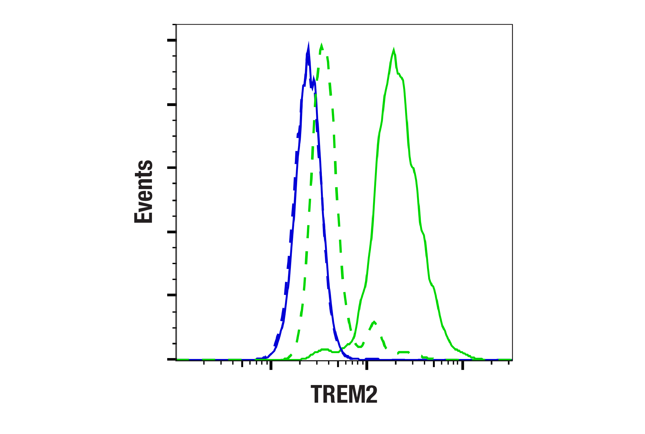
| Flow Cytometry (Fixed/Permeabilized) | 1:400 |
Storage
Specificity / Sensitivity
Species Reactivity:
Human
Source / Purification
Monoclonal antibody is produced by immunizing animals with a synthetic peptide corresponding to residues surrounding Leu221 of human TREM2 protein.
Background
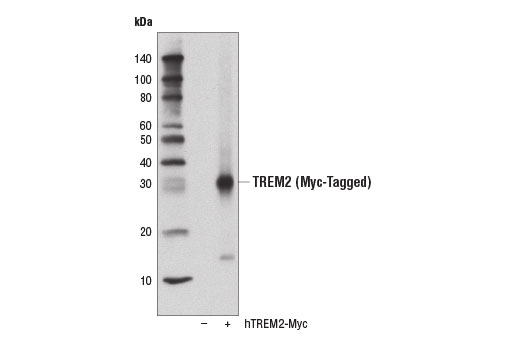
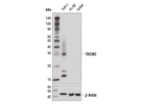
The triggering receptor expressed on myeloid cells 2 (TREM2) protein is an innate immune receptor that is expressed on the cell surface of microglia, macrophages, osteoclasts, and immature dendritic cells (1). The TREM2 receptor is a single-pass type I membrane glycoprotein that consists of an extracellular immunoglobulin-like domain, a transmembrane domain, and a cytoplasmic tail. TREM2 interacts with the tyrosine kinase-binding protein DAP12 to form a receptor-signaling complex (2). The TREM2 protein plays a role in innate immunity and a rare functional variant (R47H) of TREM2 is associated with the late-onset risk of Alzheimer’s disease (1,3). Research studies using mouse models of Alzheimer’s disease indicate that deficiency and haploinsufficiency of TREM2 can lead to increased β-amyloid (Aβ) accumulation as a result of dysfunctional microglial response (4). These results agree with the distribution of TREM2 in human brain regions (e.g., white matter, the hippocampus, and neocortex) that are involved in Alzheimer's disease pathology (2). In addition, amyloid plaque formation induces expression of TREM2 and amyloid phagocytosis (5). Loss-of-function mutations in the corresponding TREM2 or DAP12 genes can result in Nasu-Hakola disease, a rare form of progressive presenile dementia that results from polycystic osseous lesions (6). TREM2 membrane shedding occurs by cleavage at the extracellular site between H157/S158, generating an N-terminal shedded fragment and a membrane bound C-terminal fragment (7,8).
- Colonna, M. (2003) Nat Rev Immunol 3, 445-53.
- Jonsson, T. et al. (2013) N Engl J Med 368, 107-16.
- Boutajangout, A. and Wisniewski, T. (2013) Int J Cell Biol 2013, 576383.
- Wang, Y. et al. (2015) Cell 160, 1061-71.
- Melchior, B. et al. (2010) ASN Neuro 2, e00037.
- Klünemann, H.H. et al. (2005) Neurology 64, 1502-7.
- Thornton, P. et al. (2017) EMBO Mol Med 9, 1366-1378.
- Schlepckow, K. et al. (2017) EMBO Mol Med 9, 1356-1365.
Species Reactivity
Species reactivity is determined by testing in at least one approved application (e.g., western blot).
Western Blot Buffer
IMPORTANT: For western blots, incubate membrane with diluted primary antibody in 5% w/v BSA, 1X TBS, 0.1% Tween® 20 at 4°C with gentle shaking, overnight.
Applications Key
WB: Western Blotting W-S: Simple Western™ IP: Immunoprecipitation IF-IC: Immunofluorescence (Immunocytochemistry) FC-FP: Flow Cytometry (Fixed/Permeabilized)
Cross-Reactivity Key
H: human M: mouse R: rat Hm: hamster Mk: monkey Vir: virus Mi: mink C: chicken Dm: D. melanogaster X: Xenopus Z: zebrafish B: bovine Dg: dog Pg: pig Sc: S. cerevisiae Ce: C. elegans Hr: horse GP: Guinea Pig Rab: rabbit All: all species expected
Trademarks and Patents
使用に関する制限
法的な権限を与えられたCSTの担当者が署名した書面によって別途明示的に合意された場合を除き、 CST、その関連会社または代理店が提供する製品には以下の条件が適用されます。お客様が定める条件でここに定められた条件に含まれるものを超えるもの、 または、ここに定められた条件と異なるものは、法的な権限を与えられたCSTの担当者が別途書面にて受諾した場合を除き、拒絶され、 いかなる効力も効果も有しません。
研究専用 (For Research Use Only) またはこれに類似する表示がされた製品は、 いかなる目的についても FDA または外国もしくは国内のその他の規制機関により承認、認可または許可を受けていません。 お客様は製品を診断もしくは治療目的で使用してはならず、また、製品に表示された内容に違反する方法で使用してはなりません。 CST が販売または使用許諾する製品は、エンドユーザーであるお客様に対し、使途を研究および開発のみに限定して提供されるものです。 診断、予防もしくは治療目的で製品を使用することまたは製品を再販売 (単独であるか他の製品等の一部であるかを問いません) もしくはその他の商業的利用の目的で購入することについては、CST から別途許諾を得る必要があります。 お客様は以下の事項を遵守しなければなりません。(a) CST の製品 (単独であるか他の資材と一緒であるかを問いません) を販売、使用許諾、貸与、寄付もしくはその他の態様で第三者に譲渡したり使用させたりしてはなりません。また、商用の製品を製造するために CST の製品を使用してはなりません。(b) 複製、改変、リバースエンジニアリング、逆コンパイル、 分解または他の方法により製品の構造または技術を解明しようとしてはなりません。また、 CST の製品またはサービスと競合する製品またはサービスを開発する目的で CST の製品を使用してはなりません。(c) CST の製品の商標、商号、ロゴ、特許または著作権に関する通知または表示を除去したり改変したりしてはなりません。(d) CST の製品をCST 製品販売条件(CST’s Product Terms of Sale) および該当する書面のみに従って使用しなければなりません。(e) CST の製品に関連してお客様が使用する第三者の製品またはサービスに関する使用許諾条件、 サービス提供条件またはこれに類する合意事項を遵守しなければなりません。