| Product Includes | Product # | Quantity | Mol. Wt | Isotype/Source |
|---|---|---|---|---|
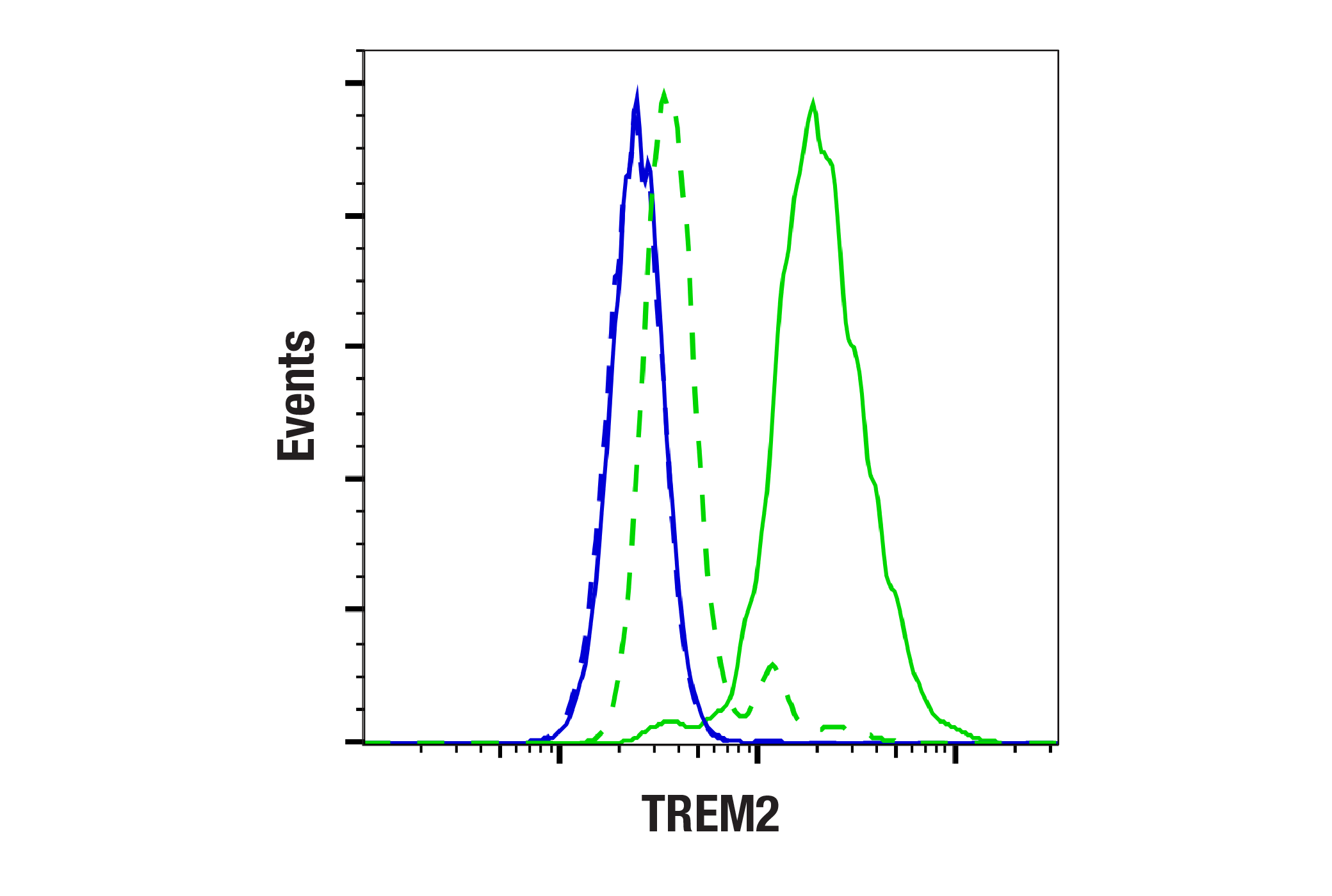
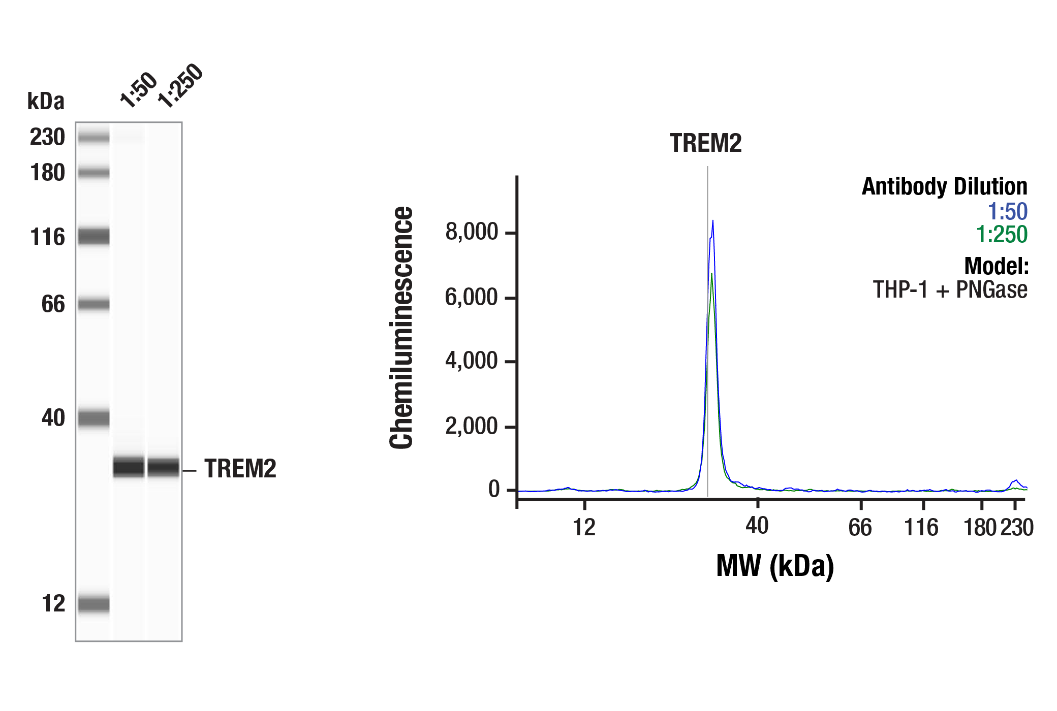
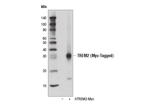
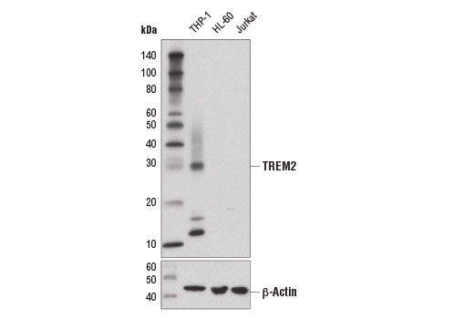
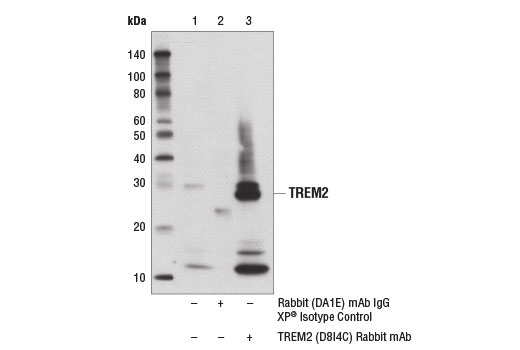
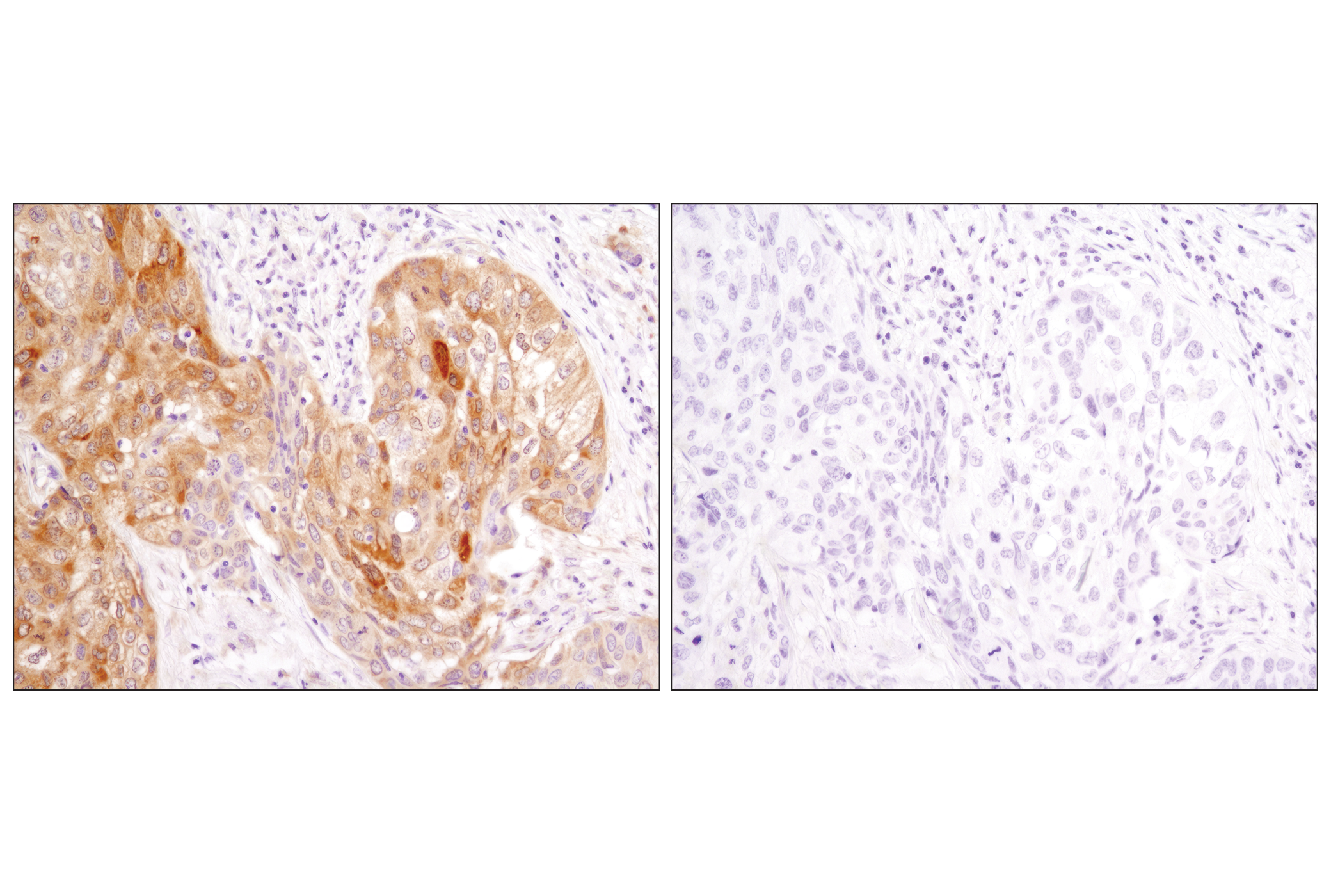
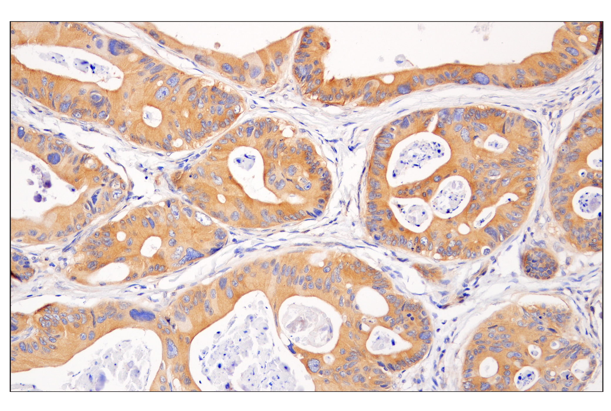
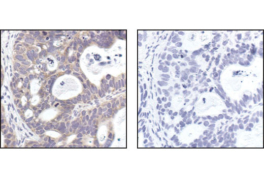
| TREM2 (D8I4C) Rabbit mAb | 91068 | 20 µl | 28 kDa | Rabbit IgG |
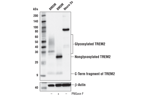
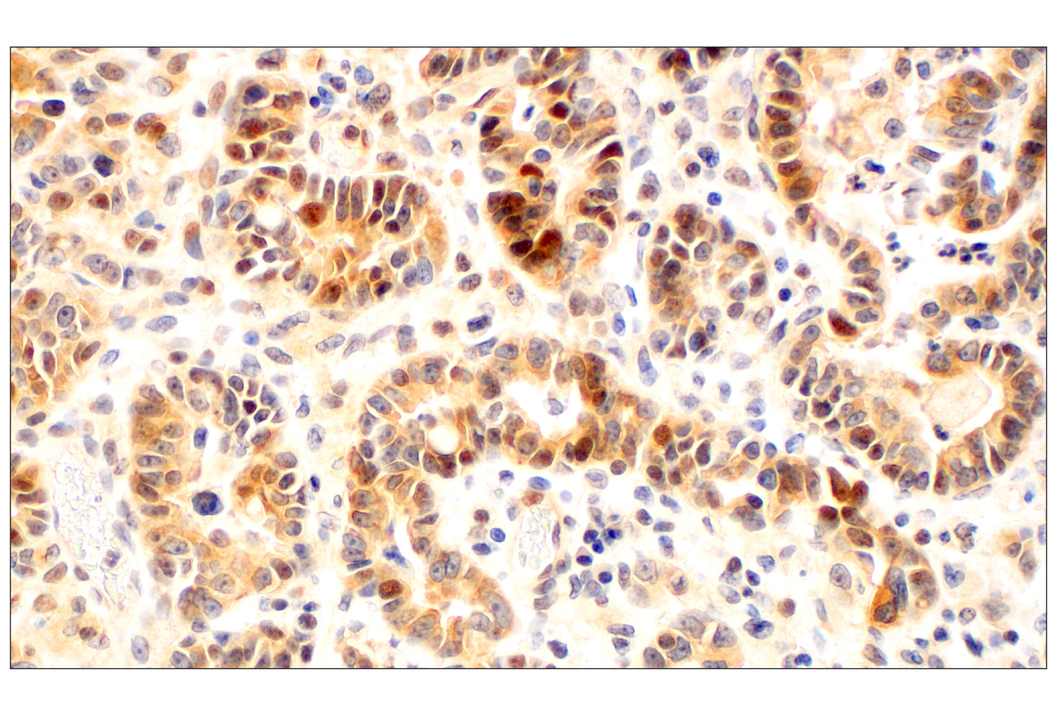
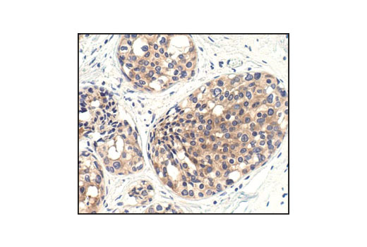
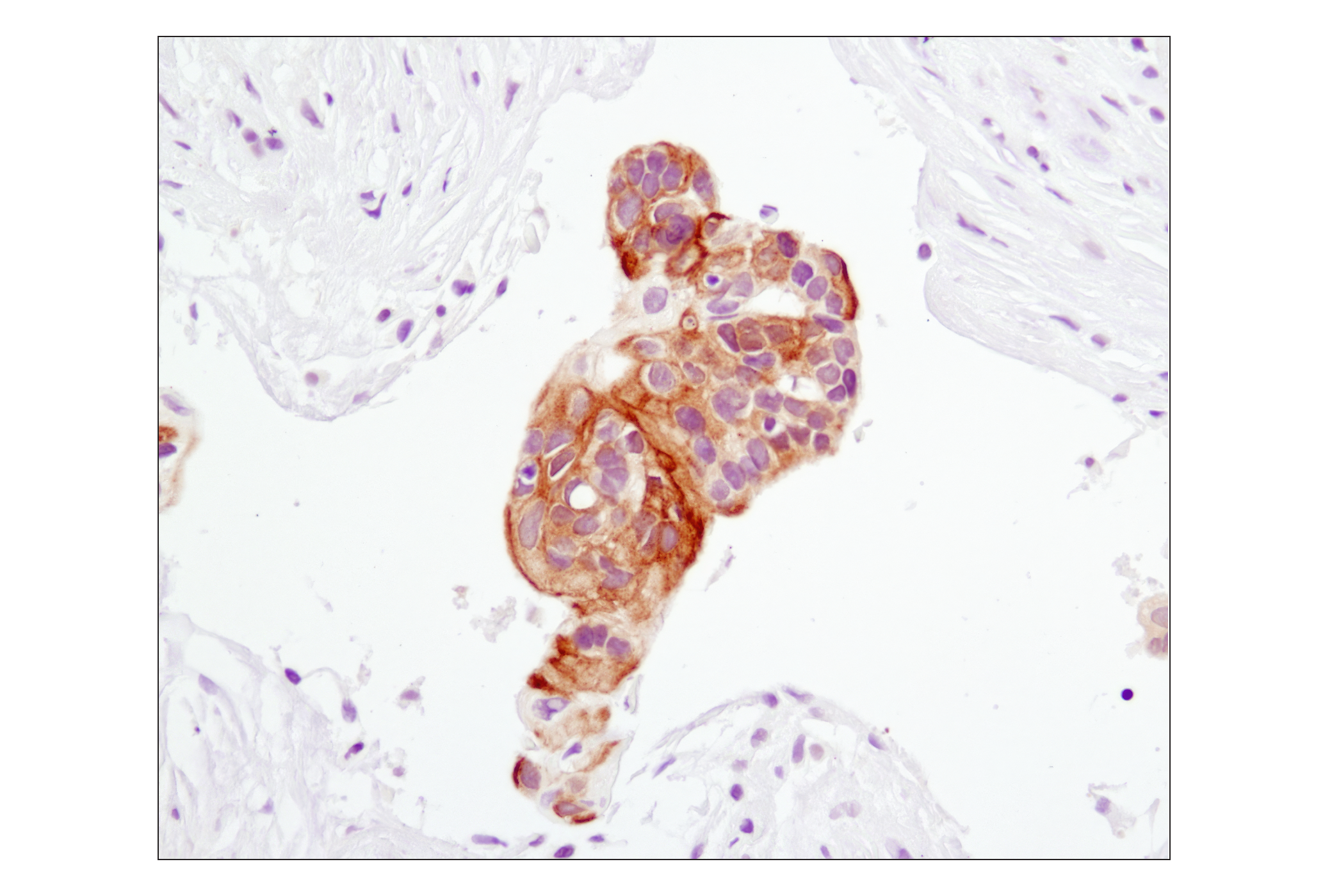
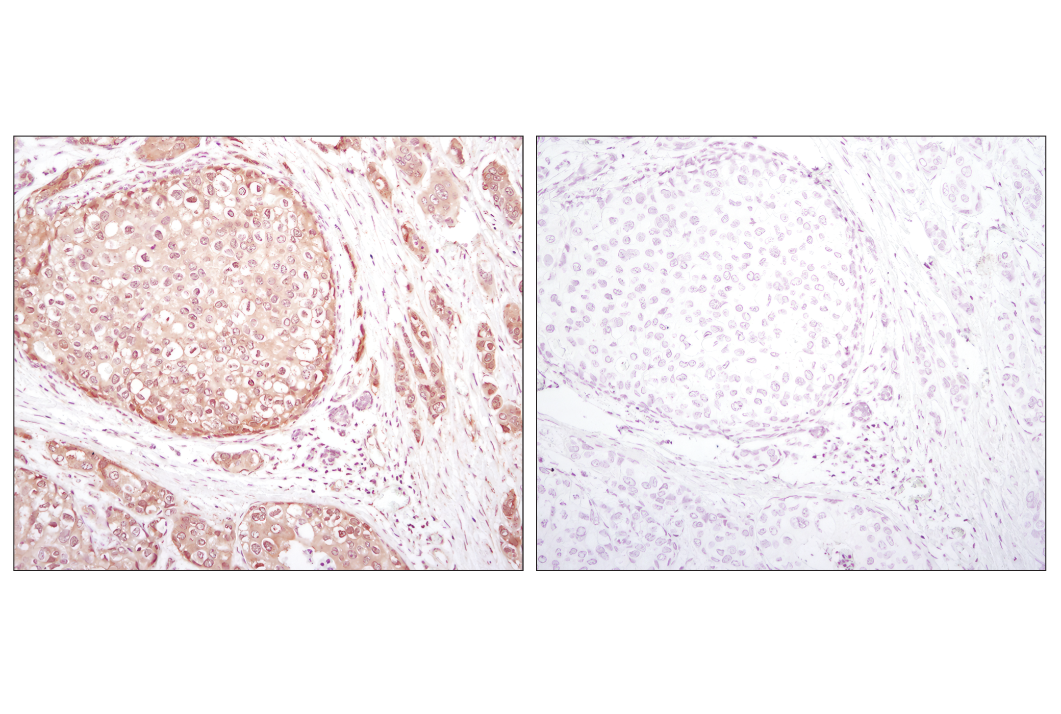
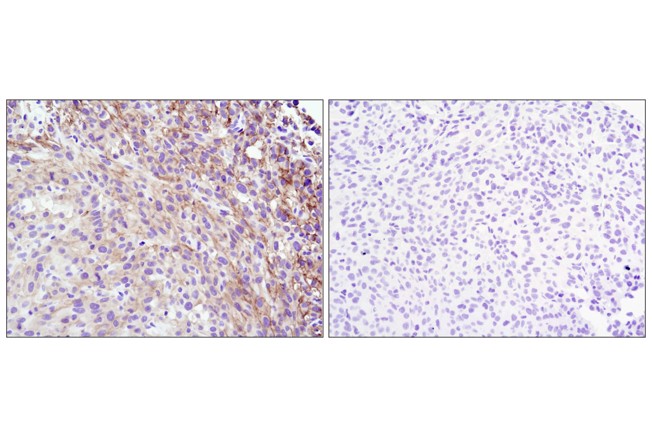
| TREM2 (E7P8J) Rabbit mAb (Carboxy-terminal Antigen) | 76765 | 20 µl | 11, 28 kDa | Rabbit IgG |
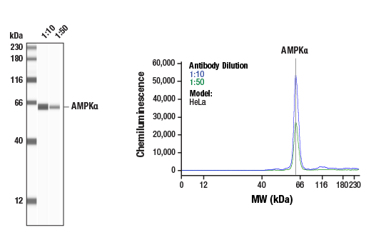
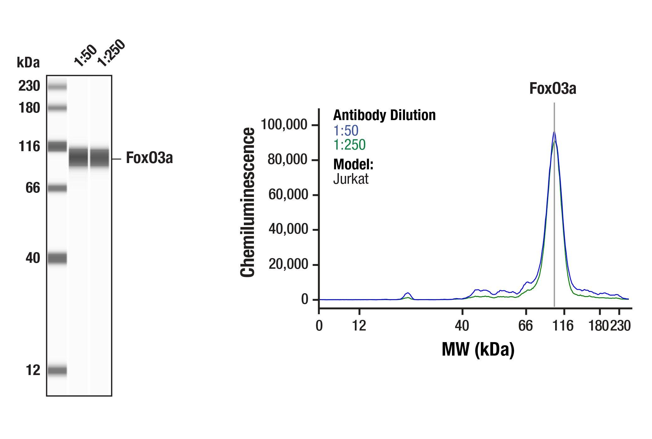
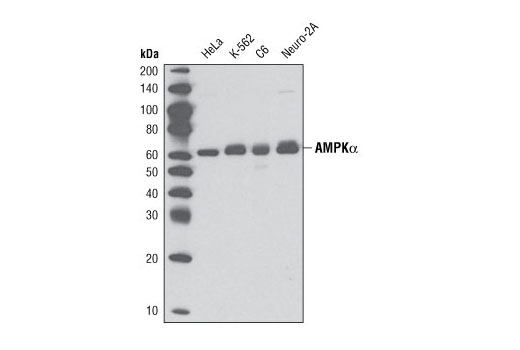
| AMPKα (D5A2) Rabbit mAb | 5831 | 20 µl | 62 kDa | Rabbit IgG |
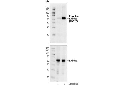
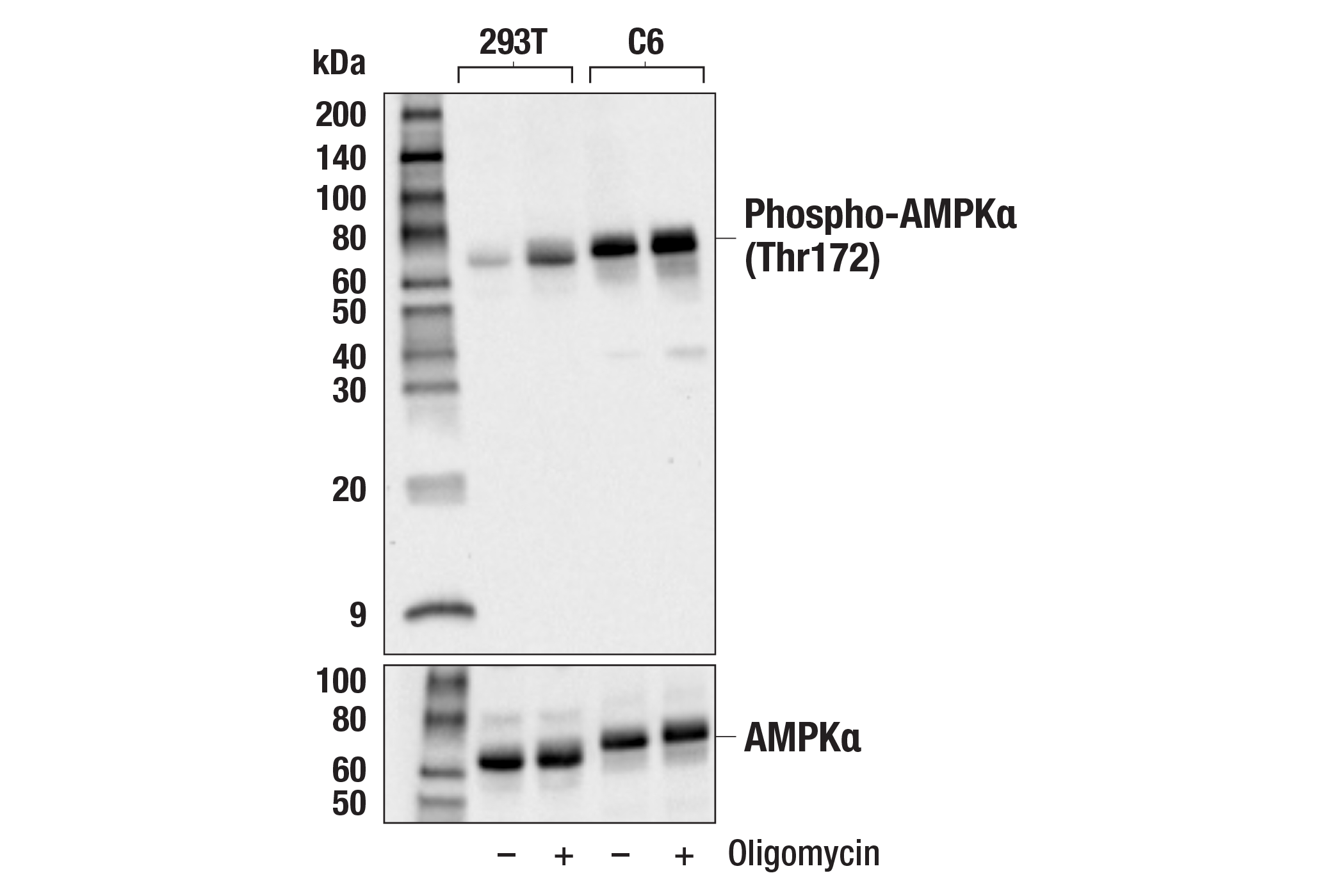
| Phospho-AMPKα (Thr172) (40H9) Rabbit mAb | 2535 | 20 µl | 62 kDa | Rabbit IgG |
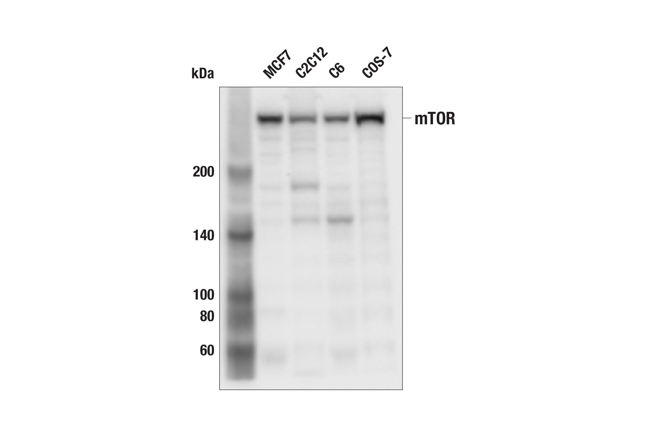
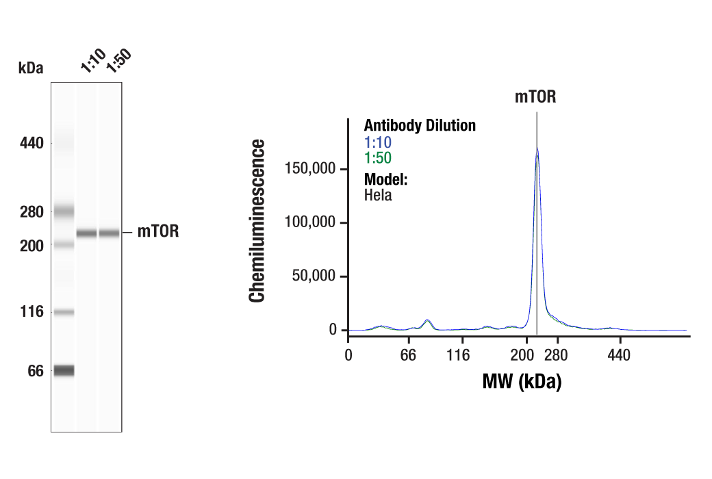
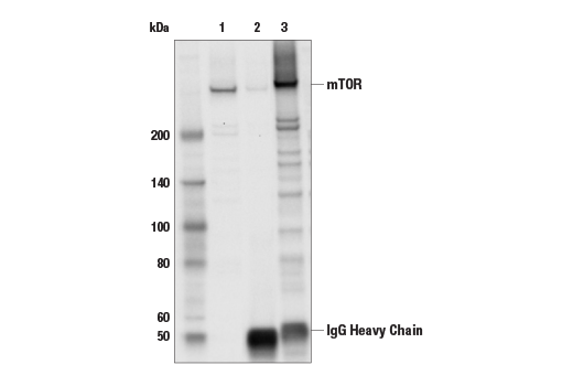
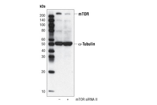
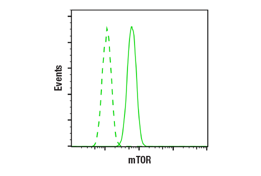
| mTOR (7C10) Rabbit mAb | 2983 | 20 µl | 289 kDa | Rabbit IgG |
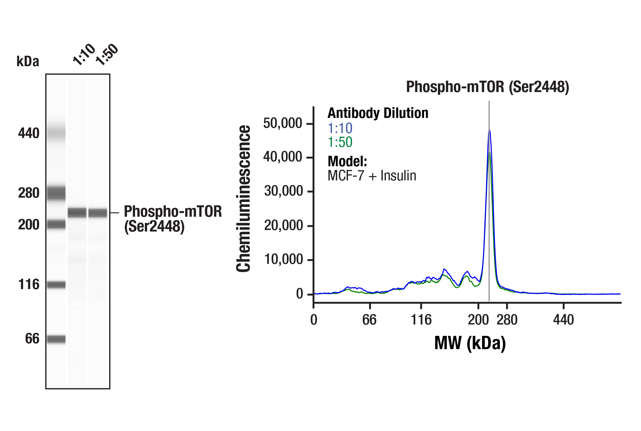
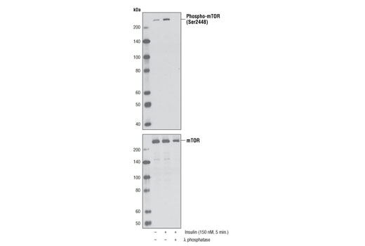
| Phospho-mTOR (Ser2448) (D9C2) XP® Rabbit mAb | 5536 | 20 µl | 289 kDa | Rabbit IgG |
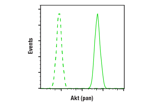
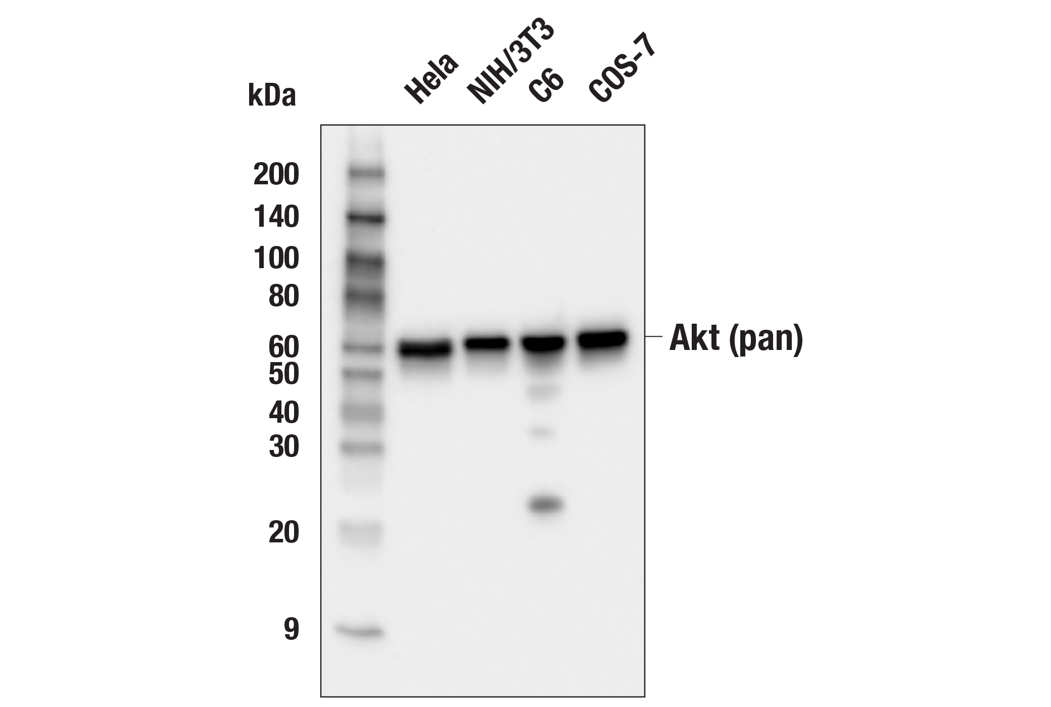
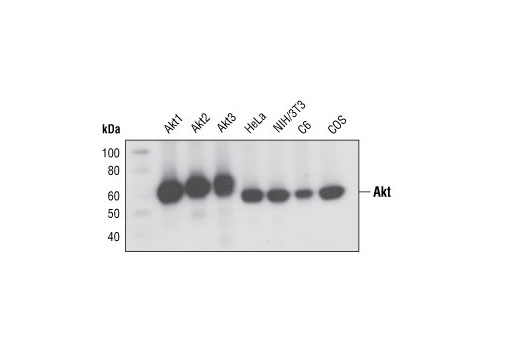
| Akt (pan) (C67E7) Rabbit mAb | 4691 | 20 µl | 60 kDa | Rabbit IgG |
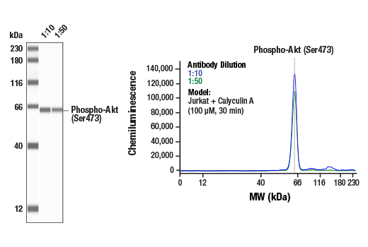
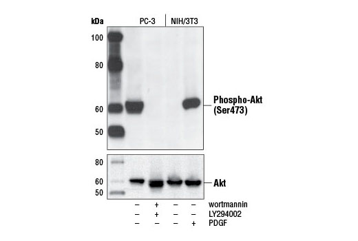
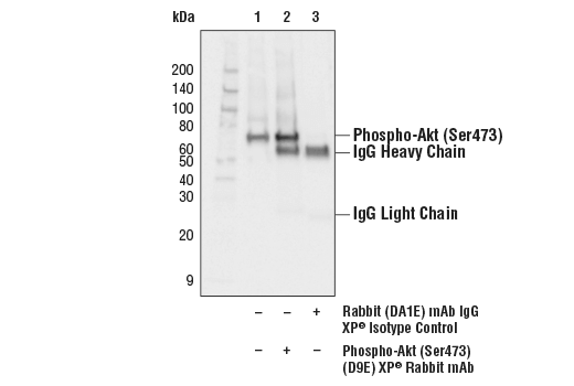
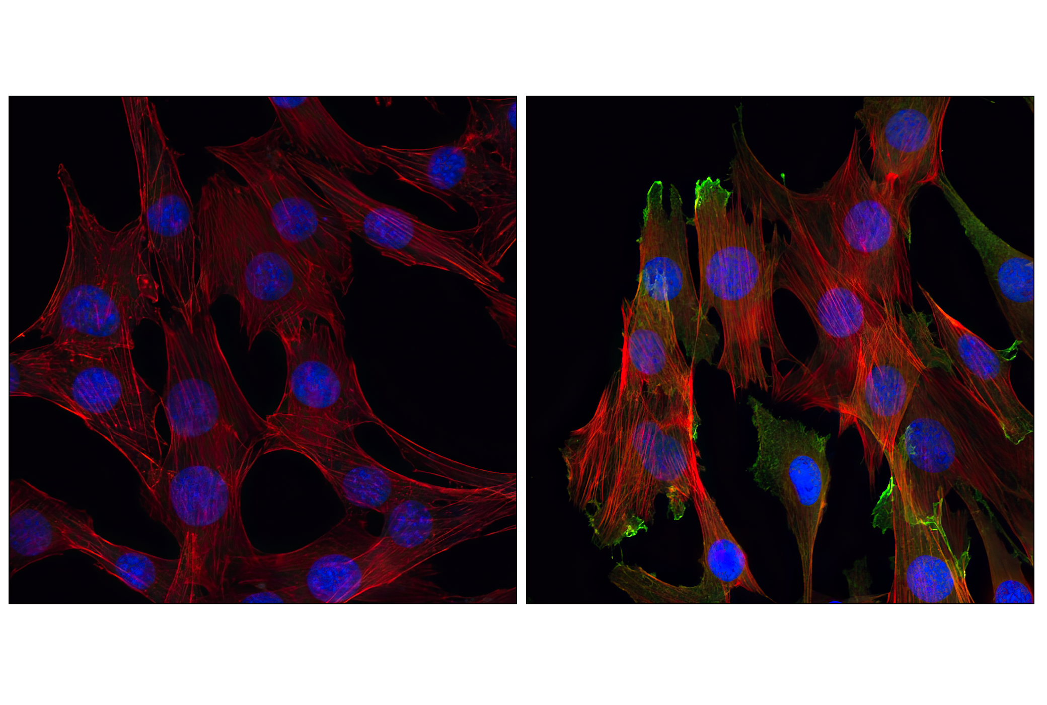
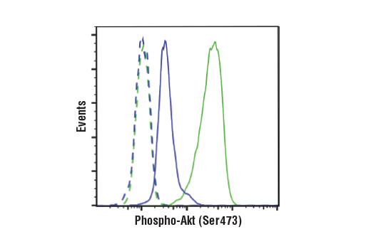
| Phospho-Akt (Ser473) (D9E) XP® Rabbit mAb | 4060 | 20 µl | 60 kDa | Rabbit IgG |
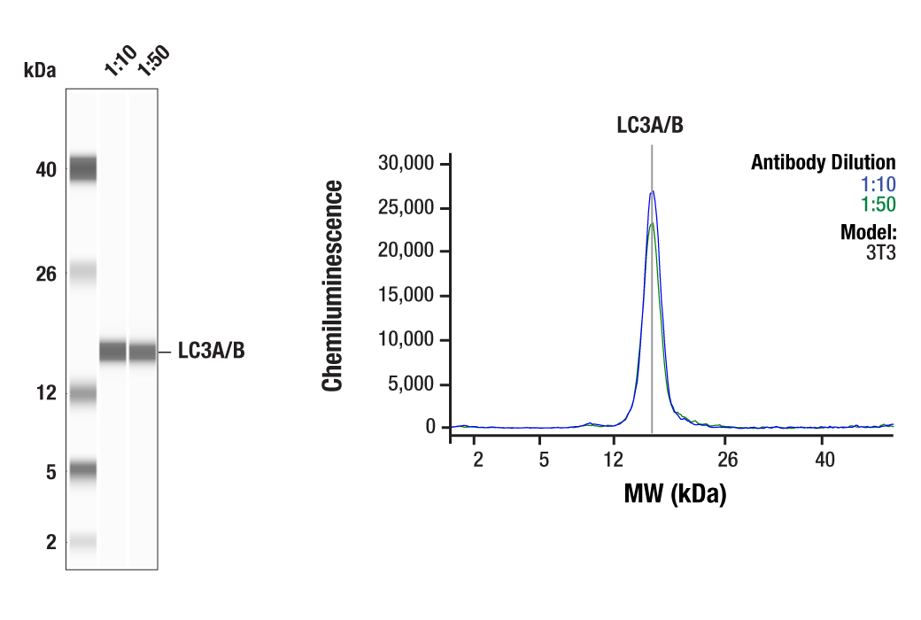
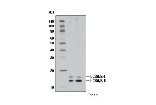
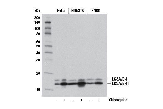
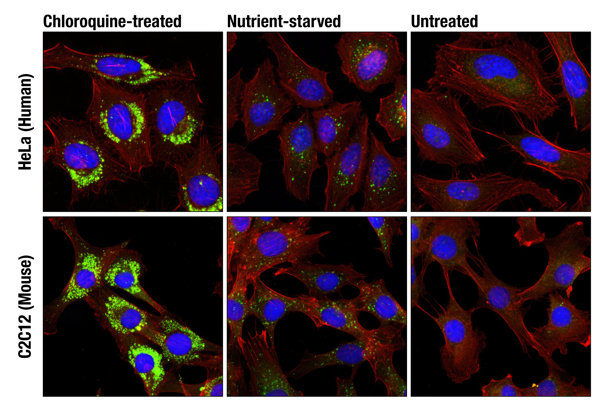
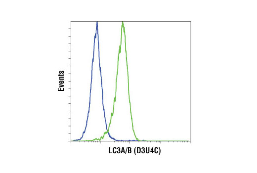
| LC3A/B (D3U4C) XP® Rabbit mAb | 12741 | 20 µl | 14, 16 kDa | Rabbit IgG |
| Anti-rabbit IgG, HRP-linked Antibody | 7074 | 100 µl | Goat |
Please visit cellsignal.com for individual component applications, species cross-reactivity, dilutions, protocols, and additional product information.
Description
The TREM2-dependent mTOR Metabolic Fitness Antibody Sampler Kit provides an economical means of detecting metabolic signaling pathways downstream of TREM2 by western blot. The kit includes enough antibodies to perform at least two western blot experiments with each primary antibody.
Storage
Background
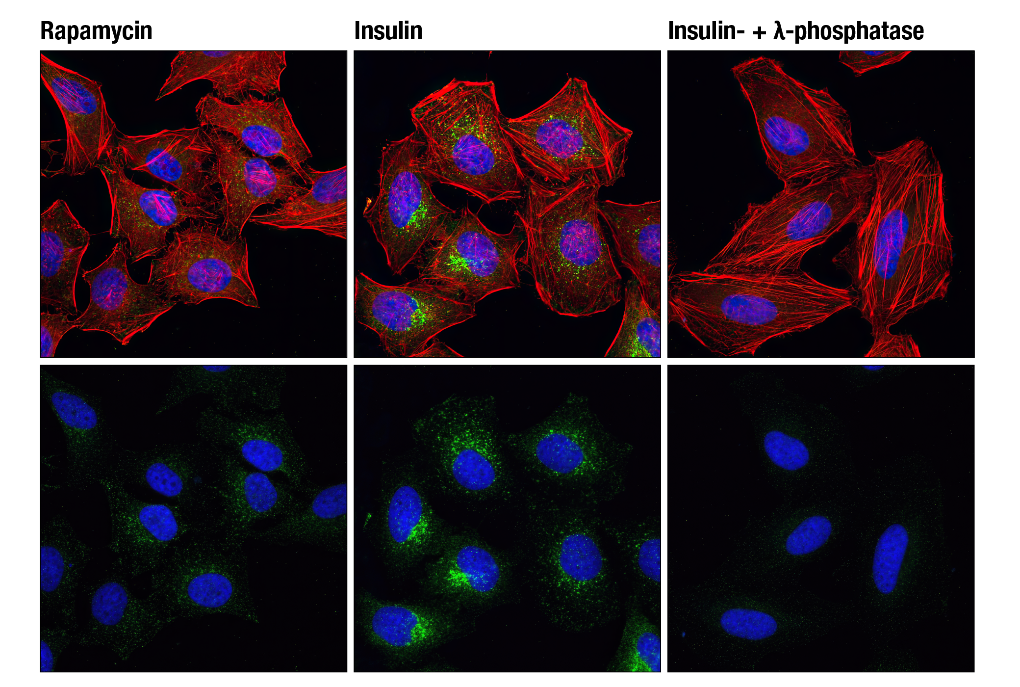
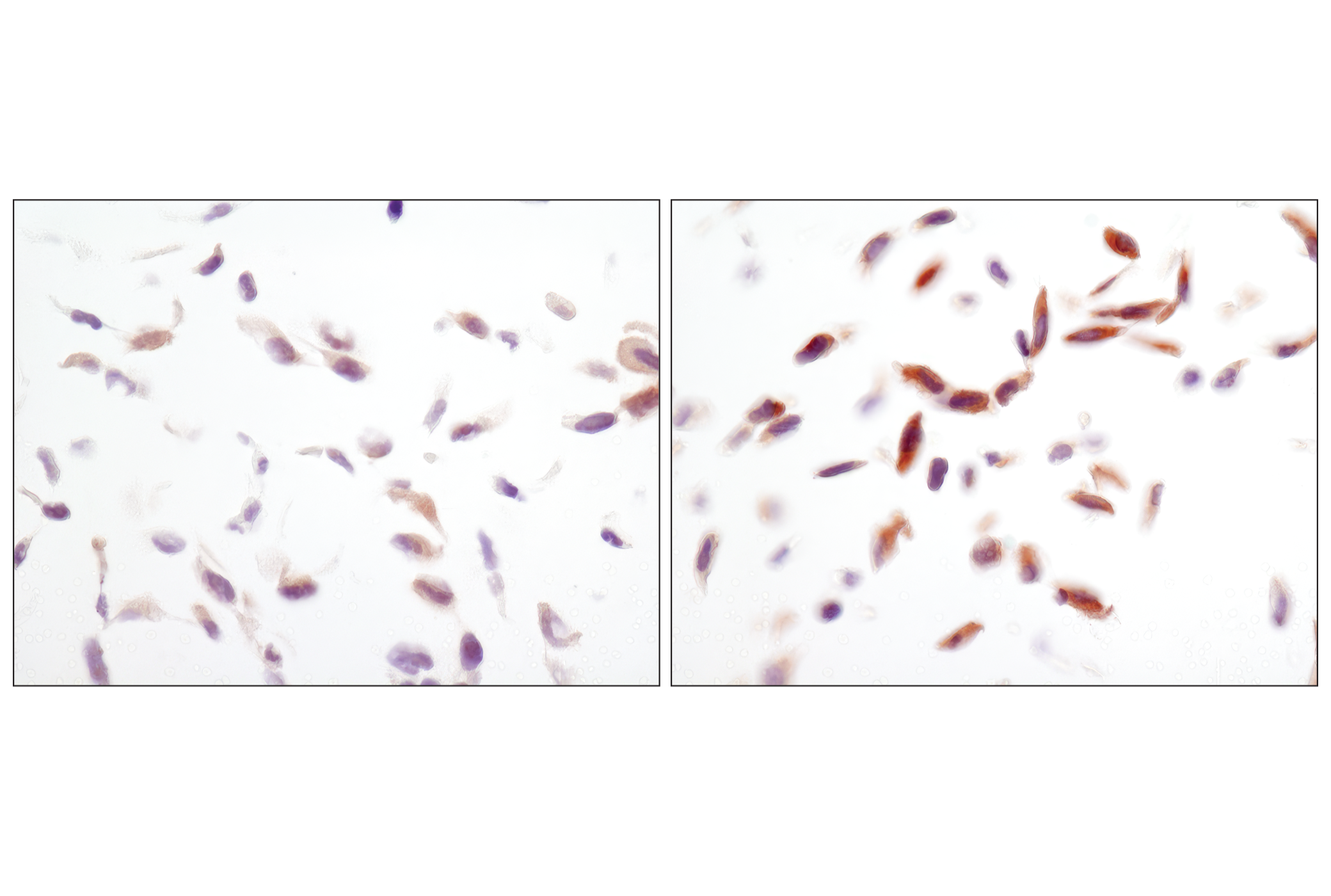
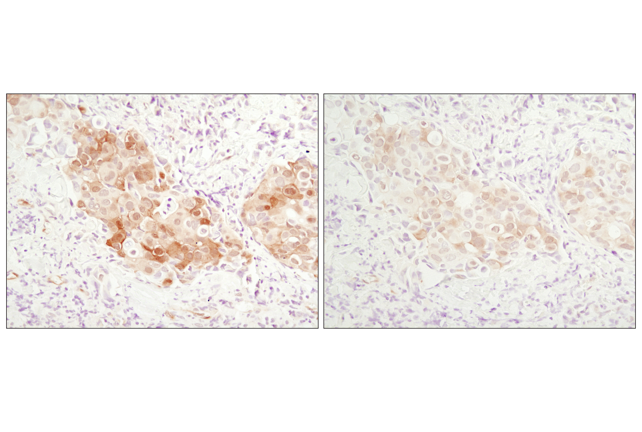
The triggering receptor expressed on myeloid cells 2 (TREM2) protein is an innate immune receptor that is expressed on the cell surface of microglia, macrophages, osteoclasts, and immature dendritic cells (1). The TREM2 protein plays a role in innate immunity and a rare functional variant (R47H) of TREM2 is associated with the late-onset risk of Alzheimer’s disease (AD) (1,2). Research studies using mouse models of AD indicate that deficiency and haploinsufficiency of TREM2 can lead to increased β-amyloid (Aβ) accumulation as a result of dysfunctional microglia response (3). Activation of TREM2 in mouse models of AD ameliorates several forms of AD pathology, likely through a microglia-specific mechanism (4,5). This mechanism is under intense investigation, but may involve TREM2-dependent maintenance microglia energetic and biosynthetic metabolism (6). Autophagy is one mechanism by which cellular metabolism is maintained and, in the absence of TREM2, several AMPK-dependent autophagy cell signaling pathways are enhanced. AMP-activated protein kinase (AMPK) is highly conserved from yeast to plants and animals and plays a key role in the regulation of energy homeostasis (7). The tumor suppressor LKB1, in association with accessory proteins STRAD and MO25, phosphorylates AMPKα at Thr172 in the activation loop, and this phosphorylation is required for AMPK activation (8-10). AMPK is further regulated by several proteins within a regulatory cell signaling pathway. The mammalian target of rapamycin (mTOR, FRAP, RAFT) is a Ser/Thr protein kinase (11) that functions as an ATP and amino acid sensor to balance nutrient availability and cell growth (12). mTOR is phosphorylated at Ser2448 via the PI3 kinase/Akt signaling pathway and autophosphorylated at Ser2481 (13). Akt, also referred to as PKB or Rac, is regulated by phosphorylation at Ser473 (14,15). The presence of autophagy marker Light Chain 3 (LC3) in autophagosomes and the conversion of LC3 to the lower migrating form, LC3-II, have been used as indicators of autophagy (16).
- Colonna, M. (2003) Nat Rev Immunol 3, 445-53.
- Boutajangout, A. and Wisniewski, T. (2013) Int J Cell Biol 2013, 576383.
- Wang, Y. et al. (2015) Cell 160, 1061-71.
- Schlepckow, K. et al. (2020) EMBO Mol Med 12, e11227.
- Wang, S. et al. (2020) J Exp Med 217, e20200785.
- Ulland, T.K. et al. (2017) Cell 170, 649-663.e13.
- Hardie, D.G. (2004) J Cell Sci 117, 5479-87.
- Hawley, S.A. et al. (1996) J Biol Chem 271, 27879-87.
- Lizcano, J.M. et al. (2004) EMBO J 23, 833-43.
- Shaw, R.J. et al. (2004) Proc Natl Acad Sci U S A 101, 3329-35.
- Sabatini, D.M. et al. (1994) Cell 78, 35-43.
- Dennis, P.B. et al. (2001) Science 294, 1102-5.
- Navé, B.T. et al. (1999) Biochem J 344 Pt 2, 427-31.
- Burgering, B.M. and Coffer, P.J. (1995) Nature 376, 599-602.
- Franke, T.F. et al. (1995) Cell 81, 727-36.
- Kabeya, Y. et al. (2004) J Cell Sci 117, 2805-12.
Background References
Trademarks and Patents
使用に関する制限
法的な権限を与えられたCSTの担当者が署名した書面によって別途明示的に合意された場合を除き、 CST、その関連会社または代理店が提供する製品には以下の条件が適用されます。お客様が定める条件でここに定められた条件に含まれるものを超えるもの、 または、ここに定められた条件と異なるものは、法的な権限を与えられたCSTの担当者が別途書面にて受諾した場合を除き、拒絶され、 いかなる効力も効果も有しません。
研究専用 (For Research Use Only) またはこれに類似する表示がされた製品は、 いかなる目的についても FDA または外国もしくは国内のその他の規制機関により承認、認可または許可を受けていません。 お客様は製品を診断もしくは治療目的で使用してはならず、また、製品に表示された内容に違反する方法で使用してはなりません。 CST が販売または使用許諾する製品は、エンドユーザーであるお客様に対し、使途を研究および開発のみに限定して提供されるものです。 診断、予防もしくは治療目的で製品を使用することまたは製品を再販売 (単独であるか他の製品等の一部であるかを問いません) もしくはその他の商業的利用の目的で購入することについては、CST から別途許諾を得る必要があります。 お客様は以下の事項を遵守しなければなりません。(a) CST の製品 (単独であるか他の資材と一緒であるかを問いません) を販売、使用許諾、貸与、寄付もしくはその他の態様で第三者に譲渡したり使用させたりしてはなりません。また、商用の製品を製造するために CST の製品を使用してはなりません。(b) 複製、改変、リバースエンジニアリング、逆コンパイル、 分解または他の方法により製品の構造または技術を解明しようとしてはなりません。また、 CST の製品またはサービスと競合する製品またはサービスを開発する目的で CST の製品を使用してはなりません。(c) CST の製品の商標、商号、ロゴ、特許または著作権に関する通知または表示を除去したり改変したりしてはなりません。(d) CST の製品をCST 製品販売条件(CST’s Product Terms of Sale) および該当する書面のみに従って使用しなければなりません。(e) CST の製品に関連してお客様が使用する第三者の製品またはサービスに関する使用許諾条件、 サービス提供条件またはこれに類する合意事項を遵守しなければなりません。