| Product Includes | Product # | Quantity | Mol. Wt | Isotype/Source |
|---|---|---|---|---|
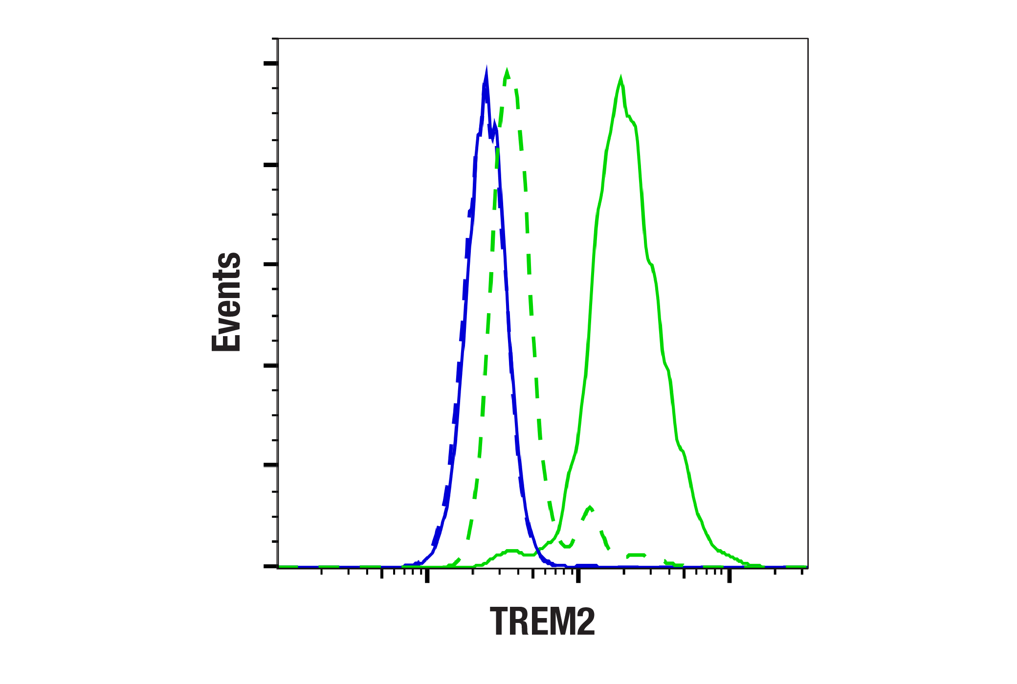
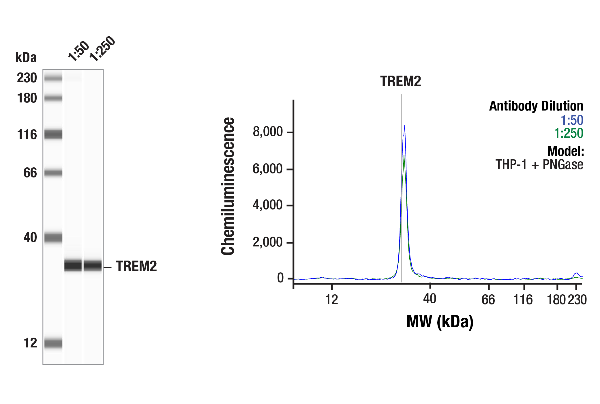
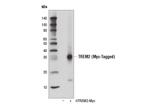
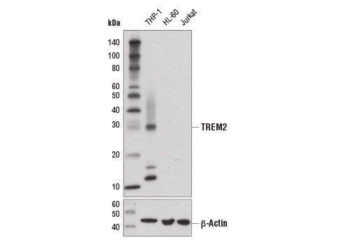
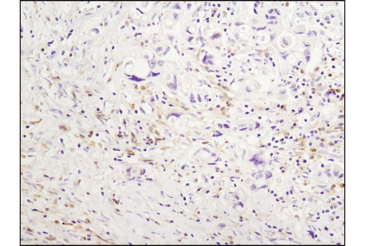
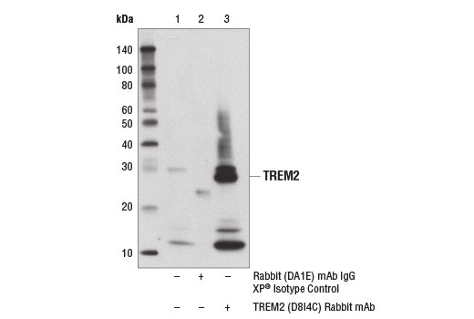
| TREM2 (D8I4C) Rabbit mAb | 91068 | 20 µl | 28 kDa | Rabbit IgG |
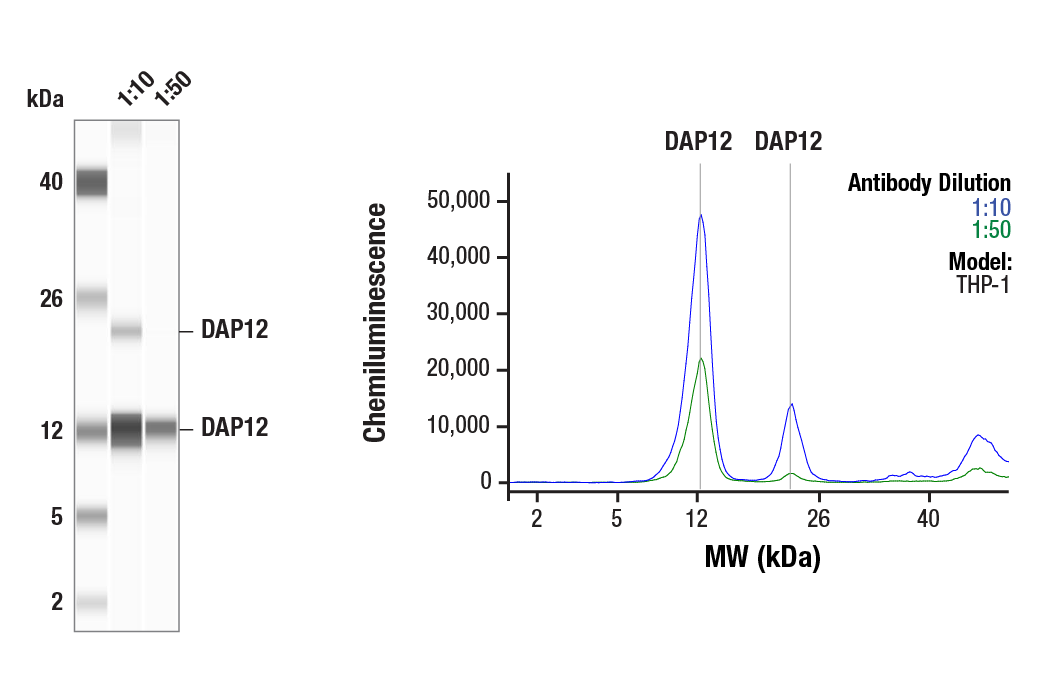
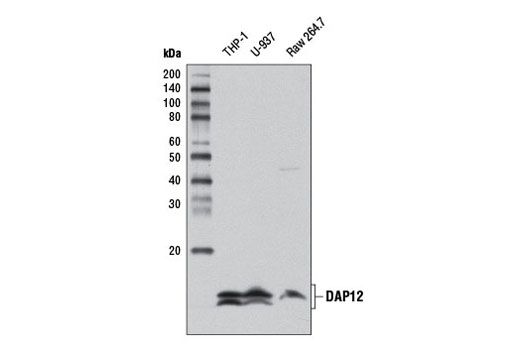
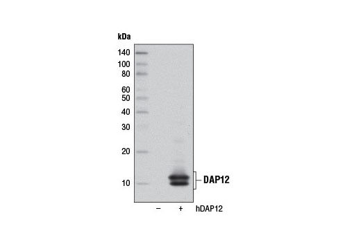
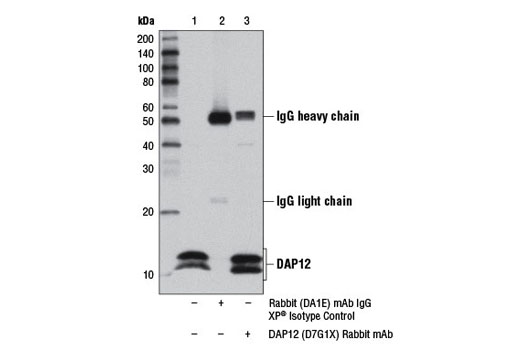
| DAP12 (D7G1X) Rabbit mAb | 12492 | 20 µl | 10, 12 kDa | Rabbit IgG |
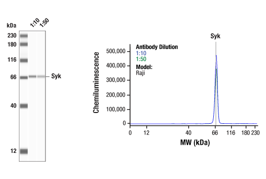
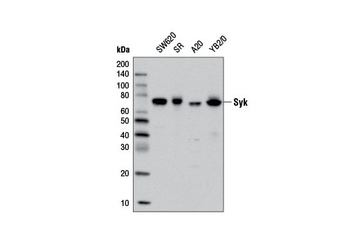
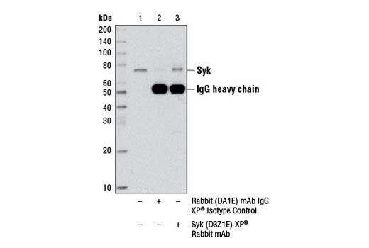
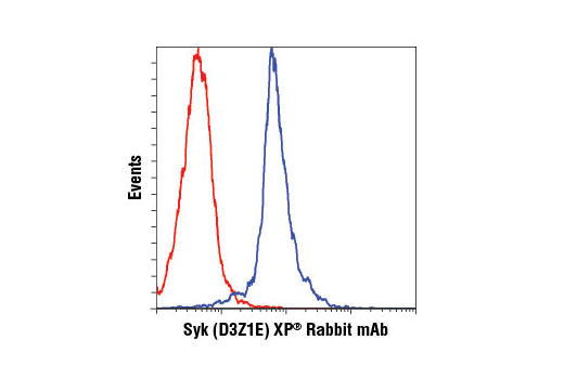
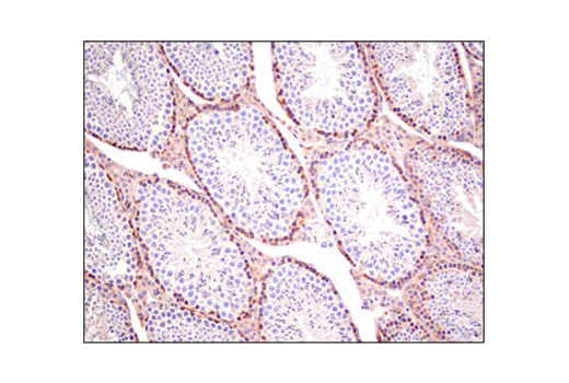
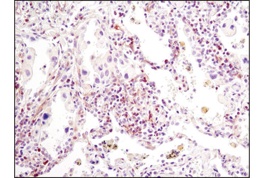
| Syk (D3Z1E) XP® Rabbit mAb | 13198 | 20 µl | 72 kDa | Rabbit IgG |
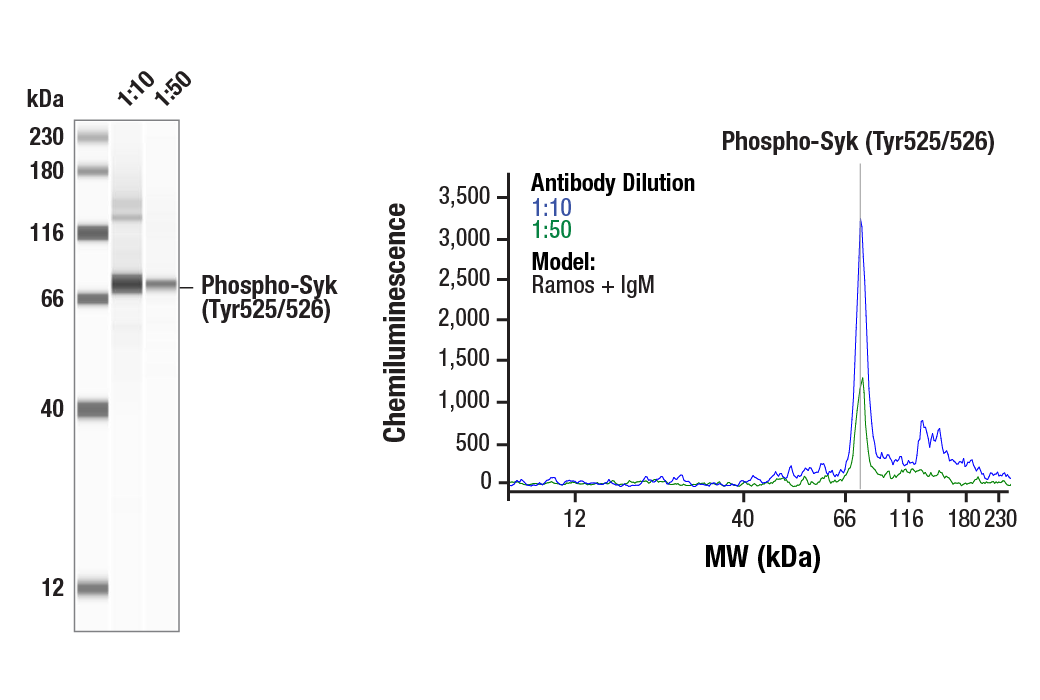
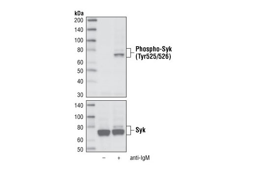
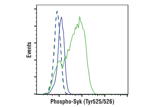
| Phospho-Syk (Tyr525/526) (C87C1) Rabbit mAb | 2710 | 20 µl | 72 kDa | Rabbit IgG |
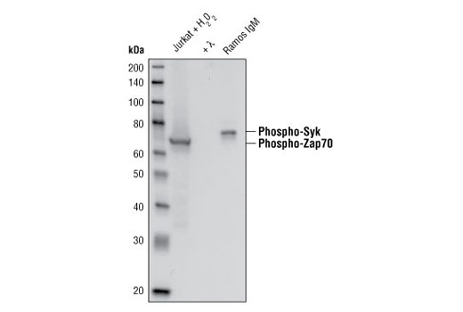
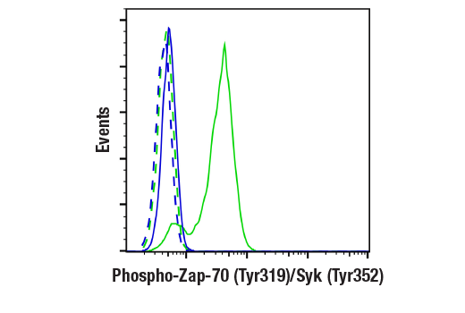
| Phospho-Zap-70 (Tyr319)/Syk (Tyr352) (65E4) Rabbit mAb | 2717 | 20 µl | 70, 72 kDa | Rabbit IgG |
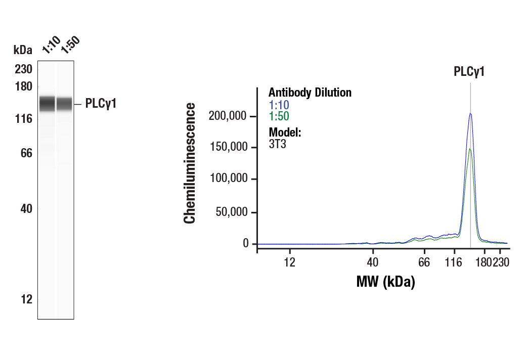
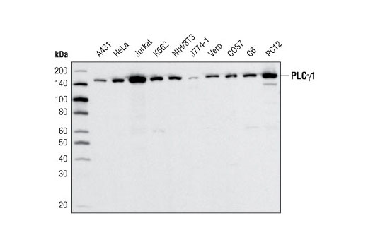
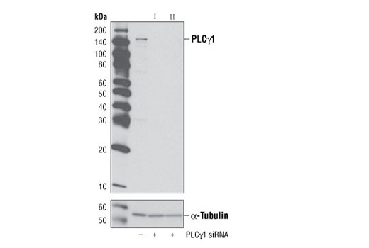
| PLCγ1 (D9H10) XP® Rabbit mAb | 5690 | 20 µl | 150 kDa | Rabbit IgG |
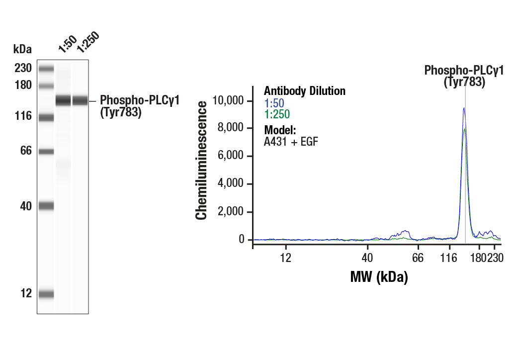
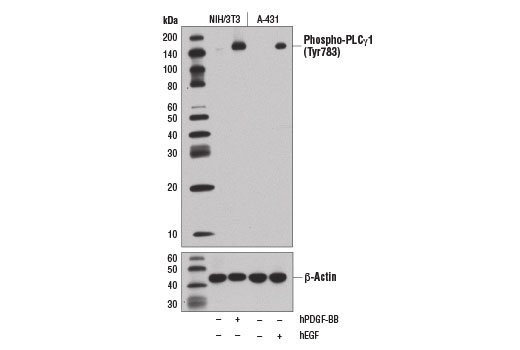
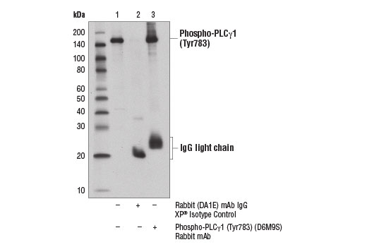
| Phospho-PLCγ1 (Tyr783) (D6M9S) Rabbit mAb | 14008 | 20 µl | 155 kDa | Rabbit IgG |
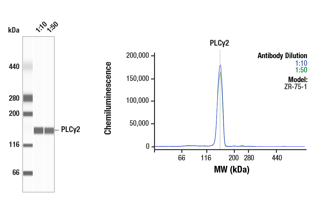
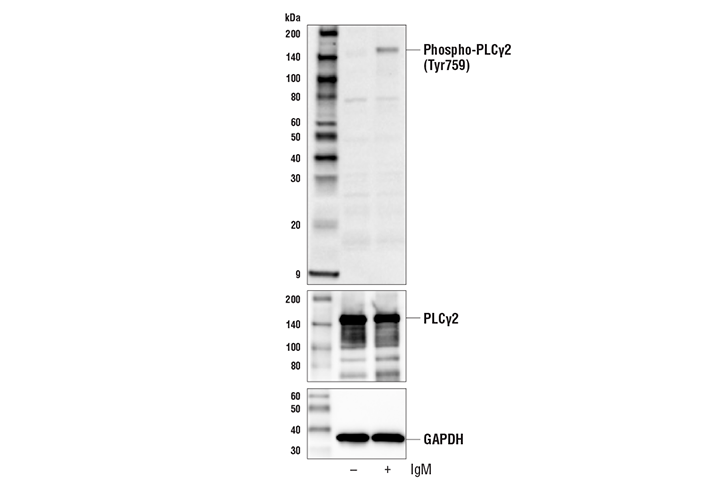
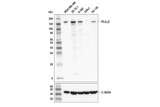
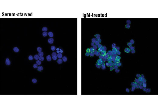
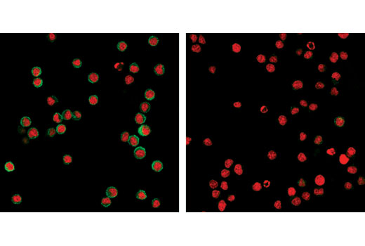
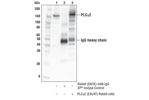
| PLCγ2 (E5U4T) Rabbit mAb | 55512 | 20 µl | 150 kDa | Rabbit IgG |
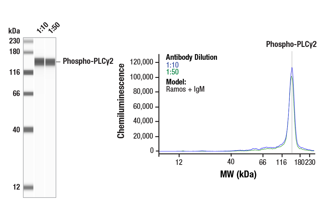
| Phospho-PLCγ2 (Tyr759) (E9E9Y) Rabbit mAb | 50535 | 20 µl | 150 kDa | Rabbit IgG |
| Anti-rabbit IgG, HRP-linked Antibody | 7074 | 100 µl | Goat |
Please visit cellsignal.com for individual component applications, species cross-reactivity, dilutions, protocols, and additional product information.
Storage
Background
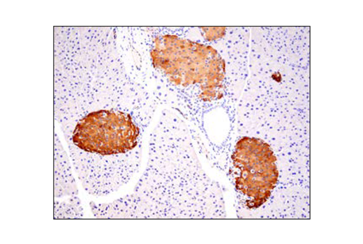
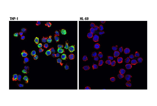
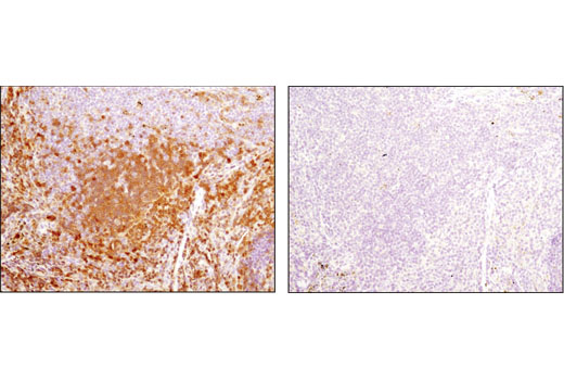
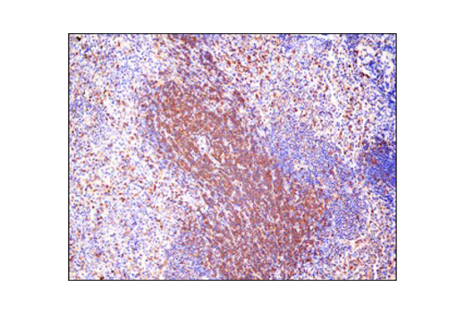
Microglia cells are resident macrophages of the brain that survey the brain environment and dynamically respond to maintain brain homeostasis. Microglial responses include phagocytosis of cellular debris, restricting sites injury or pathology, and/or releasing inflammatory signals to initiate an immune response. Such responses are important during normal development and during diseased states (1).Recently, the role of microglia in neurodegenerative disease pathology, particularly Alzheimer’s disease (AD), has been of intense investigation. Much of this work is driven by human genetic data that links microglia-enriched genes with AD progression (2). The triggering receptor expressed on myeloid cells 2 (TREM2) protein is an innate immune receptor that is expressed on the cell surface of microglia (3). TREM2 plays a role in innate immunity, and a rare functional variant (R47H) of the TREM2 gene is associated with the late-onset risk of AD (3,4). How TREM2 contributes to disease function is currently an active area of research (4,5), but might drive a number of microglial cellular functions ranging from microgliosis, phagocytosis, and cytokine release via a variety of signaling cascades triggered by TREM2.The TREM2 receptor is a single-pass type I membrane glycoprotein that consists of an extracellular immunoglobulin-like domain, a transmembrane domain, and a cytoplasmic tail. Ligands for TREM2 include phospholipids, apolipoproteins, and lipoproteins. Upon activation, TREM2 interacts with the tyrosine kinase-binding protein DNAX-activating protein 12 (DAP12, TYROBP) to form a receptor-signaling complex (6). Ligand binding by DAP12-associated receptors, including TREM2, results in phosphorylation of tyrosine residues within the DAP12 immunoreceptor tyrosine-based activation motif (ITAM) by Src family kinases; ITAM phosphorylation leads to activation of spleen tyrosine kinase (Syk) and downstream signaling cascades (7). Tyr525 and Tyr526 are located in the activation loop of the Syk kinase domain and phosphorylation at these residues (equivalent to Tyr519/520 of mouse Syk) is essential for Syk function (8). Syk phosphorylation is also a readout for β-amyloid triggered TREM2 activity (9). Phosphoinositide-specific phospholipase C γ 1/2 (PLCγ1/2) is reported to be down stream of Syk (10). Tyr352 of Syk is involved in the association of PLCγ1 (11); Syk-mediated phosphorylation PLCγ1 at Tyr783 activates PLCγ1 enzymatic activity (12). Interestingly, mutations in the microglia-enriched PLCγ2 gene are associated with AD (13,14,15).
- Kierdorf, K. and Prinz, M. (2017) J Clin Invest 127, 3201-9.
- McQuade, A. and Blurton-Jones, M. (2019) J Mol Biol.
- Colonna, M. (2003) Nat Rev Immunol 3, 445-53.
- Yeh, F.L. et al. (2017) Trends Mol Med 23, 512-33.
- Ulland, T.K. and Colonna, M. (2018) Nat Rev Neurol 14, 667-75.
- Peng, Q. et al. (2010) Sci Signal 3, ra38.
- Lanier, L.L. et al. (1998) Nature 391, 703-7.
- Zhang, J. et al. (2000) J Biol Chem 275, 35442-7.
- Zhao, Y. et al. (2018) Neuron 97, 1023-1031.e7.
- Kusuyama, J. et al. (2018) J Cell Physiol 233, 2549-59.
- Law, C.L. et al. (1996) Mol Cell Biol 16, 1305-15.
- Wang, Z. et al. (1998) Mol Cell Biol 18, 590-7.
- Magno, L. et al. (2019) Alzheimers Res Ther 11, 16.
- Sims, R. et al. (2017) Nat Genet 49, 1373-84.
- Dalmasso, M.C. et al. (2019) Transl Psychiatry 9, 55.
Background References
Trademarks and Patents
使用に関する制限
法的な権限を与えられたCSTの担当者が署名した書面によって別途明示的に合意された場合を除き、 CST、その関連会社または代理店が提供する製品には以下の条件が適用されます。お客様が定める条件でここに定められた条件に含まれるものを超えるもの、 または、ここに定められた条件と異なるものは、法的な権限を与えられたCSTの担当者が別途書面にて受諾した場合を除き、拒絶され、 いかなる効力も効果も有しません。
研究専用 (For Research Use Only) またはこれに類似する表示がされた製品は、 いかなる目的についても FDA または外国もしくは国内のその他の規制機関により承認、認可または許可を受けていません。 お客様は製品を診断もしくは治療目的で使用してはならず、また、製品に表示された内容に違反する方法で使用してはなりません。 CST が販売または使用許諾する製品は、エンドユーザーであるお客様に対し、使途を研究および開発のみに限定して提供されるものです。 診断、予防もしくは治療目的で製品を使用することまたは製品を再販売 (単独であるか他の製品等の一部であるかを問いません) もしくはその他の商業的利用の目的で購入することについては、CST から別途許諾を得る必要があります。 お客様は以下の事項を遵守しなければなりません。(a) CST の製品 (単独であるか他の資材と一緒であるかを問いません) を販売、使用許諾、貸与、寄付もしくはその他の態様で第三者に譲渡したり使用させたりしてはなりません。また、商用の製品を製造するために CST の製品を使用してはなりません。(b) 複製、改変、リバースエンジニアリング、逆コンパイル、 分解または他の方法により製品の構造または技術を解明しようとしてはなりません。また、 CST の製品またはサービスと競合する製品またはサービスを開発する目的で CST の製品を使用してはなりません。(c) CST の製品の商標、商号、ロゴ、特許または著作権に関する通知または表示を除去したり改変したりしてはなりません。(d) CST の製品をCST 製品販売条件(CST’s Product Terms of Sale) および該当する書面のみに従って使用しなければなりません。(e) CST の製品に関連してお客様が使用する第三者の製品またはサービスに関する使用許諾条件、 サービス提供条件またはこれに類する合意事項を遵守しなければなりません。