| Product Includes | Product # | Quantity | Mol. Wt | Isotype/Source |
|---|---|---|---|---|
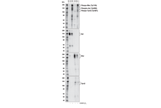
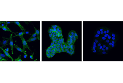
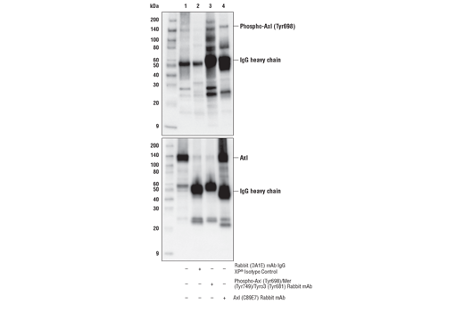
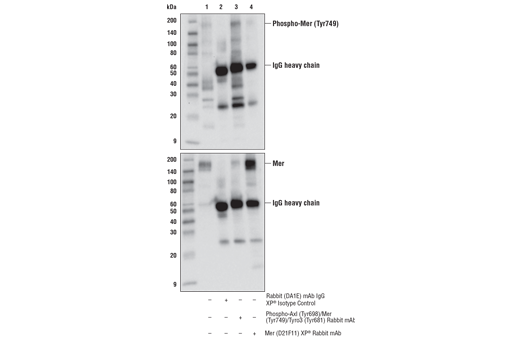
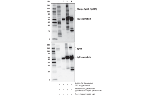
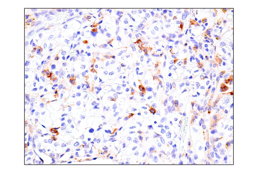
| Phospho-Axl (Tyr698)/Mer (Tyr749)/Tyro3 (Tyr681) (D6M4W) Rabbit mAb | 44463 | 20 µl | 110-130, 125-140, 180-200 kDa | Rabbit IgG |
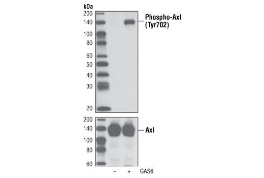
| Phospho-Axl (Tyr702) (D12B2) Rabbit mAb | 5724 | 20 µl | 138 kDa | Rabbit IgG |
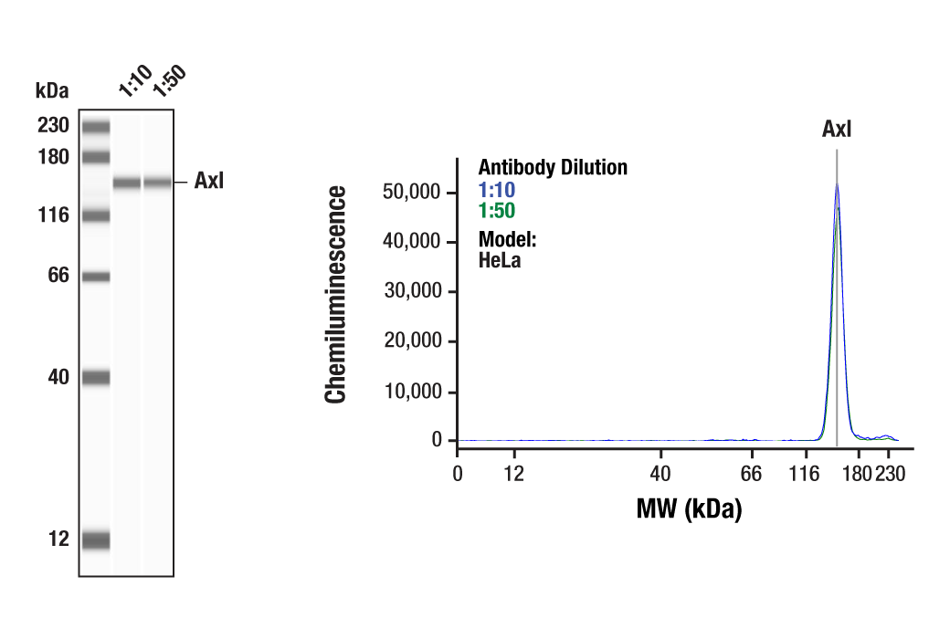
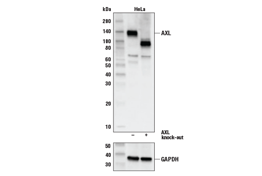
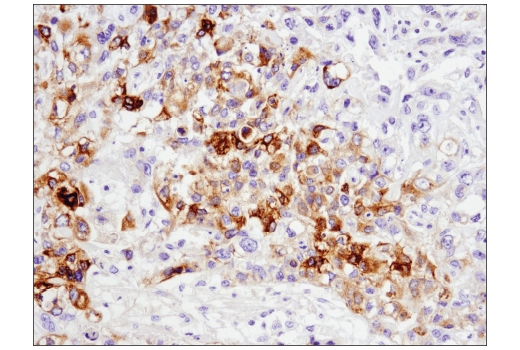
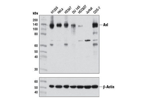
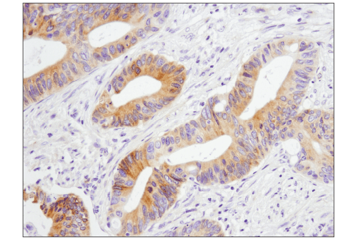
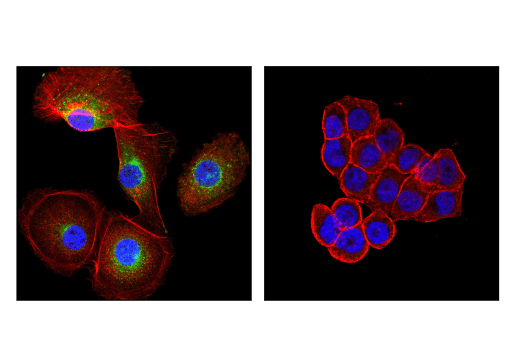
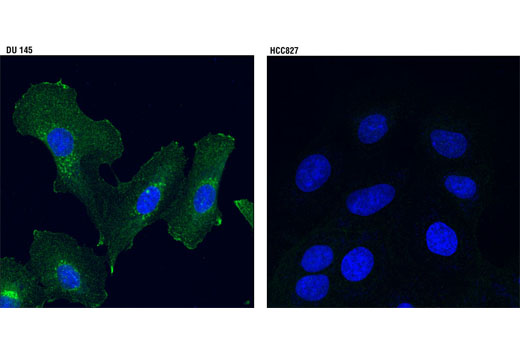
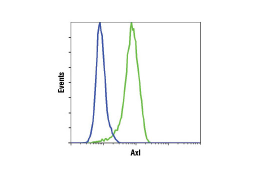
| Axl (C89E7) Rabbit mAb | 8661 | 20 µl | 138 kDa | Rabbit IgG |
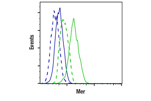
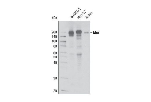
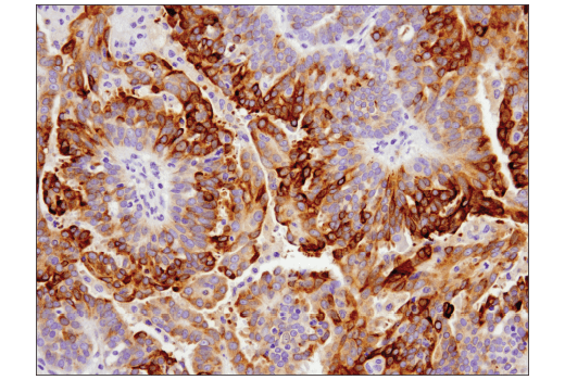
| Mer (D21F11) XP® Rabbit mAb | 4319 | 20 µl | 210 kDa | Rabbit IgG |
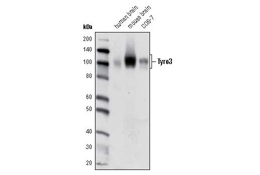
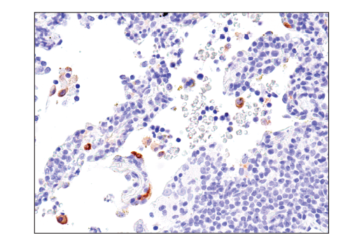
| Tyro3 (D38C6) Rabbit mAb | 5585 | 20 µl | 110, 130 kDa | Rabbit IgG |
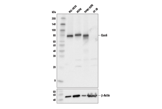
| GAS6 (D3A3G) Rabbit mAb | 67202 | 20 µl | 78 kDa | Rabbit IgG |
| Anti-rabbit IgG, HRP-linked Antibody | 7074 | 100 µl | Goat |
Please visit cellsignal.com for individual component applications, species cross-reactivity, dilutions, protocols, and additional product information.
Description
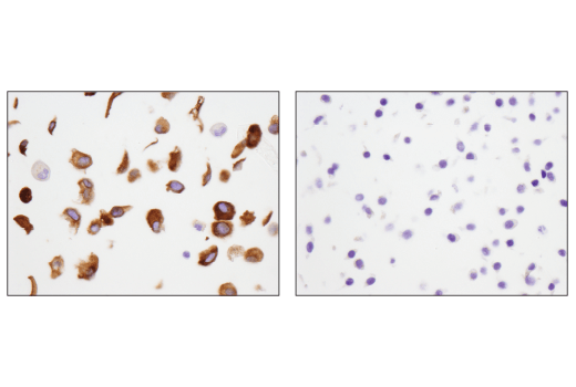
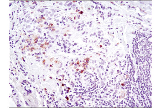
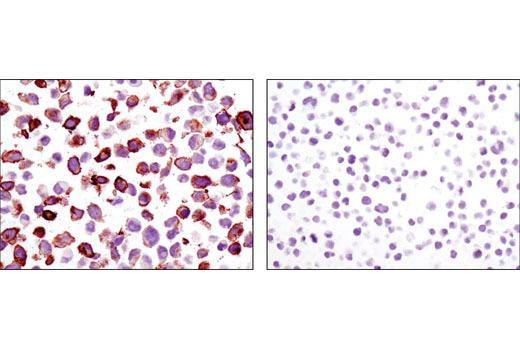
The Tyro/Axl/Mer Activation Antibody Sampler Kit provides an economical means of detecting the activation of TAM family members using phospho-specific and control antibodies. The kit includes enough antibodies to perform two western blot experiments with each primary antibody.
Storage
Background
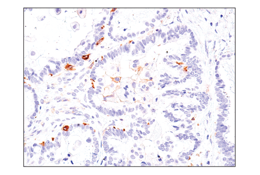
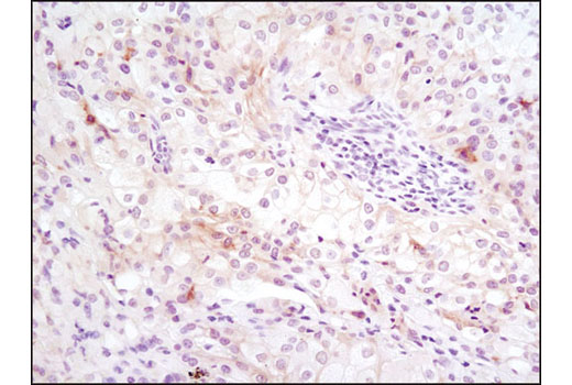
Axl, Mer and Tyro3 are three members of the TAM family receptor tyrosine kinase that share a common NCAM (neural adhesion molecule)-related extracellular domain and a conserved intracellular tyrosine kinase domain. These receptors bind common homologous vitamin K dependent protein GAS6 and protein S to activate downstream signaling pathways (1). TAM family receptors are involved in the development of immune, nervous, vascular and reproductive systems, autoimmune disease, cancer drug resistance and tumor immunity response (2-5). Axl (Tyr698), Axl (Tyr702), Mer Tyr(749) and Tyro3 (Tyr681) are conserved autophosphorylation sites located in the activation loop of the respective tyrosine kinase domains. Phosphorylation at these sites is required for full kinase activation of each of the corresponding receptors (6,7).
- Rothlin, C.V. et al. (2015) Annu Rev Immunol 33, 355-91.
- Burstyn-Cohen, T. (2017) Int J Dev Biol 61, 215-24.
- Bellan, M. et al. (2016) Int J Mol Sci 17, pii: E1807.
- Schoumacher, M. and Burbridge, M. (2017) Curr Oncol Rep 19, 19.
- Akalu, Y.T. et al. (2017) Immunol Rev 276, 165-77.
- Ling, L. et al. (1996) J Biol Chem 271, 18355-62.
- Onken, J. et al. (2017) Oncotarget 8, 50403-14.
Background References
Trademarks and Patents
使用に関する制限
法的な権限を与えられたCSTの担当者が署名した書面によって別途明示的に合意された場合を除き、 CST、その関連会社または代理店が提供する製品には以下の条件が適用されます。お客様が定める条件でここに定められた条件に含まれるものを超えるもの、 または、ここに定められた条件と異なるものは、法的な権限を与えられたCSTの担当者が別途書面にて受諾した場合を除き、拒絶され、 いかなる効力も効果も有しません。
研究専用 (For Research Use Only) またはこれに類似する表示がされた製品は、 いかなる目的についても FDA または外国もしくは国内のその他の規制機関により承認、認可または許可を受けていません。 お客様は製品を診断もしくは治療目的で使用してはならず、また、製品に表示された内容に違反する方法で使用してはなりません。 CST が販売または使用許諾する製品は、エンドユーザーであるお客様に対し、使途を研究および開発のみに限定して提供されるものです。 診断、予防もしくは治療目的で製品を使用することまたは製品を再販売 (単独であるか他の製品等の一部であるかを問いません) もしくはその他の商業的利用の目的で購入することについては、CST から別途許諾を得る必要があります。 お客様は以下の事項を遵守しなければなりません。(a) CST の製品 (単独であるか他の資材と一緒であるかを問いません) を販売、使用許諾、貸与、寄付もしくはその他の態様で第三者に譲渡したり使用させたりしてはなりません。また、商用の製品を製造するために CST の製品を使用してはなりません。(b) 複製、改変、リバースエンジニアリング、逆コンパイル、 分解または他の方法により製品の構造または技術を解明しようとしてはなりません。また、 CST の製品またはサービスと競合する製品またはサービスを開発する目的で CST の製品を使用してはなりません。(c) CST の製品の商標、商号、ロゴ、特許または著作権に関する通知または表示を除去したり改変したりしてはなりません。(d) CST の製品をCST 製品販売条件(CST’s Product Terms of Sale) および該当する書面のみに従って使用しなければなりません。(e) CST の製品に関連してお客様が使用する第三者の製品またはサービスに関する使用許諾条件、 サービス提供条件またはこれに類する合意事項を遵守しなければなりません。