| Product Includes | Product # | Quantity | Mol. Wt | Isotype/Source |
|---|---|---|---|---|
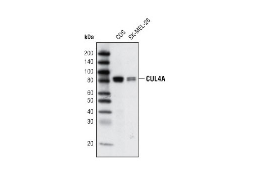
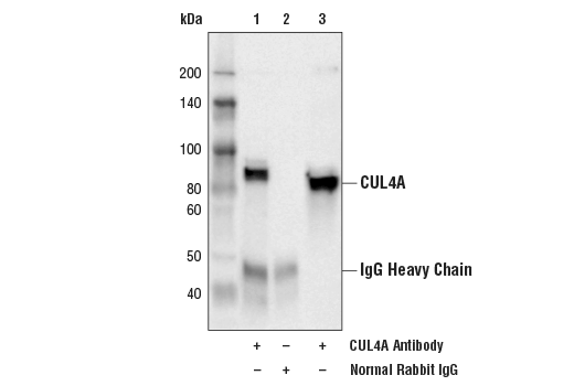
| CUL4A Antibody | 2699 | 20 µl | 80, 82 kDa | Rabbit |
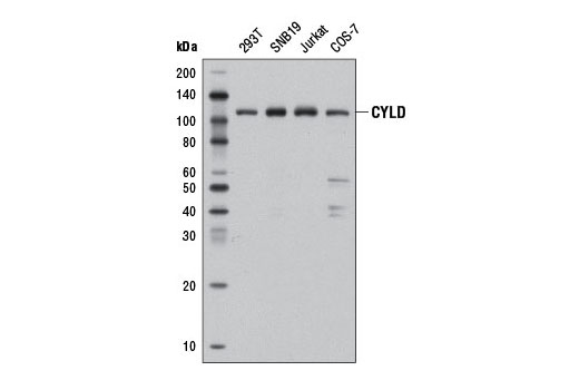
| CYLD (D6O5O) Rabbit mAb | 12797 | 20 µl | 109 kDa | Rabbit IgG |
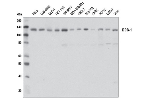
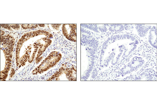
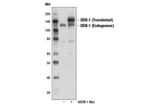
| DDB-1 (D4C8) Rabbit mAb | 6998 | 20 µl | 127 kDa | Rabbit IgG |
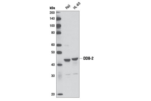
| DDB-2 (D4C4) Rabbit mAb | 5416 | 20 µl | 43 kDa | Rabbit IgG |
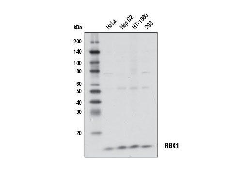
| RBX1 (D3J5I) Rabbit mAb | 11922 | 20 µl | 13 kDa | Rabbit IgG |
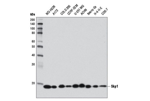
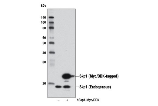
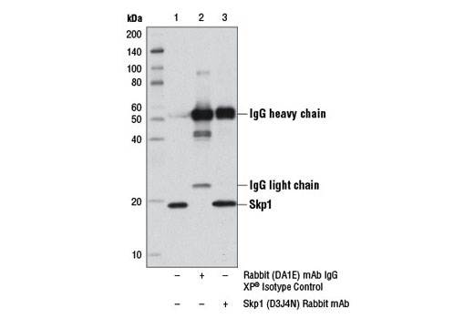
| Skp1 (D3J4N) Rabbit mAb | 12248 | 20 µl | 19 kDa | Rabbit IgG |
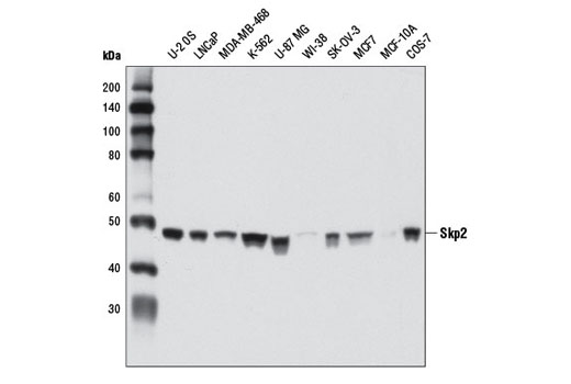
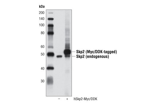
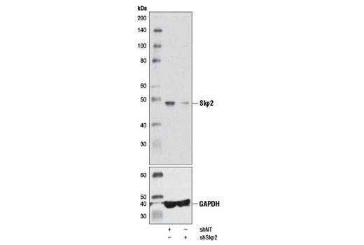
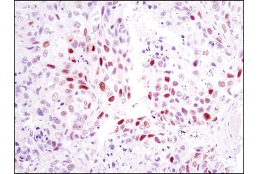
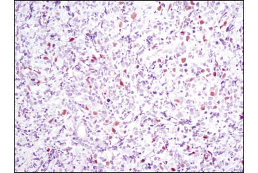
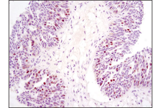
| Skp2 (D3G5) XP® Rabbit mAb | 2652 | 20 µl | 48 kDa | Rabbit IgG |
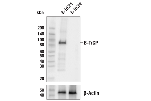
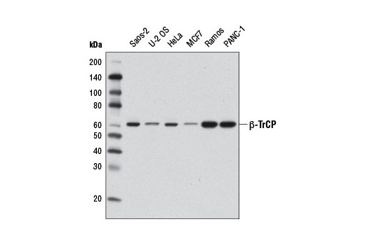
| β-TrCP (D12C8) Rabbit mAb | 11984 | 20 µl | 62 kDa | Rabbit IgG |
| Anti-rabbit IgG, HRP-linked Antibody | 7074 | 100 µl | Goat |
Please visit cellsignal.com for individual component applications, species cross-reactivity, dilutions, protocols, and additional product information.
Description
The Ubiquitin E3 Ligase Complex Antibody Sampler Kit provides an economical means to study multiple protein components of ubiquitin E3 ligase complexes. The kit includes enough antibody to perform two western blot experiments per primary antibody.
Storage
Background
Ubiquitin can be covalently linked to many cellular proteins by the ubiquitination process, which targets proteins for degradation by the 26S proteasome. Ubiquitin is first activated by forming a thiolester complex with the activation component E1. The activated ubiquitin is subsequently transferred to the ubiquitin-carrier protein E2, and then from E2 to ubiquitin ligase E3 for final delivery to the ε-NH2 of the target protein lysine residue (1-3). Research studies suggest that activated E2 associates transiently with E3, and the dissociation is a critical step for ubiquitination (4).
S phase kinase-associated protein 1 (Skp1) is a critical scaffold protein of the Skp1/CUL1/F-box (SCF) E3 ubiquitin ligase protein complex. Various F-box proteins (e.g. β-TrCP, Skp2) mediate an interaction with Skp1 via their defining and conserved domain of 40 amino acids and with substrates to be ubiquitinated (5). RING-box protein 1 (RBX1 or ROC1) is another essential component of the SCF complex (6). RBX1 mediates the neddylation of CUL1, which activates SCF E3 ligase by facilitating the ubiquitin transfer from E2 to substrates (7-9). The RING finger domain of RBX1 is required for ubiquitin ligation (10).
Cullin-4 (CUL4) is a member of the cullin family of related ubiquitin ligases (11). The carboxy-terminal domain of CUL4 interacts with Rbx1 and E2 enzyme while the amino-terminal CUL4 domain interacts with BPB domain of UV-damaged DNA binding protein DDB-1 to form a CUL4-DDB1 ubiquitin ligase complex (12). Damaged DNA-Binding Protein (DDB) consists of a 127 kDa subunit (DDB-1) and a 48 kDa subunit (DDB-2) that contribute to the formation of the UV-damaged DNA-binding protein complex (UV-DDB) (13-15). In conjunction with CUL4A and RBX1, the UV-DDB complex forms an E3 ubiquitin ligase that recognizes a broad spectrum of DNA lesions. The complex polyubiquitinates components of the nucleotide excision repair pathway (16-18).
- Ciechanover, A. (1998) EMBO J 17, 7151-60.
- Hochstrasser, M. (2000) Nat Cell Biol 2, E153-7.
- Hochstrasser, M. (2000) Science 289, 563-4.
- Deffenbaugh, A.E. et al. (2003) Cell 114, 611-22.
- DeSalle, L.M. and Pagano, M. (2001) FEBS Lett 490, 179-89.
- Zheng, N. et al. (2002) Nature 416, 703-9.
- Kamura, T. et al. (1999) Genes Dev 13, 2928-33.
- Morimoto, M. et al. (2003) Biochem Biophys Res Commun 301, 392-8.
- Pan, Z.Q. et al. (2004) Oncogene 23, 1985-97.
- Sun, Y. et al. (2001) Antioxid Redox Signal 3, 635-50.
- Petroski, M.D. and Deshaies, R.J. (2005) Nat Rev Mol Cell Biol 6, 9-20.
- Lee, J. and Zhou, P. (2007) Mol Cell 26, 775-80.
- Reardon, J.T. et al. (1993) J Biol Chem 268, 21301-8.
- Keeney, S. et al. (1993) J Biol Chem 268, 21293-300.
- Hwang, B.J. and Chu, G. (1993) Biochemistry 32, 1657-66.
- Chu, G. and Chang, E. (1990) Proc Natl Acad Sci U S A 87, 3324-7.
- Hirschfeld, S. et al. (1990) Mol Cell Biol 10, 2041-8.
- Payne, A. and Chu, G. (1994) Mutat Res 310, 89-102.
Background References
Trademarks and Patents
使用に関する制限
法的な権限を与えられたCSTの担当者が署名した書面によって別途明示的に合意された場合を除き、 CST、その関連会社または代理店が提供する製品には以下の条件が適用されます。お客様が定める条件でここに定められた条件に含まれるものを超えるもの、 または、ここに定められた条件と異なるものは、法的な権限を与えられたCSTの担当者が別途書面にて受諾した場合を除き、拒絶され、 いかなる効力も効果も有しません。
研究専用 (For Research Use Only) またはこれに類似する表示がされた製品は、 いかなる目的についても FDA または外国もしくは国内のその他の規制機関により承認、認可または許可を受けていません。 お客様は製品を診断もしくは治療目的で使用してはならず、また、製品に表示された内容に違反する方法で使用してはなりません。 CST が販売または使用許諾する製品は、エンドユーザーであるお客様に対し、使途を研究および開発のみに限定して提供されるものです。 診断、予防もしくは治療目的で製品を使用することまたは製品を再販売 (単独であるか他の製品等の一部であるかを問いません) もしくはその他の商業的利用の目的で購入することについては、CST から別途許諾を得る必要があります。 お客様は以下の事項を遵守しなければなりません。(a) CST の製品 (単独であるか他の資材と一緒であるかを問いません) を販売、使用許諾、貸与、寄付もしくはその他の態様で第三者に譲渡したり使用させたりしてはなりません。また、商用の製品を製造するために CST の製品を使用してはなりません。(b) 複製、改変、リバースエンジニアリング、逆コンパイル、 分解または他の方法により製品の構造または技術を解明しようとしてはなりません。また、 CST の製品またはサービスと競合する製品またはサービスを開発する目的で CST の製品を使用してはなりません。(c) CST の製品の商標、商号、ロゴ、特許または著作権に関する通知または表示を除去したり改変したりしてはなりません。(d) CST の製品をCST 製品販売条件(CST’s Product Terms of Sale) および該当する書面のみに従って使用しなければなりません。(e) CST の製品に関連してお客様が使用する第三者の製品またはサービスに関する使用許諾条件、 サービス提供条件またはこれに類する合意事項を遵守しなければなりません。