| Product Includes | Product # | Quantity | Mol. Wt | Isotype/Source |
|---|---|---|---|---|
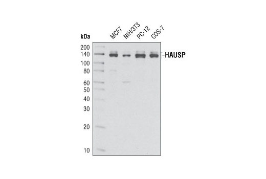
| HAUSP (D17C6) XP® Rabbit mAb | 4833 | 20 µl | 135, 140 kDa | Rabbit IgG |
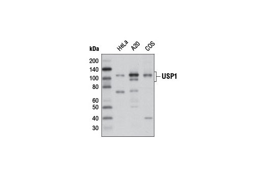
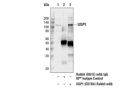
| USP1 (D37B4) Rabbit mAb | 8033 | 20 µl | 110 kDa | Rabbit IgG |
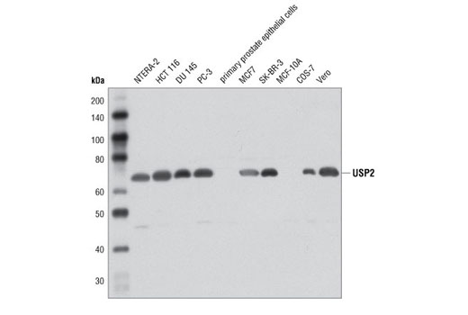
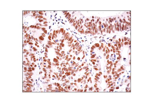
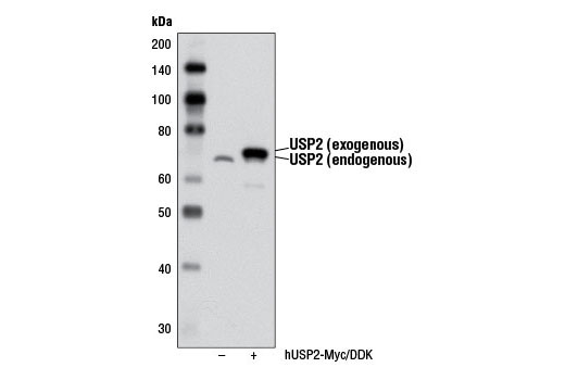
| USP2 Antibody | 8036 | 20 µl | 68 kDa | Rabbit |
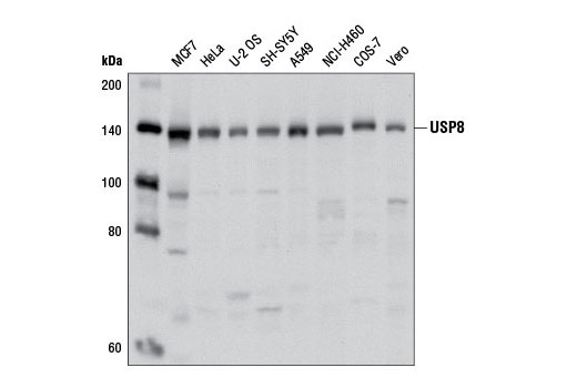
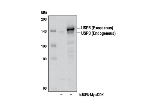
| USP8 Antibody | 8728 | 20 µl | 130 kDa | Rabbit |
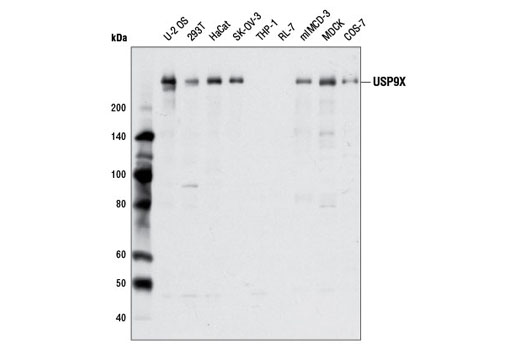
| USP9X Antibody | 5751 | 20 µl | 270 kDa | Rabbit |
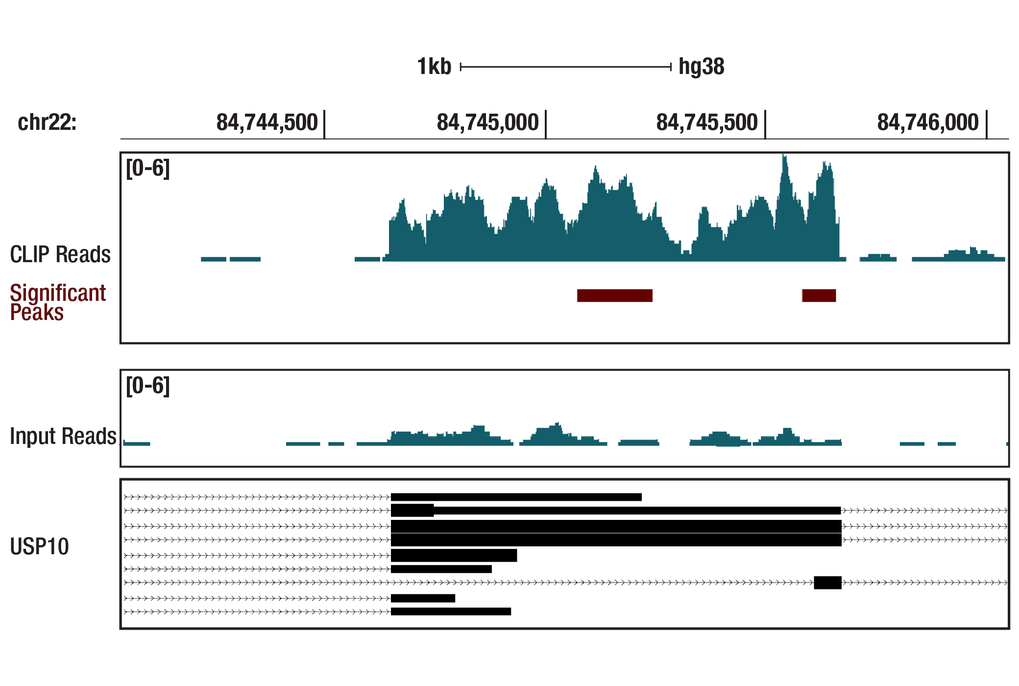
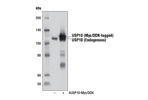
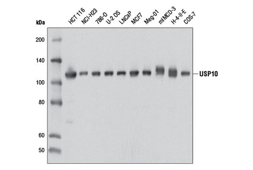
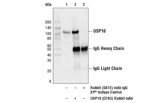
| USP10 (D7A5) Rabbit mAb | 8501 | 20 µl | 110 kDa | Rabbit IgG |
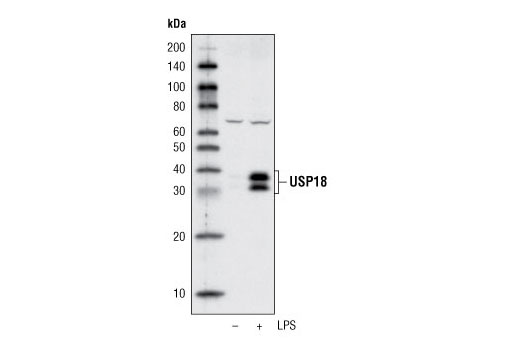
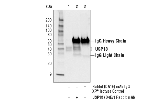
| USP18 (D4E7) Rabbit mAb | 4813 | 20 µl | 34, 39 kDa | Rabbit IgG |
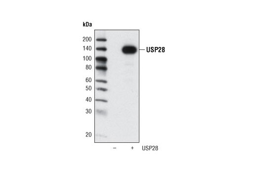
| USP28 Antibody | 4217 | 20 µl | 135 kDa | Rabbit |
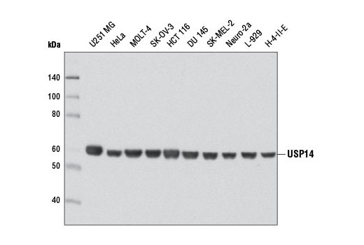
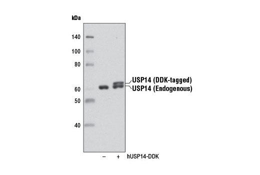
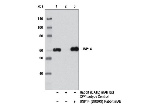
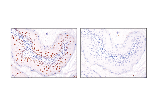
| USP14 (D8Q6S) Rabbit mAb | 11931 | 20 µl | 60 kDa | Rabbit IgG |
| Anti-rabbit IgG, HRP-linked Antibody | 7074 | 100 µl | Goat |
Please visit cellsignal.com for individual component applications, species cross-reactivity, dilutions, protocols, and additional product information.
Description
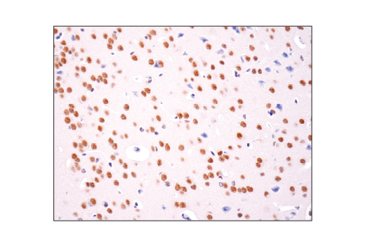
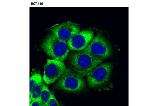
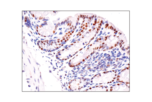
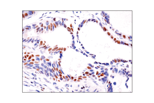
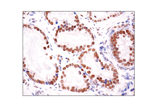
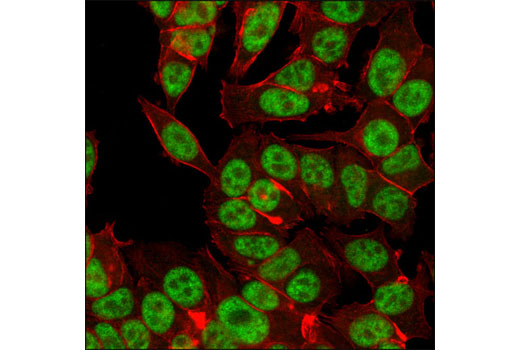
The USP Antibody Sampler Kit provides an economical means of detecting members of the ubiquitin-specific protease (USP) family. The kit includes enough primary antibody to perform two western blot experiments per primary antibody.
Storage
Background
Ubiquitinating enzymes (UBEs) catalyze protein ubiquitination, a reversible process countered by deubiquitinating enzyme (DUB) action (1,2). The ubiquitin-specific protease (USP) subfamily is one of five distinct groups of DUB enzymes. Ubiquitin-specific-processing protease 1 (USP1) is regulated in a cell cycle dependent manner by both transcriptional and ubiquitin-proteasomal mechanisms (3). Nuclear USP1 localizes to chromatin where it deubiquitinates monoubiquitinated FANCD2 and plays an important role in DNA damage repair and Chk1 protein stability (3,4). Ubiquitin-specific-processing protease 2 (USP2) contains C19 peptidase activity and is involved in ubiquitin recycling and disassembly of polymeric ubiquitin and ubiquitin-like protein complexes (5). USP2 is a putative oncoprotein that is highly over expressed in prostate cancer and drives tumor growth by binding and stabilizing fatty acid synthase through deubiquitination (6,7).
Herpesvirus-associated ubiquitin-specific protease (HAUSP, USP7) binds and deubiquitinates transcription factor p53 and regulator protein Mdm2, stabilizing both proteins (8,9). HAUSP modifies other ubiquitinated proteins, including FoxO family forkhead transcription factors and the mitotic stress checkpoint protein CHFR (10,11). Ubiquitin-specific protease 8 (USP8, UBPy) is a cysteine protease and growth-regulated enzyme that is essential for cell proliferation and survival (12,13). The catalytic domain of USP9X possesses cysteine peptidase activity that cleaves ubiquitin and polyubiquitin conjugates. USP9X may help stabilize adherens and tight junction molecules during epithelial cell polarization (14,15). USP10 is regulated at the posttranslational level through protein-protein interactions and phosphorylation. Interaction of USP10 with the Ras-GAP SH3 domain binding protein (G3BP) inhibits the ability of USP10 to catalyze ubiquitin chain disassembly (16). ATM-mediated phosphorylation of USP10 at Thr42 and Ser337 promotes USP10 stabilization and relocation from the cytoplasm to the nucleus, where it functions in p53 deubiquitination, stabilization, and activation in response to genotoxic stress (17).
USP14 is recruited to the proteasome through association with the PSMD2 (S2/hRPN1) subunit of the 19S regulatory particle, where it may antagonize substrate degradation (18,19). USP14 trims ubiquitin residues from distal polyubiquitin chain ends, decreasing chain affinity for proteasome ubiquitin receptors and allowing for enhanced substrate stability (20,21). USP18 (UBP43) catalyzes the removal of the interferon-regulated, ubiquitin-like protein ISG15 from conjugated proteins (22). Removal of ISG15 from target proteins maintains a critical balance of cellular ISG15-conjugated proteins, which is important for normal development and brain function (23,24). USP28 can bind, deubiquitinate and stabilize several DNA-damage pathway proteins, including p53BP1 and Chk2 (25). USP28 plays an important role in Myc-related signaling as it catalyzes Myc deubiquitination and promotes Myc stabilization, which contributes to tumor-cell growth (26).
- Nijman, S.M. et al. (2005) Cell 123, 773-86.
- Nalepa, G. et al. (2006) Nat Rev Drug Discov 5, 596-613.
- Nijman, S.M. et al. (2005) Mol Cell 17, 331-9.
- Guervilly, J.H. et al. (2011) Hum Mol Genet 20, 2171-81.
- Wilkinson, K.D. (1997) FASEB J 11, 1245-56.
- Graner, E. et al. (2004) Cancer Cell 5, 253-61.
- Priolo, C. et al. (2006) Cancer Res 66, 8625-32.
- Li, M. et al. (2002) Nature 416, 648-53.
- Brooks, C.L. et al. (2007) Oncogene 26, 7262-6.
- van der Horst, A. et al. (2006) Nat Cell Biol 8, 1064-73.
- Oh, Y.M. et al. (2007) Biochem Biophys Res Commun 357, 615-9.
- Naviglio, S. et al. (1998) EMBO J 17, 3241-50.
- Niendorf, S. et al. (2007) Mol Cell Biol 27, 5029-39.
- Murray, R.Z. et al. (2004) Mol Biol Cell 15, 1591-9.
- Théard, D. et al. (2010) EMBO J 29, 1499-509.
- Soncini, C. et al. (2001) Oncogene 20, 3869-79.
- Yuan, J. et al. (2010) Cell 140, 384-96.
- Lee, B.H. et al. (2010) Nature 467, 179-84.
- Koulich, E. et al. (2008) Mol Biol Cell 19, 1072-82.
- Hanna, J. et al. (2006) Cell 127, 99-111.
- Thrower, J.S. et al. (2000) EMBO J 19, 94-102.
- Malakhov, M.P. et al. (2002) J Biol Chem 277, 9976-81.
- Rempel, L.A. et al. (2007) Reprod Biol Endocrinol 5, 13.
- Ritchie, K.J. et al. (2002) Genes Dev 16, 2207-12.
- Zhang, D. et al. (2006) Cell 126, 529-42.
- Popov, N. et al. (2007) Nat Cell Biol 9, 765-74.
Background References
Trademarks and Patents
使用に関する制限
法的な権限を与えられたCSTの担当者が署名した書面によって別途明示的に合意された場合を除き、 CST、その関連会社または代理店が提供する製品には以下の条件が適用されます。お客様が定める条件でここに定められた条件に含まれるものを超えるもの、 または、ここに定められた条件と異なるものは、法的な権限を与えられたCSTの担当者が別途書面にて受諾した場合を除き、拒絶され、 いかなる効力も効果も有しません。
研究専用 (For Research Use Only) またはこれに類似する表示がされた製品は、 いかなる目的についても FDA または外国もしくは国内のその他の規制機関により承認、認可または許可を受けていません。 お客様は製品を診断もしくは治療目的で使用してはならず、また、製品に表示された内容に違反する方法で使用してはなりません。 CST が販売または使用許諾する製品は、エンドユーザーであるお客様に対し、使途を研究および開発のみに限定して提供されるものです。 診断、予防もしくは治療目的で製品を使用することまたは製品を再販売 (単独であるか他の製品等の一部であるかを問いません) もしくはその他の商業的利用の目的で購入することについては、CST から別途許諾を得る必要があります。 お客様は以下の事項を遵守しなければなりません。(a) CST の製品 (単独であるか他の資材と一緒であるかを問いません) を販売、使用許諾、貸与、寄付もしくはその他の態様で第三者に譲渡したり使用させたりしてはなりません。また、商用の製品を製造するために CST の製品を使用してはなりません。(b) 複製、改変、リバースエンジニアリング、逆コンパイル、 分解または他の方法により製品の構造または技術を解明しようとしてはなりません。また、 CST の製品またはサービスと競合する製品またはサービスを開発する目的で CST の製品を使用してはなりません。(c) CST の製品の商標、商号、ロゴ、特許または著作権に関する通知または表示を除去したり改変したりしてはなりません。(d) CST の製品をCST 製品販売条件(CST’s Product Terms of Sale) および該当する書面のみに従って使用しなければなりません。(e) CST の製品に関連してお客様が使用する第三者の製品またはサービスに関する使用許諾条件、 サービス提供条件またはこれに類する合意事項を遵守しなければなりません。