| Product Includes | Product # | Quantity | Mol. Wt | Isotype/Source |
|---|---|---|---|---|
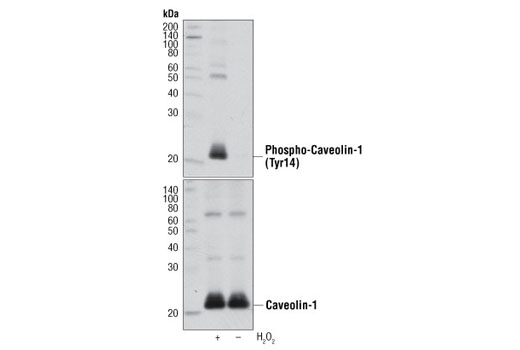
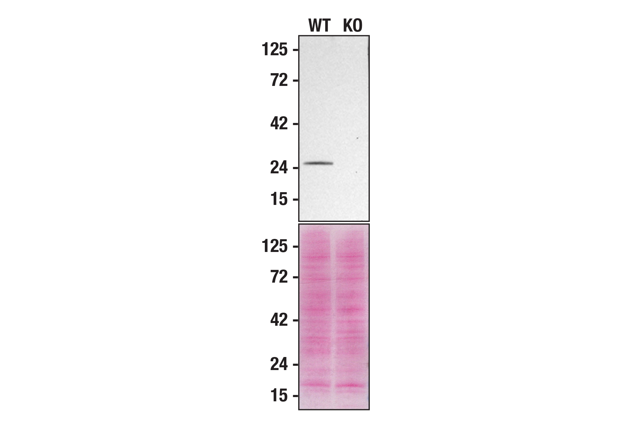
| Phospho-Caveolin-1 (Tyr14) Antibody | 3251 | 20 µl | 23, 25 kDa | Rabbit |
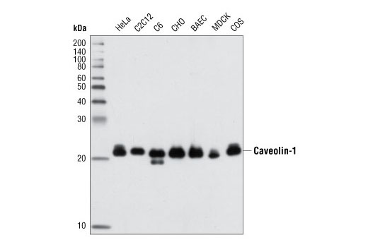
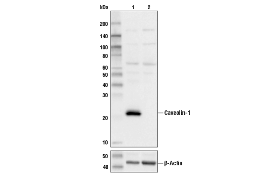
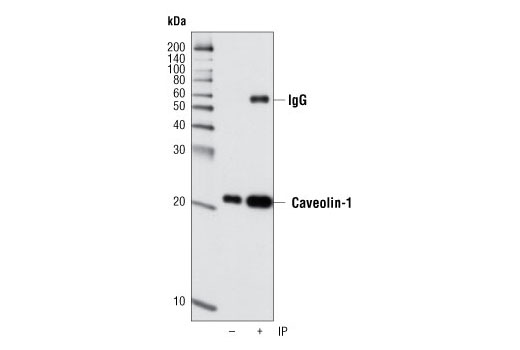
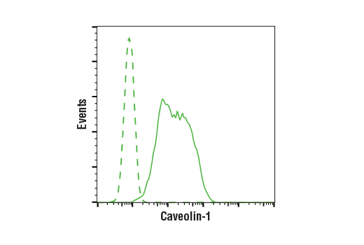
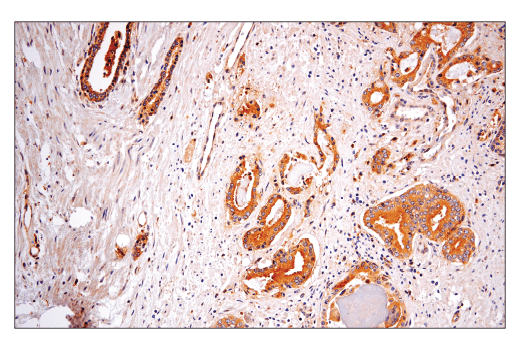
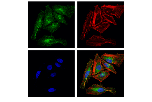
| Caveolin-1 (D46G3) XP® Rabbit mAb | 3267 | 20 µl | 21, 24 kDa | Rabbit IgG |
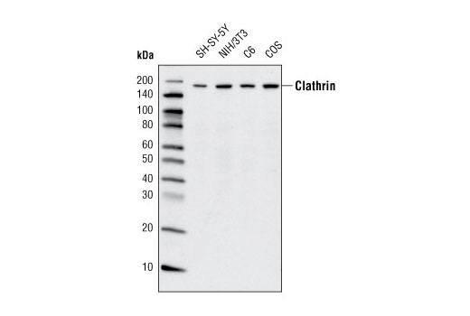
| Clathrin Heavy Chain (D3C6) XP® Rabbit mAb | 4796 | 20 µl | 190 kDa | Rabbit IgG |
| APPL1 (D83H4) XP® Rabbit mAb | 3858 | 20 µl | 82 kDa | Rabbit IgG |
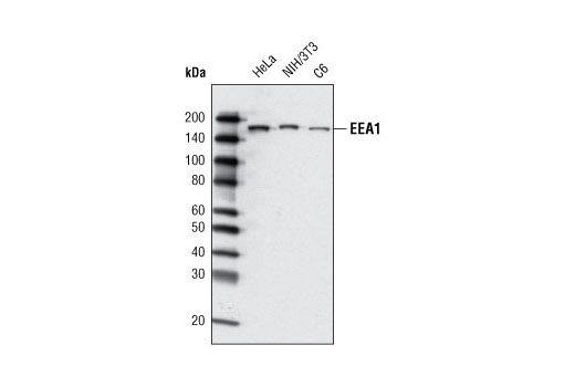
| EEA1 (C45B10) Rabbit mAb | 3288 | 20 µl | 170 kDa | Rabbit IgG |
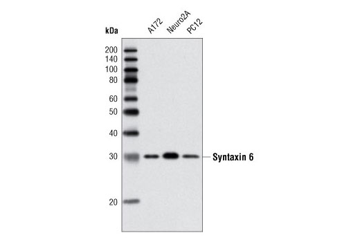
| Syntaxin 6 (C34B2) Rabbit mAb | 2869 | 20 µl | 32 kDa | Rabbit IgG |
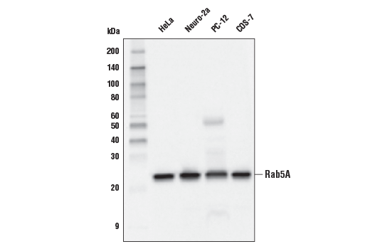
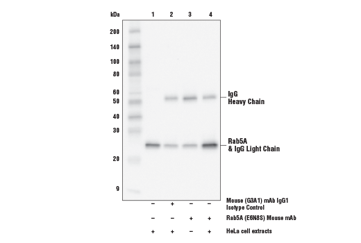
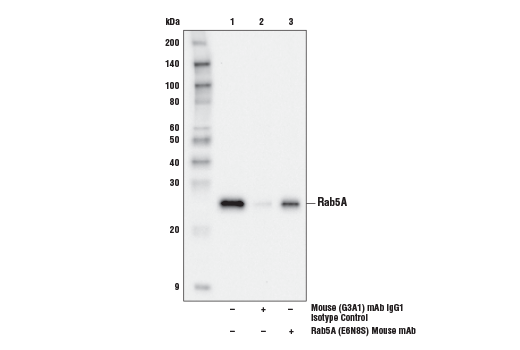
| Rab5A (E6N8S) Mouse mAb | 46449 | 20 µl | 25 kDa | Mouse IgG1 |
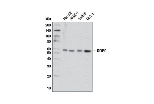
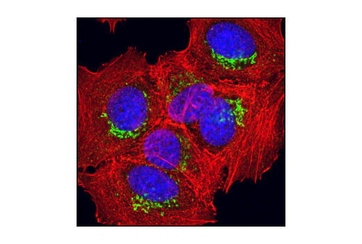
| GOPC (D10A12) Rabbit mAb | 8576 | 20 µl | 59 kDa | Rabbit IgG |
| Anti-rabbit IgG, HRP-linked Antibody | 7074 | 100 µl | Goat | |
| Anti-mouse IgG, HRP-linked Antibody | 7076 | 100 µl | Horse |
Please visit cellsignal.com for individual component applications, species cross-reactivity, dilutions, protocols, and additional product information.
Description
The Vesicle Trafficking Antibody Sampler kit provides an economical means to analyze proteins involved in the intracellular transport of cargo proteins. This kit includes enough primary and secondary antibody to perform two western blot experiments.
Storage
Background
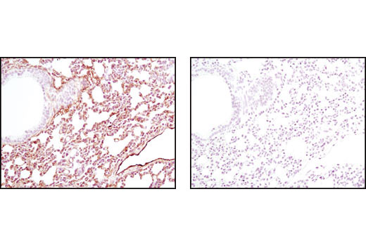
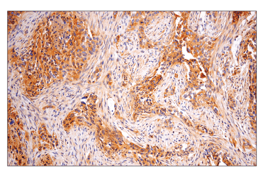
Vesicle trafficking is an integral cellular process and the associated proteins involved also play major roles in other signaling pathways. Caveolins are involved in diverse biological functions including vesicular trafficking, cholesterol homeostasis, cell adhesion, apoptosis, and are also indicated in neurodegenerative disease (1). It is believed that caveolins serve as scaffolding proteins for the integration of signal transduction. Phosphorylation at Tyr14 is essential for caveolin association with SH2 or PTB domain-containing adaptor proteins, such as GRB7 (2-4).
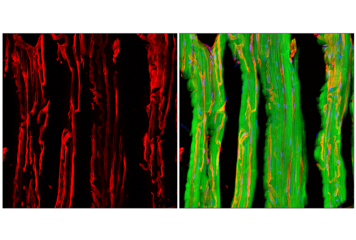
Clathrin-coated vesicles provide for the intracellular transport of proteins following endocytosis and during multiple vesicle trafficking pathways. Vesicles form at specialized areas of the cell membrane where clathrin and associated proteins form clathrin-coated pits. Invagination of these cell membrane-associated pits internalizes proteins and forms an intracellular clathrin-coated vesicle (5,6). Clathrin is the most abundant protein in these vesicles and is present as a basic assembly unit called a triskelion. Each clathrin triskelion is composed of three clathrin heavy chains and three clathrin light chains. Clathrin heavy chain proteins are composed of several functional domains that associate with other vesicle proteins (6).
The APPL1 multidomain adaptor protein is a BAR-domain protein family member that is involved in membrane trafficking within a number of signal transduction pathways (7).
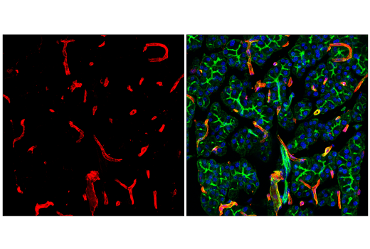
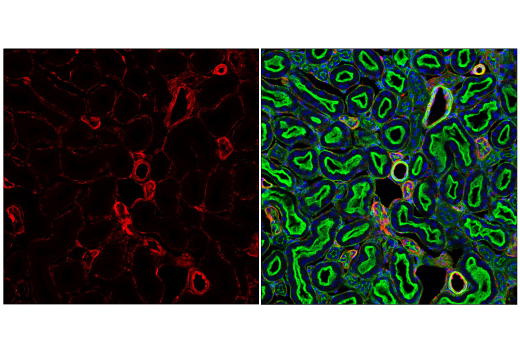
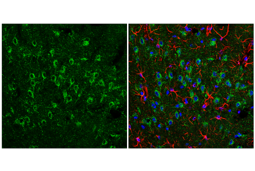
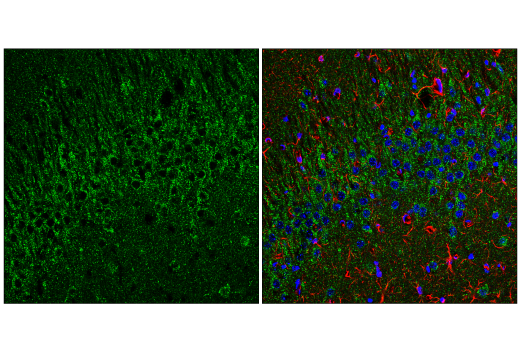
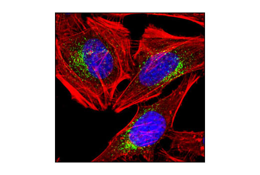
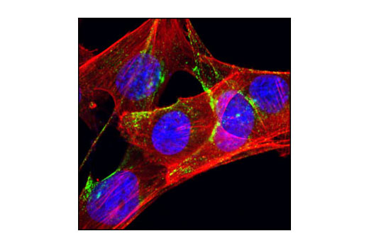
EEA1 is an early endosomal marker and a Rab5 effector protein essential for early endosomal membrane fusion and trafficking (8,9). Syntaxin 6 is a ubiquitously expressed S25C family member of the SNARE proteins (10,11). Syntaxin 6 protein is localized to the trans-Golgi and within endosomes and regulates membrane trafficking by partnering with a variety of other SNARE proteins (12-14). It has two coiled-coil domains (CC1 and CC2) located in the amino-terminal region and a PDZ domain in the carboxy-terminal region (15). The CC2 domain and its adjacent linker region mediate the association of GOPC with the Golgi protein golgin-160 and the Q-SNARE protein syntaxin 6 (15,16). The PDZ domain of GOPC interacts with the carboxy terminus of target proteins to mediate target protein vesicular trafficking and surface expression (17-20).
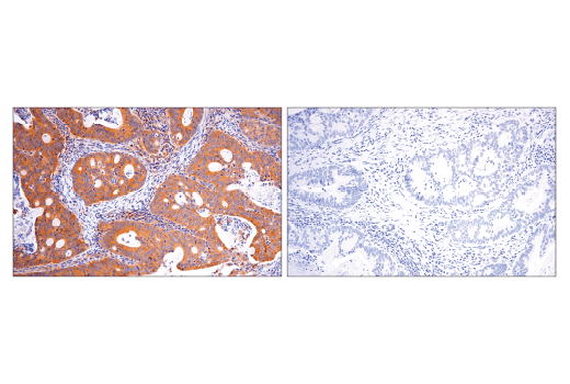
Rab5 is a member of the Ras superfamily of small Rab GTPases. Rab5 is localized at the plasma membrane and early endosomes and functions as a key regulator of vesicular trafficking during early endocytosis (21).
- Smart, E.J. et al. (1999) Mol Cell Biol 19, 7289-304.
- Nomura, R. and Fujimoto, T. (1999) Mol Biol Cell 10, 975-86.
- Volonté, D. et al. (2001) J Biol Chem 276, 8094-103.
- Lee, H. et al. (2000) Mol Endocrinol 14, 1750-75.
- Rodriguez-Boulan, E. et al. (2005) Nat Rev Mol Cell Biol 6, 233-47.
- Mousavi, S.A. et al. (2004) Biochem J 377, 1-16.
- Habermann, B. (2004) EMBO Rep 5, 250-5.
- Mu, F.T. et al. (1995) J Biol Chem 270, 13503-11.
- Christoforidis, S. et al. (1999) Nature 397, 621-5.
- Bock, J.B. et al. (2001) Nature 409, 839-41.
- Bock, J.B. et al. (1996) J Biol Chem 271, 17961-5.
- Wendler, F. and Tooze, S. (2001) Traffic 2, 606-11.
- Bock, J.B. et al. (1997) Mol Biol Cell 8, 1261-71.
- Mallard, F. et al. (2002) J Cell Biol 156, 653-64.
- Charest, A. et al. (2001) J Biol Chem 276, 29456-65.
- Hicks, S.W. and Machamer, C.E. (2005) J Biol Chem 280, 28944-51.
- Cheng, J. et al. (2002) J Biol Chem 277, 3520-9.
- He, J. et al. (2004) J Biol Chem 279, 50190-6.
- Wente, W. et al. (2005) J Biol Chem 280, 32419-25.
- Ito, H. et al. (2006) Biochem J 397, 389-98.
- Zerial, M. and McBride, H. (2001) Nat Rev Mol Cell Biol 2, 107-17.
Background References
Trademarks and Patents
使用に関する制限
法的な権限を与えられたCSTの担当者が署名した書面によって別途明示的に合意された場合を除き、 CST、その関連会社または代理店が提供する製品には以下の条件が適用されます。お客様が定める条件でここに定められた条件に含まれるものを超えるもの、 または、ここに定められた条件と異なるものは、法的な権限を与えられたCSTの担当者が別途書面にて受諾した場合を除き、拒絶され、 いかなる効力も効果も有しません。
研究専用 (For Research Use Only) またはこれに類似する表示がされた製品は、 いかなる目的についても FDA または外国もしくは国内のその他の規制機関により承認、認可または許可を受けていません。 お客様は製品を診断もしくは治療目的で使用してはならず、また、製品に表示された内容に違反する方法で使用してはなりません。 CST が販売または使用許諾する製品は、エンドユーザーであるお客様に対し、使途を研究および開発のみに限定して提供されるものです。 診断、予防もしくは治療目的で製品を使用することまたは製品を再販売 (単独であるか他の製品等の一部であるかを問いません) もしくはその他の商業的利用の目的で購入することについては、CST から別途許諾を得る必要があります。 お客様は以下の事項を遵守しなければなりません。(a) CST の製品 (単独であるか他の資材と一緒であるかを問いません) を販売、使用許諾、貸与、寄付もしくはその他の態様で第三者に譲渡したり使用させたりしてはなりません。また、商用の製品を製造するために CST の製品を使用してはなりません。(b) 複製、改変、リバースエンジニアリング、逆コンパイル、 分解または他の方法により製品の構造または技術を解明しようとしてはなりません。また、 CST の製品またはサービスと競合する製品またはサービスを開発する目的で CST の製品を使用してはなりません。(c) CST の製品の商標、商号、ロゴ、特許または著作権に関する通知または表示を除去したり改変したりしてはなりません。(d) CST の製品をCST 製品販売条件(CST’s Product Terms of Sale) および該当する書面のみに従って使用しなければなりません。(e) CST の製品に関連してお客様が使用する第三者の製品またはサービスに関する使用許諾条件、 サービス提供条件またはこれに類する合意事項を遵守しなければなりません。