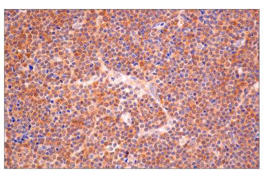
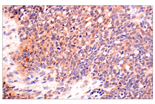
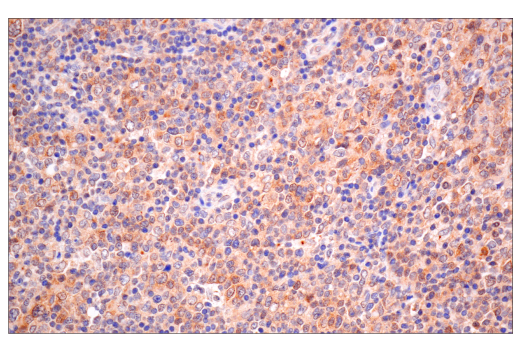
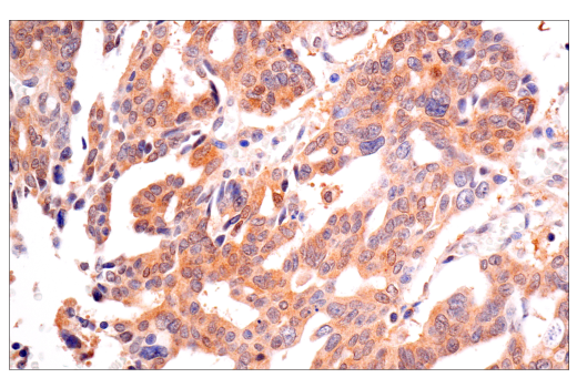
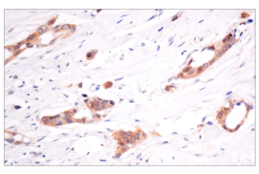
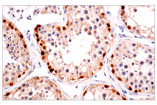
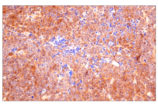
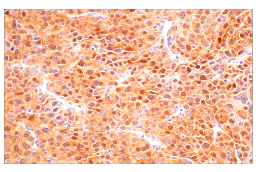
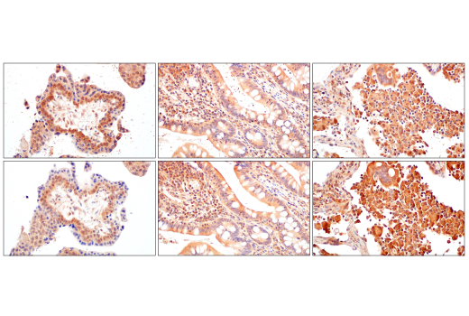
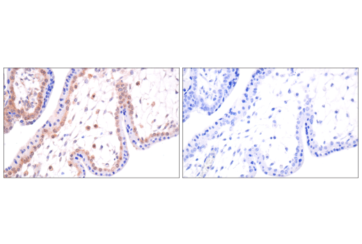
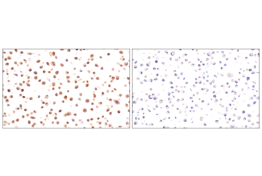
Revision 3

#81292Store at -20C

Cell Signaling Technology

Orders: 877-616-CELL (2355) [email protected]

Support: 877-678-TECH (8324)

Web: [email protected] cellsignal.com

3 Trask LaneDanversMassachusetts01923USA
For Research Use Only. Not for Use in Diagnostic Procedures.
Applications:

IHC-Bond, IHC-P

REACTIVITY:

H M

SENSITIVITY:

Endogenous

MW (kDa):

Source/Isotype:

Rabbit IgG

UniProt ID:

#P40337

Entrez-Gene Id:

7428

Product Information

Product Usage Information

Application Dilution
IHC Leica Bond 1:100 - 1:400
Immunohistochemistry (Paraffin) 1:100 - 1:400

Storage

Supplied in 10 mM sodium HEPES (pH 7.5), 150 mM NaCl, 100 µg/mL BSA, 50% glycerol, and less than 0.02% sodium azide. Store at –20°C. Do not aliquot the antibody.

For a carrier free (BSA and azide free) version of this product see product #82127.

Specificity / Sensitivity

VHL (E3X9K) Rabbit mAb recognizes endogenous levels of total VHL protein. Non-specific staining of skeletal and cardiac muscle has been observed by immunohistochemistry.

Species Reactivity:

Human, Mouse

Source / Purification

Monoclonal antibody is produced by immunizing animals with recombinant protein specific to human VHL protein.

Background

The von Hippel-Lindau (VHL) protein is a substrate recognition component of an E3 ubiquitin ligase complex containing elongin BC (TCEB1 and TCEB2), cullin 1 (CUL1), and RING-box protein 1 (RBX1) (1-3). VHL protein has been shown to exist as three distinct isoforms resulting from alternatively spliced transcript variants (4). Loss of VHL protein function results in a dominantly inherited familial cancer syndrome that manifests as angiomas of the retina, hemangioblastomas of the central nervous system, renal clear cell carcinomas, and pheochromocytomas (4). Under normoxic conditions, VHL directs the ubiquitylation and subsequent proteasomal degradation of the hypoxia-inducible factor 1α (HIF-1α), maintaining very low levels of HIF-1α in the cell. Cellular exposure to hypoxic conditions, or loss of VHL protein function, results in increased HIF-1α protein levels and increased expression of HIF-induced gene products, many of which are angiogenesis factors such as vascular endothelial growth factor (VEGF). Thus, loss of VHL protein function is believed to contribute to the formation of highly vascular neoplasias (4). In addition to HIF-1α, VHL is known to regulate the ubiquitylation of several other proteins, including tat-binding protein-1 (TBP-1), the atypical protein kinase C (aPKC) lambda, and two subunits of the multiprotein RNA polymerase II complex (RPB1 and RPB7) (5-8). Interactions with elongin BC, RPB1, RPB7, and the pVHL-associated KRAB-A domain-containing protein (VHLaK) suggest that VHL may also play a more direct role in transcriptional repression.

  1. Kibel, A. et al. (1995) Science 269, 1444-6.
  2. Pause, A. et al. (1997) Proc Natl Acad Sci U S A 94, 2156-61.
  3. Kamura, T. et al. (2000) Proc Natl Acad Sci U S A 97, 10430-5.
  4. Czyzyk-Krzeska, M.F. and Meller, J. (2004) Trends Mol Med 10, 146-9.
  5. Corn, P.G. et al. (2003) Nat Genet 35, 229-37.
  6. Na, X. et al. (2003) EMBO J 22, 4249-59.
  7. Kuznetsova, A.V. et al. (2003) Proc Natl Acad Sci U S A 100, 2706-11.
  8. Li, Z. et al. (2003) EMBO J 22, 1857-67.

Species Reactivity

Species reactivity is determined by testing in at least one approved application (e.g., western blot).

Applications Key

IHC-Bond: IHC Leica Bond IHC-P: Immunohistochemistry (Paraffin)

Cross-Reactivity Key

H: human M: mouse R: rat Hm: hamster Mk: monkey Vir: virus Mi: mink C: chicken Dm: D. melanogaster X: Xenopus Z: zebrafish B: bovine Dg: dog Pg: pig Sc: S. cerevisiae Ce: C. elegans Hr: horse GP: Guinea Pig Rab: rabbit All: all species expected

Trademarks and Patents

Cell Signaling Technology is a trademark of Cell Signaling Technology, Inc.
XP is a registered trademark of Cell Signaling Technology, Inc.
All other trademarks are the property of their respective owners. Visit cellsignal.com/trademarks for more information.

使用に関する制限

法的な権限を与えられたCSTの担当者が署名した書面によって別途明示的に合意された場合を除き、 CST、その関連会社または代理店が提供する製品には以下の条件が適用されます。お客様が定める条件でここに定められた条件に含まれるものを超えるもの、 または、ここに定められた条件と異なるものは、法的な権限を与えられたCSTの担当者が別途書面にて受諾した場合を除き、拒絶され、 いかなる効力も効果も有しません。

研究専用 (For Research Use Only) またはこれに類似する表示がされた製品は、 いかなる目的についても FDA または外国もしくは国内のその他の規制機関により承認、認可または許可を受けていません。 お客様は製品を診断もしくは治療目的で使用してはならず、また、製品に表示された内容に違反する方法で使用してはなりません。 CST が販売または使用許諾する製品は、エンドユーザーであるお客様に対し、使途を研究および開発のみに限定して提供されるものです。 診断、予防もしくは治療目的で製品を使用することまたは製品を再販売 (単独であるか他の製品等の一部であるかを問いません) もしくはその他の商業的利用の目的で購入することについては、CST から別途許諾を得る必要があります。 お客様は以下の事項を遵守しなければなりません。(a) CST の製品 (単独であるか他の資材と一緒であるかを問いません) を販売、使用許諾、貸与、寄付もしくはその他の態様で第三者に譲渡したり使用させたりしてはなりません。また、商用の製品を製造するために CST の製品を使用してはなりません。(b) 複製、改変、リバースエンジニアリング、逆コンパイル、 分解または他の方法により製品の構造または技術を解明しようとしてはなりません。また、 CST の製品またはサービスと競合する製品またはサービスを開発する目的で CST の製品を使用してはなりません。(c) CST の製品の商標、商号、ロゴ、特許または著作権に関する通知または表示を除去したり改変したりしてはなりません。(d) CST の製品をCST 製品販売条件(CST’s Product Terms of Sale) および該当する書面のみに従って使用しなければなりません。(e) CST の製品に関連してお客様が使用する第三者の製品またはサービスに関する使用許諾条件、 サービス提供条件またはこれに類する合意事項を遵守しなければなりません。

Revision 3
#81292

VHL (E3X9K) Rabbit mAb

Immunohistochemistry Image 1: VHL (E3X9K) Rabbit mAb Expand Image
Immunohistochemical analysis of paraffin-embedded human esophageal adenocarcinoma using VHL (E3X9K) Rabbit mAb performed on the Leica BOND Rx.
Immunohistochemistry Image 2: VHL (E3X9K) Rabbit mAb Expand Image
Immunohistochemical analysis of paraffin-embedded human colon carcinoma using VHL (E3X9K) Rabbit mAb performed on the Leica BOND Rx.
Immunohistochemistry Image 1: VHL (E3X9K) Rabbit mAb Expand Image
Immunohistochemical analysis of paraffin-embedded human clear cell renal cell carcinoma using VHL (E3X9K) Rabbit mAb performed on the Leica BOND Rx.
Immunohistochemistry Image 2: VHL (E3X9K) Rabbit mAb Expand Image
Immunohistochemical analysis of paraffin-embedded human small cell carcinoma of the salivary gland using VHL (E3X9K) Rabbit mAb.
Immunohistochemistry Image 3: VHL (E3X9K) Rabbit mAb Expand Image
Immunohistochemical analysis of paraffin-embedded human B-cell non-Hodgkin lymphoma using VHL (E3X9K) Rabbit mAb.
Immunohistochemistry Image 4: VHL (E3X9K) Rabbit mAb Expand Image
Immunohistochemical analysis of paraffin-embedded human serous adenocarcinoma of the ovary using VHL (E3X9K) Rabbit mAb.
Immunohistochemistry Image 5: VHL (E3X9K) Rabbit mAb Expand Image
Immunohistochemical analysis of paraffin-embedded human T-cell lymphoma using VHL (E3X9K) Rabbit mAb.
Immunohistochemistry Image 6: VHL (E3X9K) Rabbit mAb Expand Image
Immunohistochemical analysis of paraffin-embedded human serous papillary carcinoma of the ovary using VHL (E3X9K) Rabbit mAb.
Immunohistochemistry Image 7: VHL (E3X9K) Rabbit mAb Expand Image

Immunohistochemical analysis of paraffin-embedded human ductal breast carcinoma using VHL (E3X9K) Rabbit mAb.

Immunohistochemistry Image 8: VHL (E3X9K) Rabbit mAb Expand Image
Immunohistochemical analysis of paraffin-embedded normal human testis using VHL (E3X9K) Rabbit mAb.
Immunohistochemistry Image 9: VHL (E3X9K) Rabbit mAb Expand Image
Immunohistochemical analysis of paraffin-embedded A20 syngeneic tumor using VHL (E3X9K) Rabbit mAb.
Immunohistochemistry Image 10: VHL (E3X9K) Rabbit mAb Expand Image

Immunohistochemical analysis of paraffin-embedded Renca syngeneic tumor using VHL (E3X9K) Rabbit mAb.

Immunohistochemistry Image 11: VHL (E3X9K) Rabbit mAb Expand Image
Immunohistochemical analysis of paraffin-embedded normal human placenta (left), small intestine (middle), or lung (right) using VHL (E3X9K) Rabbit mAb (top) or a VHL Rabbit mAb (bottom). These two antibodies detect unique, non-overlapping epitopes on human VHL. The similar staining patterns obtained with both antibodies help to confirm the specificity of the staining.
Immunohistochemistry Image 12: VHL (E3X9K) Rabbit mAb Expand Image
Immunohistochemical analysis of paraffin-embedded normal human placenta using VHL (E3X9K) Rabbit mAb (left) compared to concentration-matched Rabbit (DA1E) mAb XP® IgG Isotype Control #3900 (right).
Immunohistochemistry Image 13: VHL (E3X9K) Rabbit mAb Expand Image
Immunohistochemical analysis of paraffin-embedded HeLa cell pellet (left, positive) or 786-O cell pellet (right, negative) using VHL (E3X9K) Rabbit mAb.