Revision 2

#51754Store at -20C

1 Kit

(10 slides)

UniProt ID:

#P07766

Entrez-Gene Id:

#916

Cell Signaling Technology

Orders: 877-616-CELL (2355) [email protected]

Support: 877-678-TECH (8324)

Web: [email protected] cellsignal.com

3 Trask LaneDanversMassachusetts01923USA
For Research Use Only. Not for Use in Diagnostic Procedures.
Product Includes Item # Volume Reactivity Isotype
CD3ε (D7A6E) XP® Rabbit mAb (SignalStar Conjugate 0001) 27460 50 µl H Rabbit IgG
Complementary Oligo (CO-0001-750) 19832 22 µl

Storage

SignalStar conjugates are supplied in PBS (pH 7.2), less than 0.1% sodium azide, 2 mM EDTA, 0.05% Triton X-100, 2 mg/mL BSA, and 50% glycerol. Complementary oligos are supplied in nuclease-free water. Store at -20°C. Do not aliquot the antibody. All components in this kit are stable for at least 12 months when stored at the recommended temperature.

Description

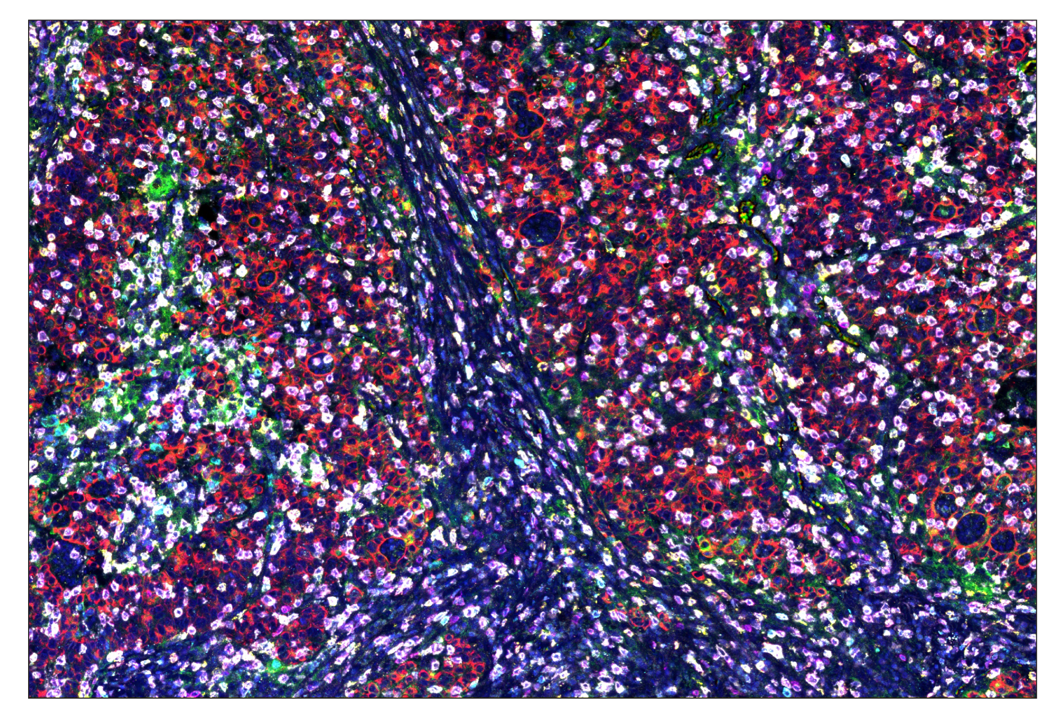
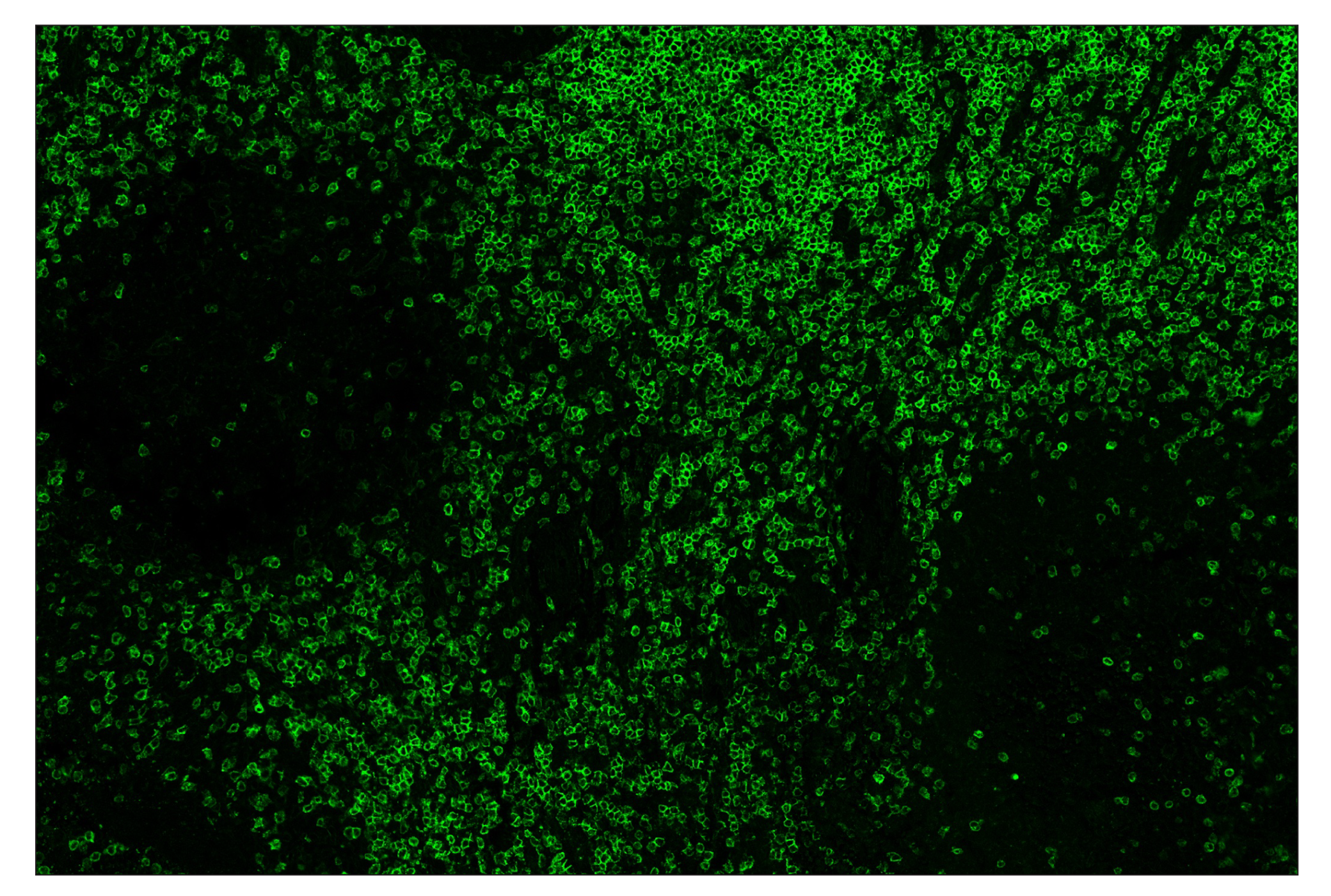
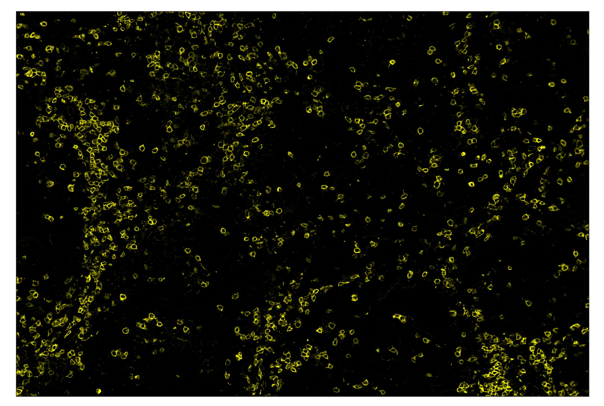
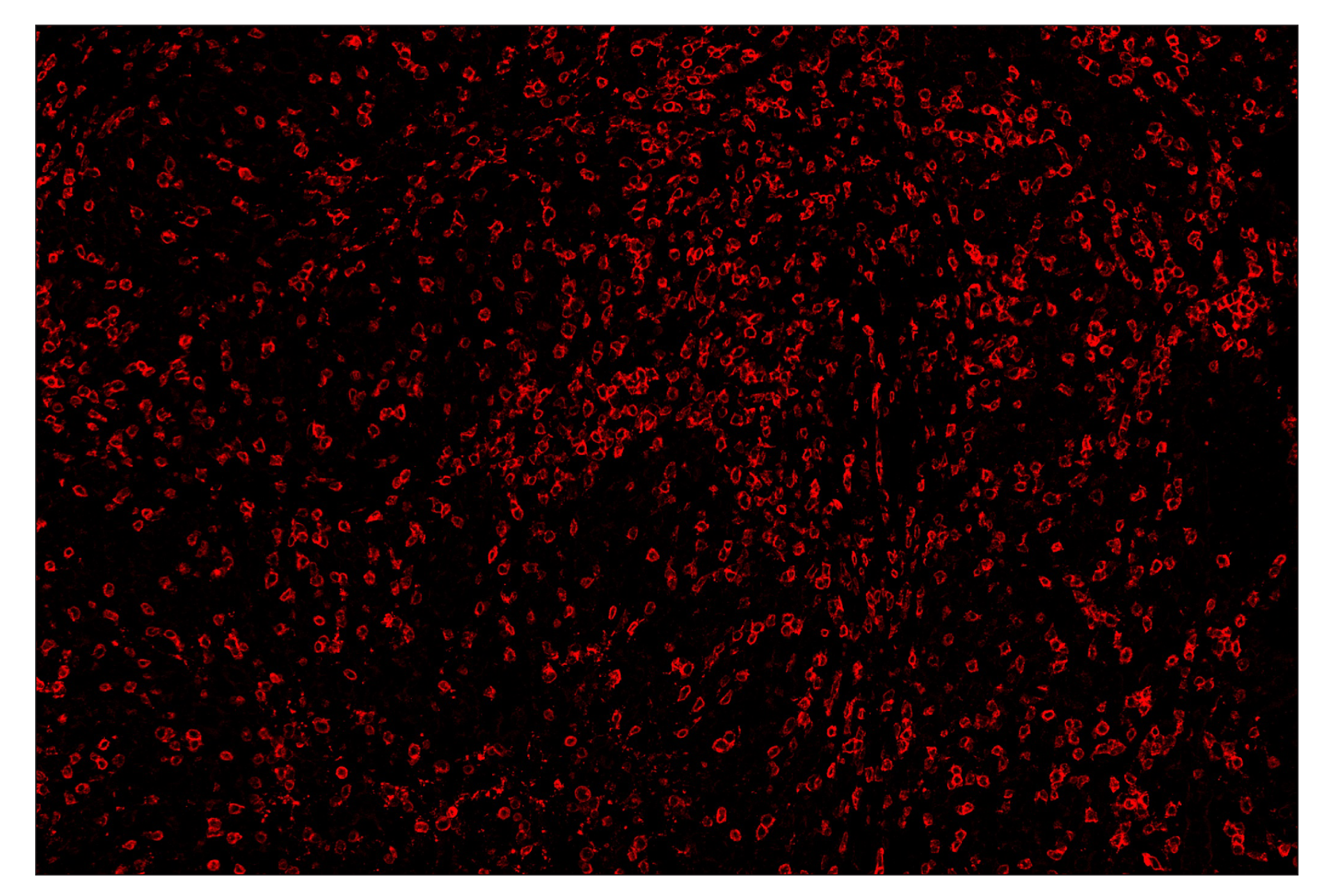
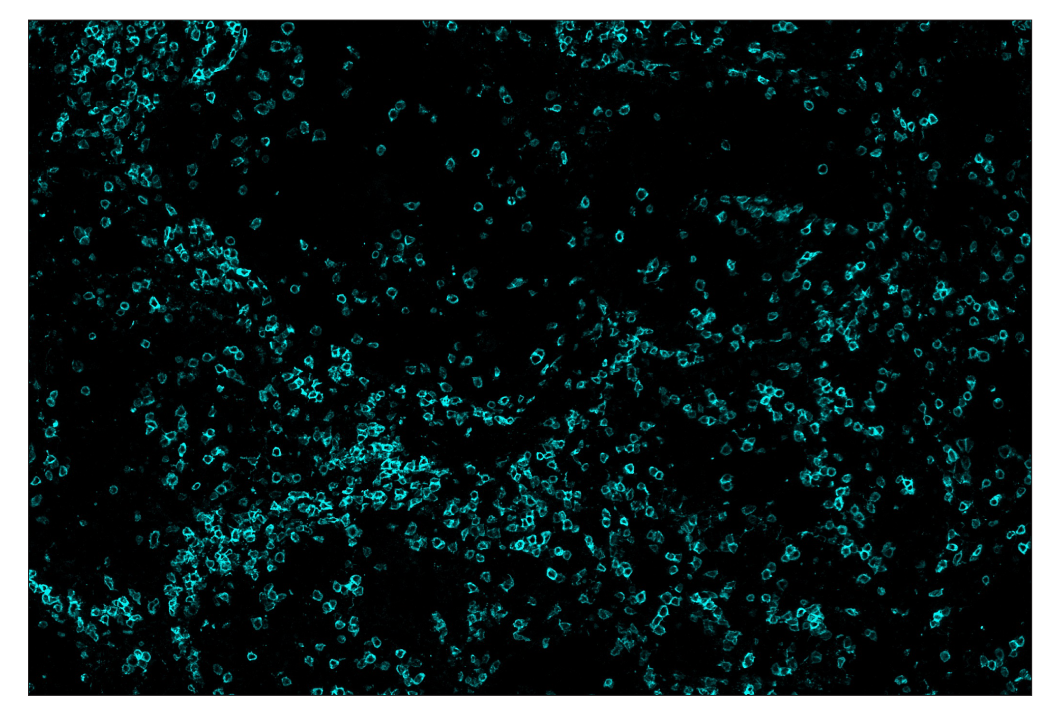
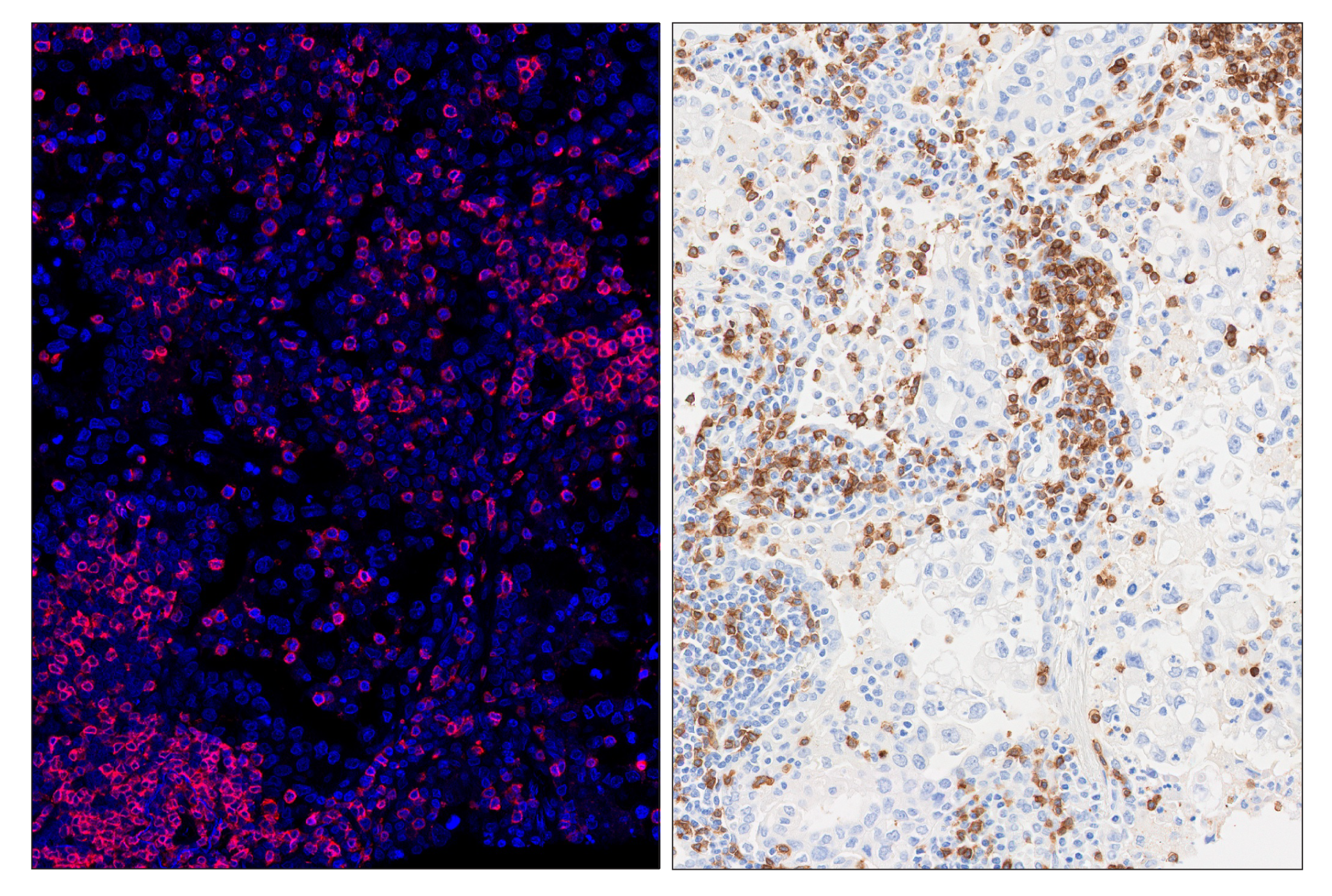
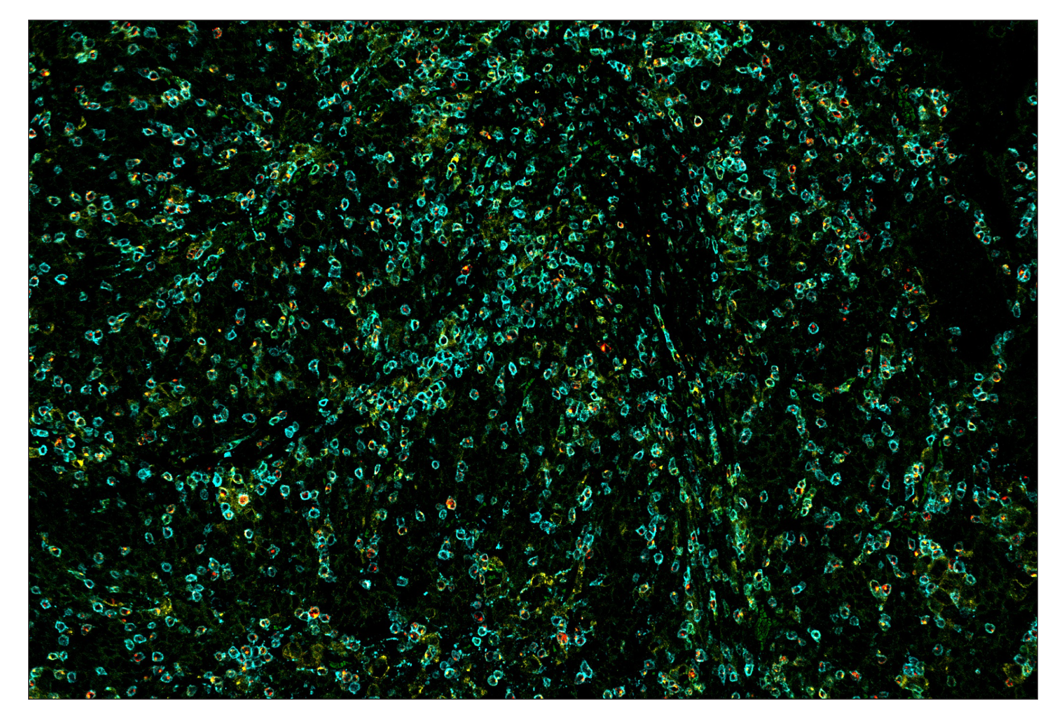
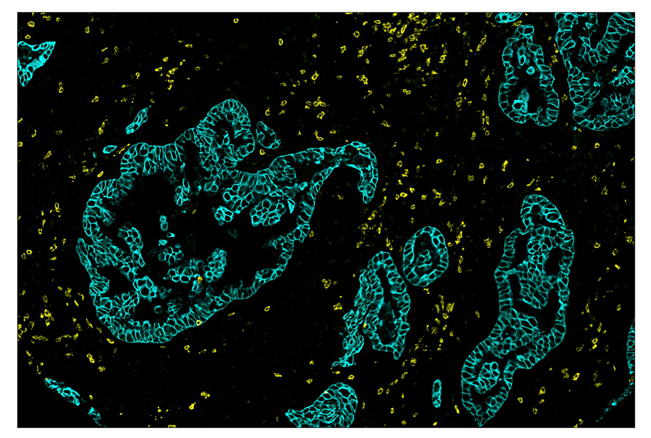
SignalStar multiplex immunohistochemistry (IHC) is an advanced technology for labeling multiple proteins simultaneously in tissue samples using specific primary antibodies and fluorescent detection reagents. This technology offers accuracy and reliability in visualizing and analyzing protein expression while maintaining spatial context and tissue architecture.

SignalStar Oligo-Antibody Pairs are compatible with the SignalStar Multiplex IHC Buffer Kits for use in fluorescent multiplex imaging experiments. This product includes the oligo-conjugated antibodies and complementary oligos required for labeling your target protein on up to 10 slides. SignalStar Multiplex IHC Buffer Kits are required to amplify and image the target signal. Multiple oligo-antibody pairs can be conveniently combined into a multiplex panel using the SignalStar Multiplex IHC Panel Builder. SignalStar Multiplex IHC Kits & Reagents are not compatible with all of Cell Signaling Technology® products and protocols that are recommended for use in immunohistochemical assays.

Specificity/Sensitivity

CD3ε (D7A6E) XP® Rabbit mAb (SignalStar Conjugate 0001) recognizes endogenous levels of total CD3ε protein. This antibody recognizes human CD3ε protein and is also reactive with mouse CD3ε; however, this antibody is not suggested for immunohistochemical analysis of mouse tissues. Monkey species reactivity is Immunohistochemistry (Paraffin) only.

Source / Purification

Monoclonal antibody is produced by immunizing animals with a synthetic peptide corresponding to residues surrounding Glu178 of human CD3ε protein.

Background

When T cells encounter antigens via the T cell receptor (TCR), information about the quantity and quality of antigens is relayed to the intracellular signal transduction machinery (1). This activation process depends mainly on CD3 (Cluster of Differentiation 3), a multiunit protein complex that directly associates with the TCR. CD3 is composed of four polypeptides: ζ, γ, ε, and δ. Each of these polypeptides contains at least one immunoreceptor tyrosine-based activation motif (ITAM) (2). Engagement of the TCR complex with foreign antigens induces tyrosine phosphorylation in the ITAM motifs and phosphorylated ITAMs function as docking sites for signaling molecules such as ZAP-70 and the p85 subunit of PI-3 kinase (3,4). TCR ligation also induces a conformational change in CD3ε, such that a proline region is exposed and then associates with the adaptor protein Nck (5).

  1. Kuhns, M.S. et al. (2006) Immunity 24, 133-139.
  2. Pitcher, L.A. and van Oers, N.S. (2003) Trends Immunol. 24, 554-560.
  3. Osman, N. et al. (1996) Eur. J. Immunol. 26, 1063-1068.
  4. Hatada, M.H. et al. (1995) Nature 377, 32-38.
  5. Gil, D. et al. (2002) Cell 109, 901-912.

Species Reactivity

Species reactivity is determined by testing in at least one approved application (e.g., western blot).

Cross-Reactivity Key

H: human M: mouse R: rat Hm: hamster Mk: monkey Vir: virus Mi: mink C: chicken Dm: D. melanogaster X: Xenopus Z: zebrafish B: bovine Dg: dog Pg: pig Sc: S. cerevisiae Ce: C. elegans Hr: horse GP: Guinea Pig Rab: rabbit All: all species expected

Trademarks and Patents

Cell Signaling Technology is a trademark of Cell Signaling Technology, Inc.
SignalStar is a trademark of Cell Signaling Technology, Inc.
XP is a registered trademark of Cell Signaling Technology, Inc.
All other trademarks are the property of their respective owners. Visit cellsignal.com/trademarks for more information.

使用に関する制限

法的な権限を与えられたCSTの担当者が署名した書面によって別途明示的に合意された場合を除き、 CST、その関連会社または代理店が提供する製品には以下の条件が適用されます。お客様が定める条件でここに定められた条件に含まれるものを超えるもの、 または、ここに定められた条件と異なるものは、法的な権限を与えられたCSTの担当者が別途書面にて受諾した場合を除き、拒絶され、 いかなる効力も効果も有しません。

研究専用 (For Research Use Only) またはこれに類似する表示がされた製品は、 いかなる目的についても FDA または外国もしくは国内のその他の規制機関により承認、認可または許可を受けていません。 お客様は製品を診断もしくは治療目的で使用してはならず、また、製品に表示された内容に違反する方法で使用してはなりません。 CST が販売または使用許諾する製品は、エンドユーザーであるお客様に対し、使途を研究および開発のみに限定して提供されるものです。 診断、予防もしくは治療目的で製品を使用することまたは製品を再販売 (単独であるか他の製品等の一部であるかを問いません) もしくはその他の商業的利用の目的で購入することについては、CST から別途許諾を得る必要があります。 お客様は以下の事項を遵守しなければなりません。(a) CST の製品 (単独であるか他の資材と一緒であるかを問いません) を販売、使用許諾、貸与、寄付もしくはその他の態様で第三者に譲渡したり使用させたりしてはなりません。また、商用の製品を製造するために CST の製品を使用してはなりません。(b) 複製、改変、リバースエンジニアリング、逆コンパイル、 分解または他の方法により製品の構造または技術を解明しようとしてはなりません。また、 CST の製品またはサービスと競合する製品またはサービスを開発する目的で CST の製品を使用してはなりません。(c) CST の製品の商標、商号、ロゴ、特許または著作権に関する通知または表示を除去したり改変したりしてはなりません。(d) CST の製品をCST 製品販売条件(CST’s Product Terms of Sale) および該当する書面のみに従って使用しなければなりません。(e) CST の製品に関連してお客様が使用する第三者の製品またはサービスに関する使用許諾条件、 サービス提供条件またはこれに類する合意事項を遵守しなければなりません。

Revision 2
#51754

CD3ε (D7A6E) & CO-0001-750 SignalStar Oligo-Antibody Pair

Immunohistochemistry Image 1: CD3ε (D7A6E™) & CO-0001-750 SignalStar™ Oligo-Antibody Pair Expand Image
SignalStar multiplex immunohistochemical analysis of paraffin-embedded human gastric adenocarcinoma using CD3ε (D7A6E) & CO-0001-750 SignalStar Oligo-Antibody Pair #51754 (magenta), Pan-Keratin (C11) & CO-0003-488 SignalStar Oligo-Antibody Pair #63566 (red), PD-L1 (E1L3N®) & CO-0005-488 SignalStar Oligo-Antibody Pair #85646 (green), PD-1 (D4W2J) & CO-0008-594 SignalStar Oligo-Antibody Pair #35347 (yellow), Granzyme B (D6E9W) & CO-0009-594 SignalStar Oligo-Antibody Pair #15194 (orange), TIGIT (E5Y1W) & CO-0002-647 SignalStar Oligo-Antibody Pair #18288 (cyan), CD20 (E7B7T) & CO-0011-647 SignalStar Oligo-Antibody Pair #36775 (pink), CD8α (D8A8Y) & CO-0004-750 SignalStar Oligo-Antibody Pair #62750 (white), and ProLong® Gold Antifade Reagent with DAPI #8961 (blue). All fluorophores have been assigned a pseudocolor, as indicated. Staining was performed on the Leica BOND Rx.
Immunohistochemistry Image 2: CD3ε (D7A6E™) & CO-0001-750 SignalStar™ Oligo-Antibody Pair Expand Image
SignalStar immunohistochemical analysis of paraffin-embedded human tonsil using CD3ε (D7A6E) & CO-0001-488 SignalStar Oligo-Antibody Pair #92856 (green). All fluorophores have been assigned a pseudocolor, as indicated. Staining was performed on the Leica BOND Rx.
Immunohistochemistry Image 3: CD3ε (D7A6E™) & CO-0001-750 SignalStar™ Oligo-Antibody Pair Expand Image
SignalStar immunohistochemical analysis of paraffin-embedded human non-small cell lung carcinoma using CD3ε (D7A6E) & CO-0001-594 SignalStar Oligo-Antibody Pair #84634 (yellow). All fluorophores have been assigned a pseudocolor, as indicated. Staining was performed on the Leica BOND Rx.
Immunohistochemistry Image 4: CD3ε (D7A6E™) & CO-0001-750 SignalStar™ Oligo-Antibody Pair Expand Image
SignalStar immunohistochemical analysis of paraffin-embedded human gastric adenocarcinoma using CD3ε (D7A6E) & CO-0001-647 SignalStar Oligo-Antibody Pair #33888 (red). All fluorophores have been assigned a pseudocolor, as indicated. Staining was performed on the Leica BOND Rx.
Immunohistochemistry Image 5: CD3ε (D7A6E™) & CO-0001-750 SignalStar™ Oligo-Antibody Pair Expand Image
SignalStar immunohistochemical analysis of paraffin-embedded human non-small cell lung carcinoma using CD3ε (D7A6E) & CO-0001-750 SignalStar Oligo-Antibody Pair #51754 (cyan). All fluorophores have been assigned a pseudocolor, as indicated. Staining was performed on the Leica BOND Rx.
Immunohistochemistry Image 6: CD3ε (D7A6E™) & CO-0001-750 SignalStar™ Oligo-Antibody Pair Expand Image
SignalStar multiplex immunohistochemical analysis of paraffin-embedded human non-small cell lung carcinoma using CD3ε (D7A6E) & CO-0001-647 SignalStar Oligo-Antibody Pair #33888 (red, left) and ProLong® Gold Antifade Reagent with DAPI #8961 (blue) compared to chromogenic immunohistochemical analysis of a serial section of paraffin-embedded human non-small cell lung carcinoma using CD3ε (D7A6E) XP® Rabbit mAb #85061 (right). All fluorophores have been assigned a pseudocolor, as indicated. Staining was performed on the Leica BOND Rx.
Immunohistochemistry Image 7: CD3ε (D7A6E™) & CO-0001-750 SignalStar™ Oligo-Antibody Pair Expand Image
SignalStar multiplex immunohistochemical analysis of paraffin-embedded human gastric adenocarcinoma using CD3ε (D7A6E) & CO-0001-750 SignalStar Oligo-Antibody Pair #51754 (cyan), PD-1 (D4W2J) & CO-0008-488 SignalStar Oligo-Antibody Pair #17942 (green), TIM-3 (D5D5R) & CO-0010-594 SignalStar Oligo-Antibody Pair #55508 (yellow), and LAG3 (D2G4O) & CO-0026-647 SignalStar Oligo-Antibody Pair #40966 (red). All fluorophores have been assigned a pseudocolor, as indicated. Staining was performed on the Leica BOND Rx.
Immunohistochemistry Image 1: CD3ε (D7A6E™) & CO-0001-750 SignalStar™ Oligo-Antibody Pair Expand Image
SignalStar multiplex immunohistochemical analysis of paraffin-embedded human non-small cell lung carcinoma using CD3ε (D7A6E) & CO-0001-594 SignalStar Oligo-Antibody Pair #84634 (yellow) and Pan-Keratin (C11) & CO-0003-750 SignalStar Oligo-Antibody Pair #97227 (cyan). All fluorophores have been assigned a pseudocolor, as indicated.