WB, IP, IF-IC
H
Transfected Only
87 (Akt-GFP)
Rabbit IgG
#P31751, #Q9Y243, #P31749
208, 10000, 207
Product Information
Product Usage Information
| Application | Dilution |
|---|---|
| Western Blotting | 1:1000 |
| Immunoprecipitation | 1:50 |
| Immunofluorescence (Immunocytochemistry) | 1:800 |
Storage
Specificity / Sensitivity
Species Reactivity:
Human
Species predicted to react based on 100% sequence homology
The antigen sequence used to produce this antibody shares
100% sequence homology with the species listed here, but
reactivity has not been tested or confirmed to work by CST.
Use of this product with these species is not covered under
our
Product Performance Guarantee.
Mouse, Rat
Source / Purification
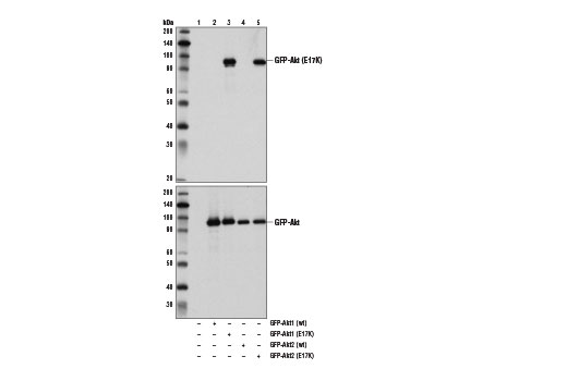
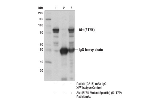
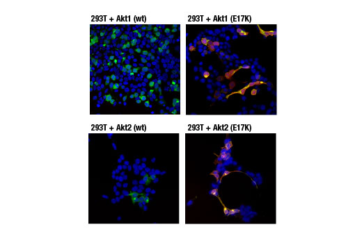
Monoclonal antibody is produced by immunizing animals with a synthetic peptide corresponding to E17K mutant sequence of human Akt1 protein.
Background
Akt, also referred to as PKB or Rac, plays a critical role in controlling cell survival and apoptosis (1-3). This protein kinase is activated by insulin and various growth and survival factors to function in a wortmannin-sensitive pathway involving PI3 kinase (2,3). Akt is activated by phospholipid binding and activation loop phosphorylation at Thr308 by PDK1 (4) and by phosphorylation within the carboxy terminus at Ser473. The previously elusive PDK2 responsible for phosphorylation of Akt at Ser473 has been identified as mammalian target of rapamycin (mTOR) in a rapamycin-insensitive complex with rictor and Sin1 (5,6). Akt promotes cell survival by inhibiting apoptosis through phosphorylation and inactivation of several targets, including Bad (7), forkhead transcription factors (8), c-Raf (9), and caspase-9. PTEN phosphatase is a major negative regulator of the PI3K/Akt signaling pathway (10). LY294002 is a specific PI3 kinase inhibitor (11). Another essential Akt function is the regulation of glycogen synthesis through phosphorylation and inactivation of GSK-3α and β (12,13). Akt may also play a role in insulin stimulation of glucose transport (12). In addition to its role in survival and glycogen synthesis, Akt is involved in cell cycle regulation by preventing GSK-3β-mediated phosphorylation and degradation of cyclin D1 (14) and by negatively regulating the cyclin-dependent kinase inhibitors p27 Kip1 (15) and p21 Waf1/Cip1 (16). Akt also plays a critical role in cell growth by directly phosphorylating mTOR in a rapamycin-sensitive complex containing raptor (17). More importantly, Akt phosphorylates and inactivates tuberin (TSC2), an inhibitor of mTOR within the mTOR-raptor complex (18,19).
Mutation of the glutamic acid at residue 17 to lysine (E17K) of Akt was initially identified in human breast, colorectal, and ovarian cancers (20). This conserved glutamic acid residue is located at the lipid-binding pocket of the Akt1 plextrin homology domain. The E17K mutation increases the affinity between Akt1 and phospholipids at the plasma membrane, leading to increased Akt1 recruitment, super-activation of the Akt pathway, cellular transformation, and tumor formation (20,21). Additional studies detect the presence of the Akt1 (E17K) mutation in multiple cancers, including lung cancer, prostate cancer, and endometrial carcinoma (22,23). The presence of mutant Akt3 (E17K) protein has also been seen in cases of melanoma (24).
- Franke, T.F. et al. (1997) Cell 88, 435-7.
- Burgering, B.M. and Coffer, P.J. (1995) Nature 376, 599-602.
- Franke, T.F. et al. (1995) Cell 81, 727-36.
- Alessi, D.R. et al. (1996) EMBO J 15, 6541-51.
- Sarbassov, D.D. et al. (2005) Science 307, 1098-101.
- Jacinto, E. et al. (2006) Cell 127, 125-37.
- Cardone, M.H. et al. (1998) Science 282, 1318-21.
- Brunet, A. et al. (1999) Cell 96, 857-68.
- Zimmermann, S. and Moelling, K. (1999) Science 286, 1741-4.
- Cantley, L.C. and Neel, B.G. (1999) Proc Natl Acad Sci USA 96, 4240-5.
- Vlahos, C.J. et al. (1994) J Biol Chem 269, 5241-8.
- Hajduch, E. et al. (2001) FEBS Lett 492, 199-203.
- Cross, D.A. et al. (1995) Nature 378, 785-9.
- Diehl, J.A. et al. (1998) Genes Dev 12, 3499-511.
- Gesbert, F. et al. (2000) J Biol Chem 275, 39223-30.
- Zhou, B.P. et al. (2001) Nat Cell Biol 3, 245-52.
- Navé, B.T. et al. (1999) Biochem J 344 Pt 2, 427-31.
- Inoki, K. et al. (2002) Nat Cell Biol 4, 648-57.
- Manning, B.D. et al. (2002) Mol Cell 10, 151-62.
- Carpten, J.D. et al. (2007) Nature 448, 439-44.
- Landgraf, K.E. et al. (2008) Biochemistry 47, 12260-9.
- Malanga, D. et al. (2008) Cell Cycle 7, 665-9.
- Cohen, Y. et al. (2010) Gynecol Oncol 116, 88-91.
- Davies, M.A. et al. (2008) Br J Cancer 99, 1265-8.
Species Reactivity
Species reactivity is determined by testing in at least one approved application (e.g., western blot).
Western Blot Buffer
IMPORTANT: For western blots, incubate membrane with diluted primary antibody in 5% w/v BSA, 1X TBS, 0.1% Tween® 20 at 4°C with gentle shaking, overnight.
Applications Key
WB: Western Blotting IP: Immunoprecipitation IF-IC: Immunofluorescence (Immunocytochemistry)
Cross-Reactivity Key
H: human M: mouse R: rat Hm: hamster Mk: monkey Vir: virus Mi: mink C: chicken Dm: D. melanogaster X: Xenopus Z: zebrafish B: bovine Dg: dog Pg: pig Sc: S. cerevisiae Ce: C. elegans Hr: horse GP: Guinea Pig Rab: rabbit All: all species expected
Trademarks and Patents
使用に関する制限
法的な権限を与えられたCSTの担当者が署名した書面によって別途明示的に合意された場合を除き、 CST、その関連会社または代理店が提供する製品には以下の条件が適用されます。お客様が定める条件でここに定められた条件に含まれるものを超えるもの、 または、ここに定められた条件と異なるものは、法的な権限を与えられたCSTの担当者が別途書面にて受諾した場合を除き、拒絶され、 いかなる効力も効果も有しません。
研究専用 (For Research Use Only) またはこれに類似する表示がされた製品は、 いかなる目的についても FDA または外国もしくは国内のその他の規制機関により承認、認可または許可を受けていません。 お客様は製品を診断もしくは治療目的で使用してはならず、また、製品に表示された内容に違反する方法で使用してはなりません。 CST が販売または使用許諾する製品は、エンドユーザーであるお客様に対し、使途を研究および開発のみに限定して提供されるものです。 診断、予防もしくは治療目的で製品を使用することまたは製品を再販売 (単独であるか他の製品等の一部であるかを問いません) もしくはその他の商業的利用の目的で購入することについては、CST から別途許諾を得る必要があります。 お客様は以下の事項を遵守しなければなりません。(a) CST の製品 (単独であるか他の資材と一緒であるかを問いません) を販売、使用許諾、貸与、寄付もしくはその他の態様で第三者に譲渡したり使用させたりしてはなりません。また、商用の製品を製造するために CST の製品を使用してはなりません。(b) 複製、改変、リバースエンジニアリング、逆コンパイル、 分解または他の方法により製品の構造または技術を解明しようとしてはなりません。また、 CST の製品またはサービスと競合する製品またはサービスを開発する目的で CST の製品を使用してはなりません。(c) CST の製品の商標、商号、ロゴ、特許または著作権に関する通知または表示を除去したり改変したりしてはなりません。(d) CST の製品をCST 製品販売条件(CST’s Product Terms of Sale) および該当する書面のみに従って使用しなければなりません。(e) CST の製品に関連してお客様が使用する第三者の製品またはサービスに関する使用許諾条件、 サービス提供条件またはこれに類する合意事項を遵守しなければなりません。