| Product Includes | Product # | Quantity | Mol. Wt | Isotype/Source |
|---|---|---|---|---|
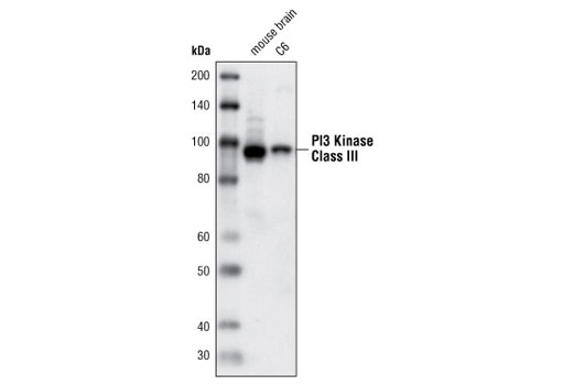
| PI3 Kinase Class III (D9A5) Rabbit mAb | 4263 | 20 µl | 100 kDa | Rabbit |
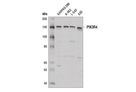
| PIK3R4 Antibody | 14580 | 20 µl | 153 kDa | Rabbit |
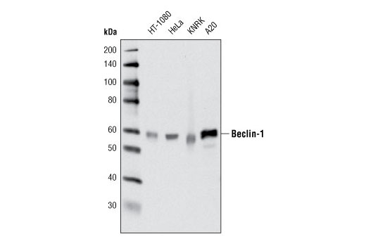
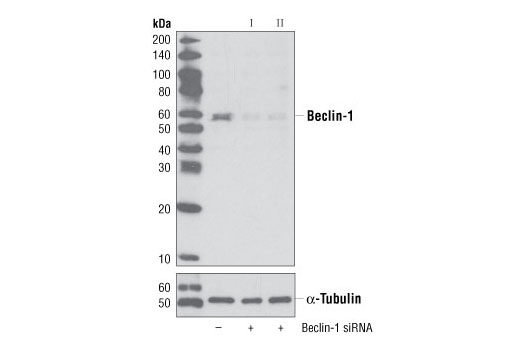
| Beclin-1 (D40C5) Rabbit mAb | 3495 | 20 µl | 60 kDa | Rabbit IgG |
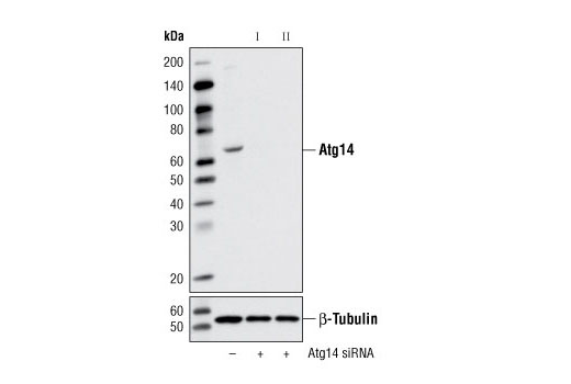
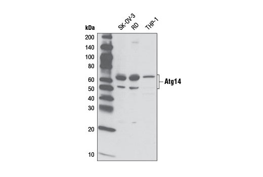
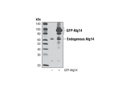
| Atg14 Antibody | 5504 | 20 µl | 65 kDa | Rabbit |
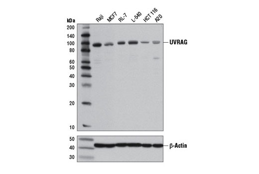
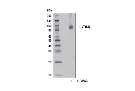
| UVRAG (D2Q1Z) Rabbit mAb | 13115 | 20 µl | 90 kDa | Rabbit IgG |
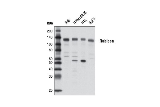
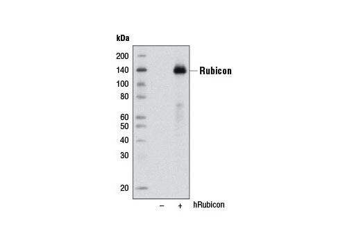
| Rubicon (D9F7) Rabbit mAb | 8465 | 20 µl | 130 kDa | Rabbit IgG |
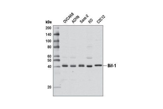
| Bif-1 Antibody | 4467 | 20 µl | 42 kDa | Rabbit |
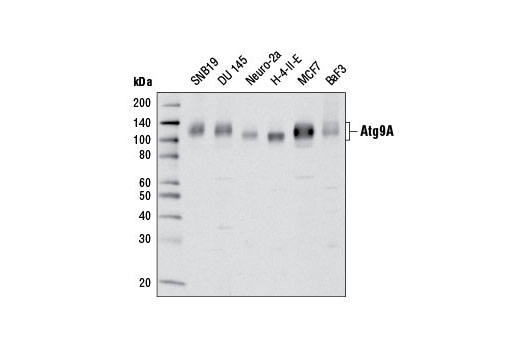
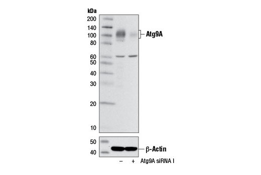
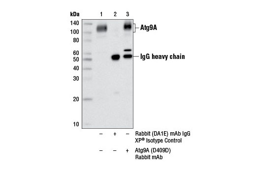
| Atg9A (D4O9D) Rabbit mAb | 13509 | 20 µl | 100-110 kDa | Rabbit IgG |
| Anti-rabbit IgG, HRP-linked Antibody | 7074 | 100 µl | Goat |
Please visit cellsignal.com for individual component applications, species cross-reactivity, dilutions, protocols, and additional product information.
Description
The Autophagy Vesicle Nucleation Antibody Sampler Kit provides an economical means of detecting target proteins involved in autophagosome formation and maturation. The kit contains enough antibody to perform two western blot experiments per primary antibody.
Storage
Background
Autophagy is a catabolic process for the autophagosomic-lysosomal degradation of bulk cytoplasmic contents (1,2). Autophagy is generally activated by conditions of nutrient deprivation but is also associated with a number of physiological processes including development, differentiation, neurodegeneration, infection and cancer (3). The molecular machinery of autophagy was largely discovered in yeast and is directed by a number of autophagy-related (Atg) genes. These proteins are involved in the formation of autophagosomes, cytoplasmic vacuoles that are delivered to lysosomes for degradation. The PIK3R4/PI3K class III complex interacts with Beclin-1 to play a role during several stages of autophagy. Autophagosome formation is stimulated when Atg14 complexes with PIK3R4, PI3K class III, and Beclin-1. The UVRAG protein competes with Atg14 for Beclin-1 binding, forming a mutually exclusive complex with PIK3R4, PI3K class III, and Beclin-1 that regulates autophagosome maturation. Autophagosome maturation is impaired in the presence of the Beclin-1-binding protein Rubicon (4,5). Co-expression of PIK3R4 is required for PI3K class III activation and regulation by both Beclin-1/UVRAG and nutrient levels (6). Bif-1 directly binds to UVRAG, forming a complex with Beclin-1, resulting in increased PI3-kinase class III/Vps34 activity required for autophagosome maturation (7). Inhibition of GSK-3β, as seen during nutrient deprivation, results in increased expression of Bif-1, and can contribute to autophagic cell death (8). Atg9A is an integral membrane protein that is required for both the initiation and the expansion of the autophagosome (9,10). Recruitment of Atg9A to the autophagosomal membrane is dynamic and transient as Atg9A also cycles between autophagy-related structures known as omegasomes, the trans-Golgi network (TGN), and endosomes, and at no point becomes a stable component of the autophagosomal membrane (9,11). The precise regulation of Atg9A trafficking is not fully clarified, yet it is suggested to involve p38 mitogen-activated protein kinase (MAPK)-binding protein p38IP and the Beclin-1-binding protein Bif-1 (12,13).
- Reggiori, F. and Klionsky, D.J. (2002) Eukaryot Cell 1, 11-21.
- Codogno, P. and Meijer, A.J. (2005) Cell Death Differ 12 Suppl 2, 1509-18.
- Levine, B. and Yuan, J. (2005) J Clin Invest 115, 2679-88.
- Zhong, Y. et al. (2009) Nat Cell Biol 11, 468-76.
- Sun, Q. et al. (2008) Proc Natl Acad Sci U S A 105, 19211-6.
- Yan, Y. et al. (2009) Biochem J 417, 747-55.
- Takahashi, Y. et al. (2007) Nat Cell Biol 9, 1142-51.
- Yang, J. et al. (2010) J Cell Sci 123, 861-70.
- Young, A.R. et al. (2006) J Cell Sci 119, 3888-900.
- Yamada, T. et al. (2005) J Biol Chem 280, 18283-90.
- Orsi, A. et al. (2012) Mol Biol Cell 23, 1860-73.
- Webber, J.L. and Tooze, S.A. (2010) EMBO J 29, 27-40.
Background References
Trademarks and Patents
使用に関する制限
法的な権限を与えられたCSTの担当者が署名した書面によって別途明示的に合意された場合を除き、 CST、その関連会社または代理店が提供する製品には以下の条件が適用されます。お客様が定める条件でここに定められた条件に含まれるものを超えるもの、 または、ここに定められた条件と異なるものは、法的な権限を与えられたCSTの担当者が別途書面にて受諾した場合を除き、拒絶され、 いかなる効力も効果も有しません。
研究専用 (For Research Use Only) またはこれに類似する表示がされた製品は、 いかなる目的についても FDA または外国もしくは国内のその他の規制機関により承認、認可または許可を受けていません。 お客様は製品を診断もしくは治療目的で使用してはならず、また、製品に表示された内容に違反する方法で使用してはなりません。 CST が販売または使用許諾する製品は、エンドユーザーであるお客様に対し、使途を研究および開発のみに限定して提供されるものです。 診断、予防もしくは治療目的で製品を使用することまたは製品を再販売 (単独であるか他の製品等の一部であるかを問いません) もしくはその他の商業的利用の目的で購入することについては、CST から別途許諾を得る必要があります。 お客様は以下の事項を遵守しなければなりません。(a) CST の製品 (単独であるか他の資材と一緒であるかを問いません) を販売、使用許諾、貸与、寄付もしくはその他の態様で第三者に譲渡したり使用させたりしてはなりません。また、商用の製品を製造するために CST の製品を使用してはなりません。(b) 複製、改変、リバースエンジニアリング、逆コンパイル、 分解または他の方法により製品の構造または技術を解明しようとしてはなりません。また、 CST の製品またはサービスと競合する製品またはサービスを開発する目的で CST の製品を使用してはなりません。(c) CST の製品の商標、商号、ロゴ、特許または著作権に関する通知または表示を除去したり改変したりしてはなりません。(d) CST の製品をCST 製品販売条件(CST’s Product Terms of Sale) および該当する書面のみに従って使用しなければなりません。(e) CST の製品に関連してお客様が使用する第三者の製品またはサービスに関する使用許諾条件、 サービス提供条件またはこれに類する合意事項を遵守しなければなりません。