WB, IP, FC-FP
M
Endogenous
17, 14
Rabbit IgG
#Q62386
16171
Product Information
Product Usage Information
| Application | Dilution |
|---|---|
| Western Blotting | 1:1000 |
| Immunoprecipitation | 1:200 |
| Flow Cytometry (Fixed/Permeabilized) | 1:400 - 1:1600 |
Storage
Specificity / Sensitivity
Species Reactivity:
Mouse
Species predicted to react based on 100% sequence homology
The antigen sequence used to produce this antibody shares
100% sequence homology with the species listed here, but
reactivity has not been tested or confirmed to work by CST.
Use of this product with these species is not covered under
our
Product Performance Guarantee.
Rat
Source / Purification
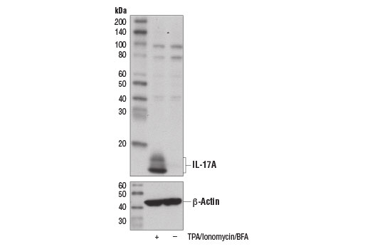
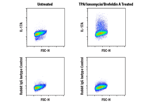
Monoclonal antibody is produced by immunizing animals with a synthetic peptide corresponding to residues surrounding Val49 of mouse IL-17A protein.
Background
The IL-17 family of cytokines consists of IL-17A-F, and their receptors include IL-17RA-RE (1). IL-17 cytokines are produced by a variety of cell types including the Th17 subset of CD4+ T cells, as well as subsets of γδ T cells, NK cells, and NKT cells (2). IL-17A and IL-17F, the most well-studied of the IL-17 cytokines, contribute to fungal and bacterial immunity by inducing expression of proinflammatory cytokines, chemokines, and antimicrobial peptides (2). In addition, IL-17A contributes to the pathogenesis of several autoimmune diseases (3). IL-17E promotes Th2 cell responses (4). The roles of IL-17B, IL-17C, and IL-17D are less clear, however these family members also appear to have the capacity to induce proinflammatory cytokines (1,5,6). IL-17 receptors have an extracellular domain, a transmembrane domain, and a SEFIR domain. They are believed to signal as homodimers, heterodimers, or multimers through their SEFIR domain by recruiting the SEFIR domain-containing adaptor Act1 (7). Unlike most cytokines that signal through Jak/STAT pathways, IL-17 signaling results in NF-κB activation (8).
IL-17A is a cysteine-linked, homodimeric, pro-inflammatory cytokine produced by Th17 cells, a distinct CD4+ T cell lineage (9,10). IL-17A stimulates the production of the pro-inflammatory cytokines IL-1β, TNFα, and IL-6. IL-17A also induces production of the neutrophil chemoattractants IL-8, CXCL1, and CXCL6 thereby bridging adaptive and innate immunity (9,10). IL-17A is intimately involved in mucosal immunity against bacterial infections (9,11) and has a putative role in some autoimmune disorders (9,12). IL-17A effects appear to be exerted primarily through binding to one of the IL-17 receptor subunits, IL-17RA (13). IL-17 binding induces production of cytokines, chemokines, and other proteins through activation of the Erk1/2 MAP kinase, PI3K/Akt, p38, and NF-κB pathways (11,12,14). Phosphorylation of some Jaks and Stats has been observed.
- Gaffen, S.L. (2009) Nat Rev Immunol 9, 556-67.
- Iwakura, Y. et al. (2011) Immunity 34, 149-62.
- Hu, Y. et al. (2011) Ann N Y Acad Sci 1217, 60-76.
- Fort, M.M. et al. (2001) Immunity 15, 985-95.
- Yamaguchi, Y. et al. (2007) J Immunol 179, 7128-36.
- Li, H. et al. (2000) Proc Natl Acad Sci U S A 97, 773-8.
- Chang, S.H. et al. (2006) J Biol Chem 281, 35603-7.
- Shalom-Barak, T. et al. (1998) J Biol Chem 273, 27467-73.
- Kolls, J.K. and Lindén, A. (2004) Immunity 21, 467-76.
- Liang, S.C. et al. (2006) J Exp Med 203, 2271-9.
- Dubin, P.J. and Kolls, J.K. (2008) Immunol Rev 226, 160-71.
- Zrioual, S. et al. (2009) J Immunol 182, 3112-20.
- Wright, J.F. et al. (2008) J Immunol 181, 2799-805.
- Rahman, M.S. et al. (2006) J Immunol 177, 4064-71.
Species Reactivity
Species reactivity is determined by testing in at least one approved application (e.g., western blot).
Western Blot Buffer
IMPORTANT: For western blots, incubate membrane with diluted primary antibody in 5% w/v BSA, 1X TBS, 0.1% Tween® 20 at 4°C with gentle shaking, overnight.
Applications Key
WB: Western Blotting IP: Immunoprecipitation FC-FP: Flow Cytometry (Fixed/Permeabilized)
Cross-Reactivity Key
H: human M: mouse R: rat Hm: hamster Mk: monkey Vir: virus Mi: mink C: chicken Dm: D. melanogaster X: Xenopus Z: zebrafish B: bovine Dg: dog Pg: pig Sc: S. cerevisiae Ce: C. elegans Hr: horse GP: Guinea Pig Rab: rabbit All: all species expected
Trademarks and Patents
使用に関する制限
法的な権限を与えられたCSTの担当者が署名した書面によって別途明示的に合意された場合を除き、 CST、その関連会社または代理店が提供する製品には以下の条件が適用されます。お客様が定める条件でここに定められた条件に含まれるものを超えるもの、 または、ここに定められた条件と異なるものは、法的な権限を与えられたCSTの担当者が別途書面にて受諾した場合を除き、拒絶され、 いかなる効力も効果も有しません。
研究専用 (For Research Use Only) またはこれに類似する表示がされた製品は、 いかなる目的についても FDA または外国もしくは国内のその他の規制機関により承認、認可または許可を受けていません。 お客様は製品を診断もしくは治療目的で使用してはならず、また、製品に表示された内容に違反する方法で使用してはなりません。 CST が販売または使用許諾する製品は、エンドユーザーであるお客様に対し、使途を研究および開発のみに限定して提供されるものです。 診断、予防もしくは治療目的で製品を使用することまたは製品を再販売 (単独であるか他の製品等の一部であるかを問いません) もしくはその他の商業的利用の目的で購入することについては、CST から別途許諾を得る必要があります。 お客様は以下の事項を遵守しなければなりません。(a) CST の製品 (単独であるか他の資材と一緒であるかを問いません) を販売、使用許諾、貸与、寄付もしくはその他の態様で第三者に譲渡したり使用させたりしてはなりません。また、商用の製品を製造するために CST の製品を使用してはなりません。(b) 複製、改変、リバースエンジニアリング、逆コンパイル、 分解または他の方法により製品の構造または技術を解明しようとしてはなりません。また、 CST の製品またはサービスと競合する製品またはサービスを開発する目的で CST の製品を使用してはなりません。(c) CST の製品の商標、商号、ロゴ、特許または著作権に関する通知または表示を除去したり改変したりしてはなりません。(d) CST の製品をCST 製品販売条件(CST’s Product Terms of Sale) および該当する書面のみに従って使用しなければなりません。(e) CST の製品に関連してお客様が使用する第三者の製品またはサービスに関する使用許諾条件、 サービス提供条件またはこれに類する合意事項を遵守しなければなりません。