| Product Includes | Product # | Quantity | Mol. Wt | Isotype/Source |
|---|---|---|---|---|
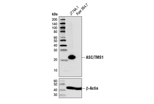
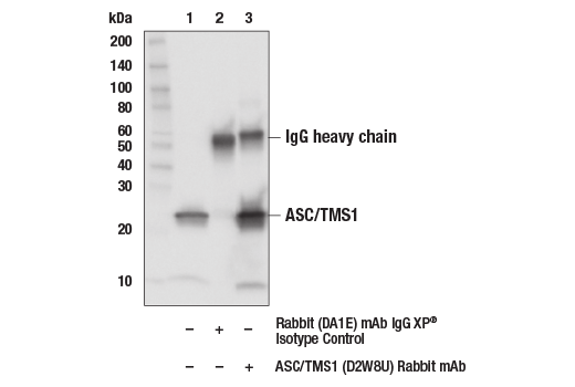
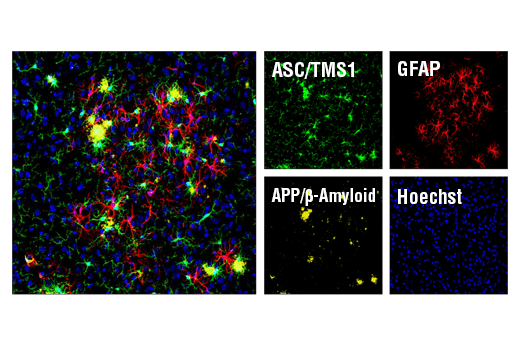
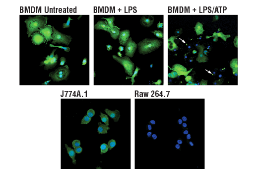
| ASC/TMS1 (D2W8U) Rabbit mAb | 67824 | 20 µl | 22 kDa | Rabbit IgG |
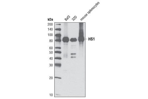
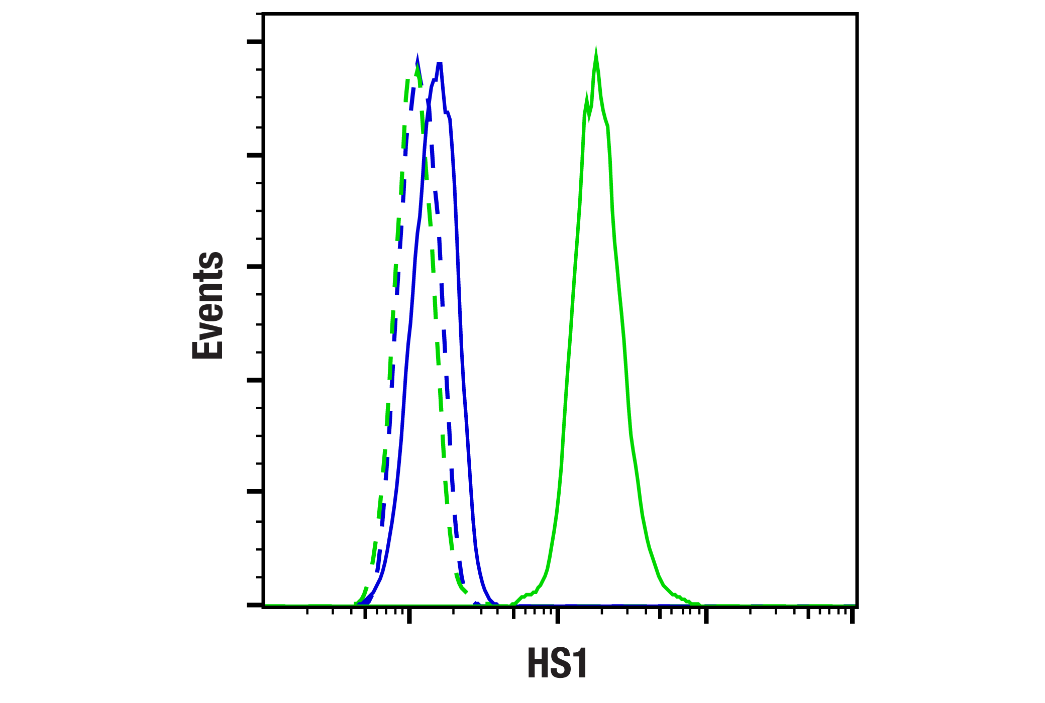
| HS1 (D5A9) XP® Rabbit mAb | 3892 | 20 µl | 80 kDa | Rabbit IgG |
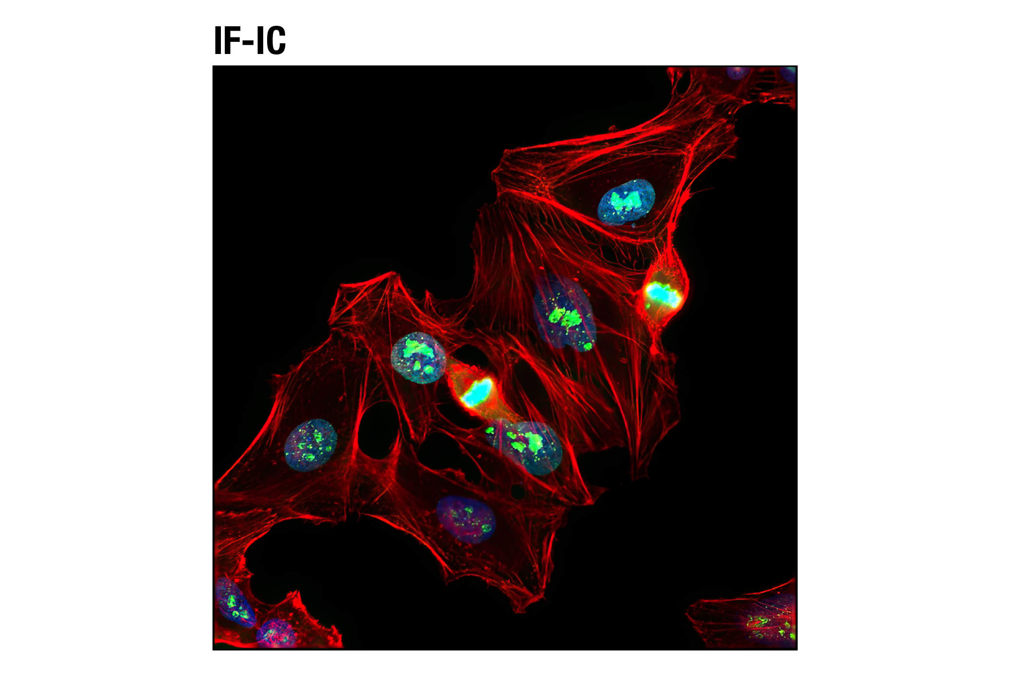
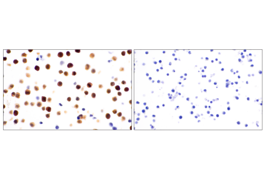
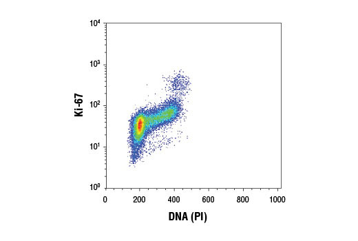
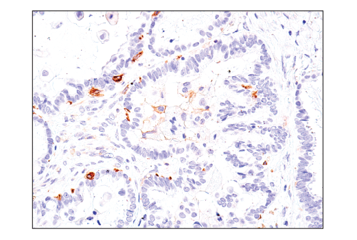
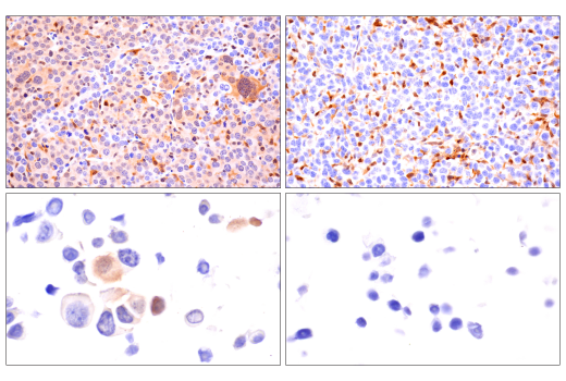
| Ki-67 (D3B5) Rabbit mAb | 9129 | 20 µl | Rabbit IgG | |
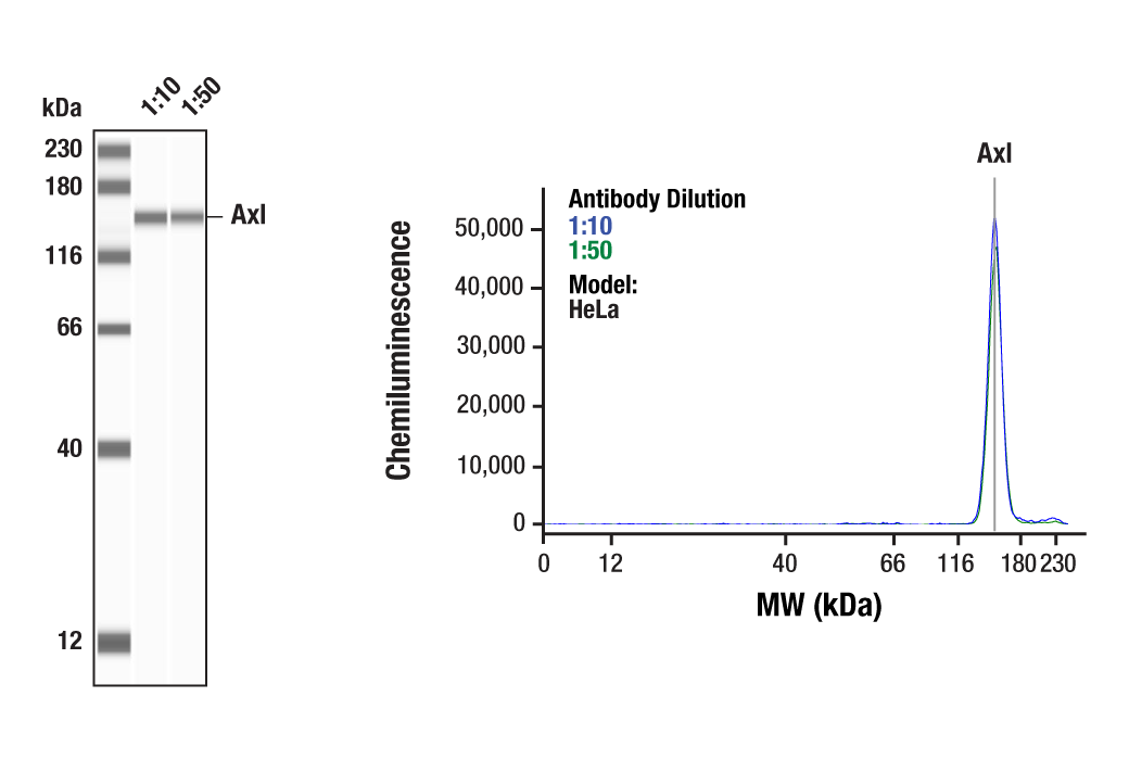
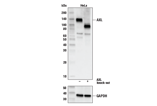
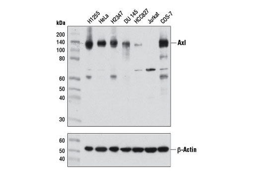
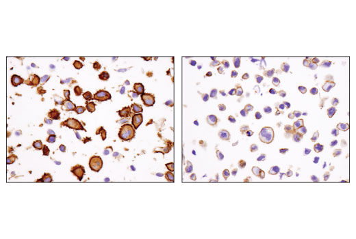
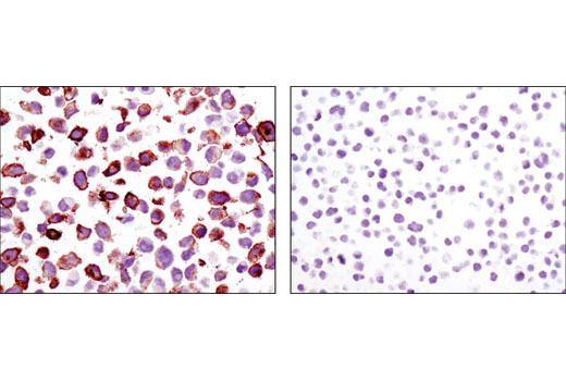
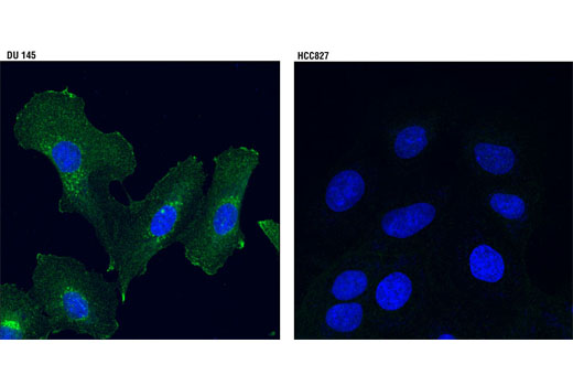
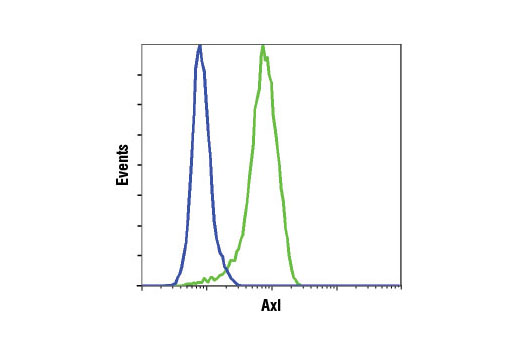
| Axl (C89E7) Rabbit mAb | 8661 | 20 µl | 138 kDa | Rabbit IgG |
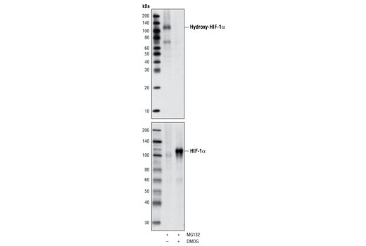
| Hydroxy-HIF-1α (Pro564) (D43B5) XP® Rabbit mAb | 3434 | 20 µl | 120 kDa | Rabbit IgG |
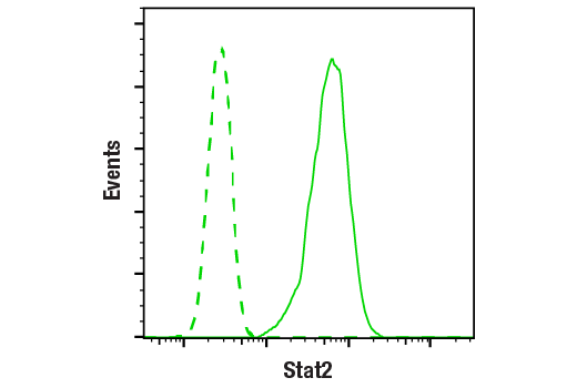
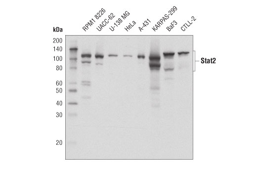
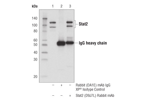
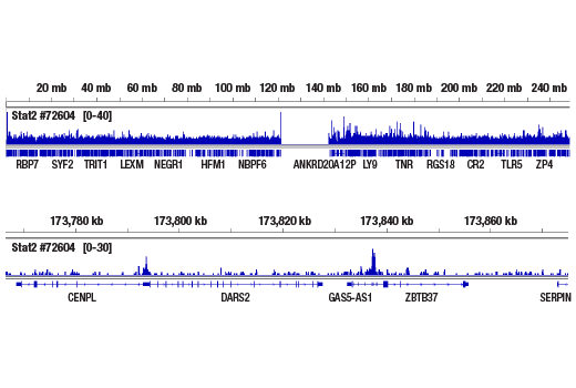
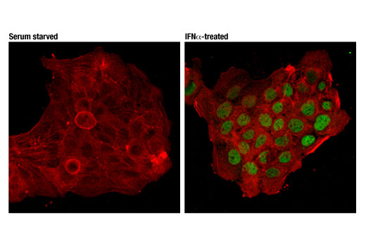
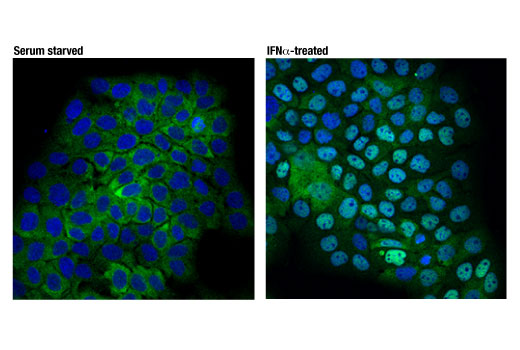
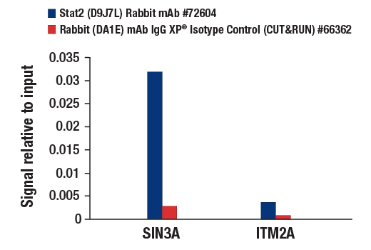
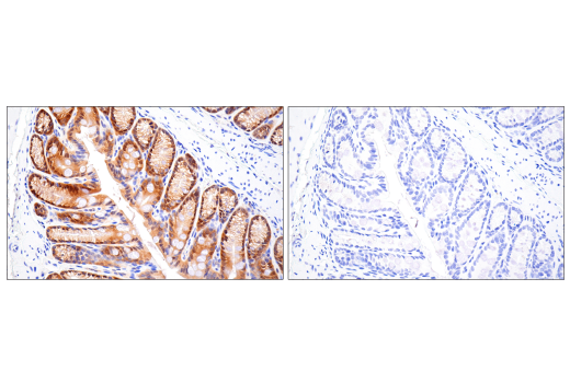
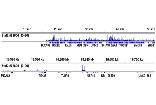
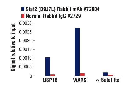
| Stat2 (D9J7L) Rabbit mAb | 72604 | 20 µl | 97, 113 kDa | Rabbit IgG |
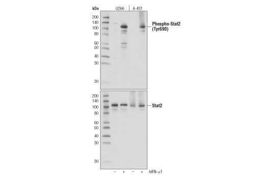
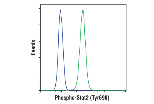
| Phospho-Stat2 (Tyr690) (D3P2P) Rabbit mAb | 88410 | 20 µl | 97, 113 kDa | Rabbit IgG |
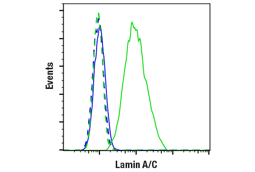
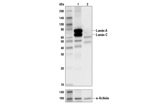
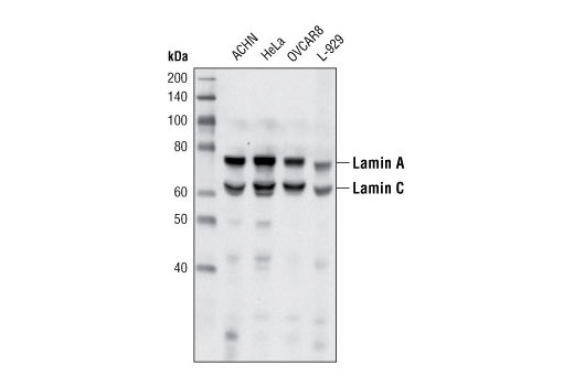
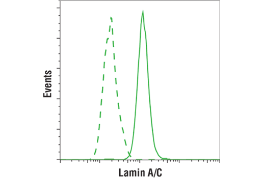
| Lamin A/C (4C11) Mouse mAb | 4777 | 20 µl | 74 (Lamin A), 63 (Lamin C) kDa | Mouse IgG2a |
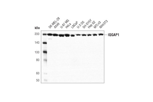
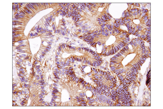
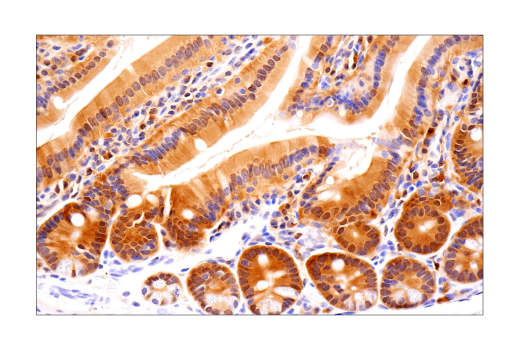
| IQGAP1 (D8K4X) XP® Rabbit mAb | 20648 | 20 µl | 195 kDa | Rabbit IgG |
| Anti-rabbit IgG, HRP-linked Antibody | 7074 | 100 µl | Goat |
Please visit cellsignal.com for individual component applications, species cross-reactivity, dilutions, protocols, and additional product information.
Description
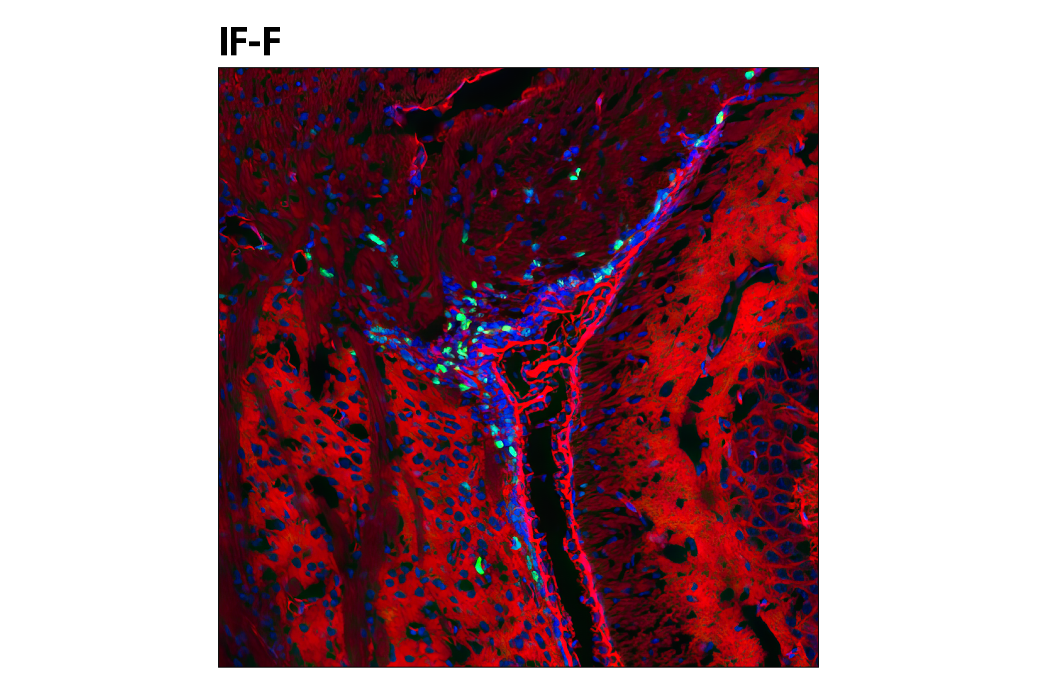
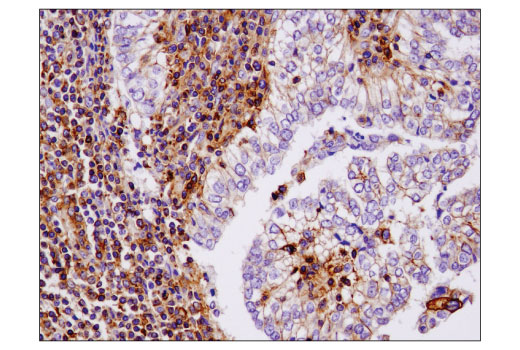
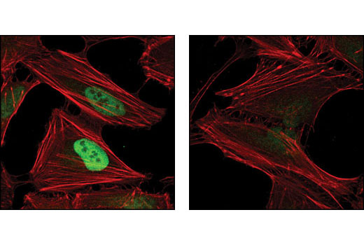
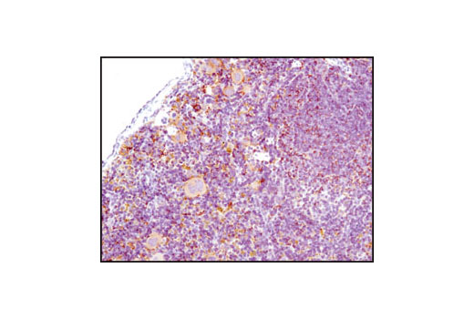
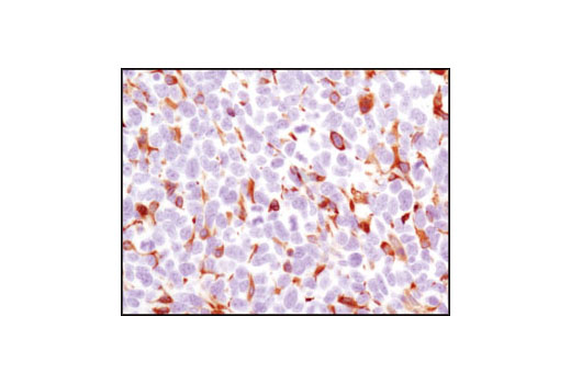
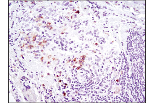
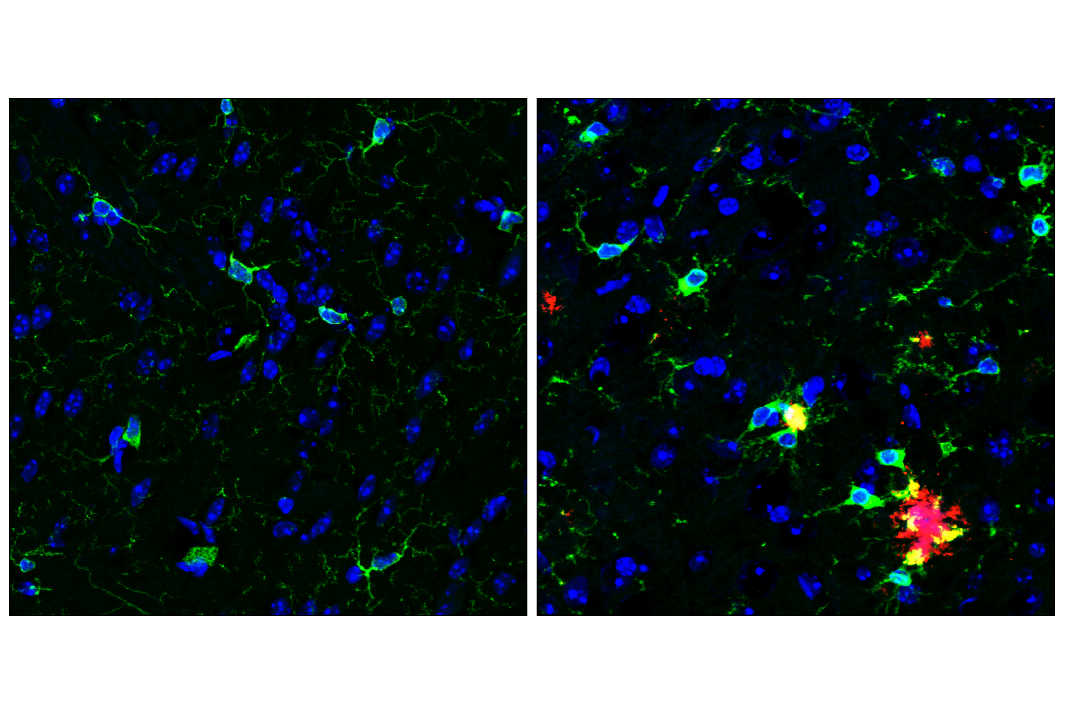
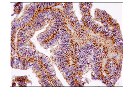
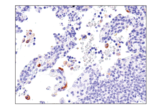
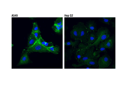
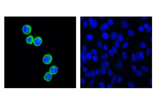
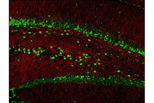
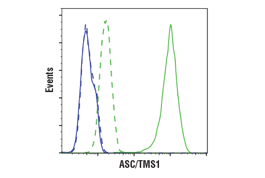
The Microglia Cross Module Antibody Sampler Kit provides an economical means of detecting proteins identified as markers of microglial activity corresponding to proliferation, neurodegeneration, interferon and LPS-relation by western blot and/or immunofluorescence.
Storage
Background
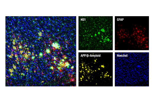
Distinct microglial activation states have been identified using RNA-seq data from a vast array of neurological disease and aging models. These activation states have been categorized into modules corresponding to proliferation, neurodegeneration, interferon-relation, LPS-relation, and many others (1). Previous work identifying markers of specific brain cell types using RNA-seq has shown HS1 and ASC/TMS1 to be useful and specific tools to study microglia (2). HS1 is a protein kinase substrate that is expressed only in tissues and cells of hematopoietic origin (3) and ASC/TMS1 has been found to be a critical component of inflammatory signaling where it associates with and activates caspase-1 in response to pro-inflammatory signals (4).
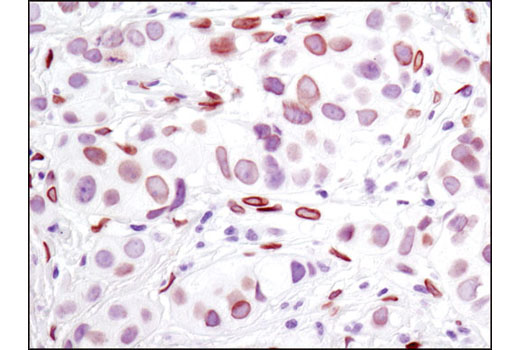
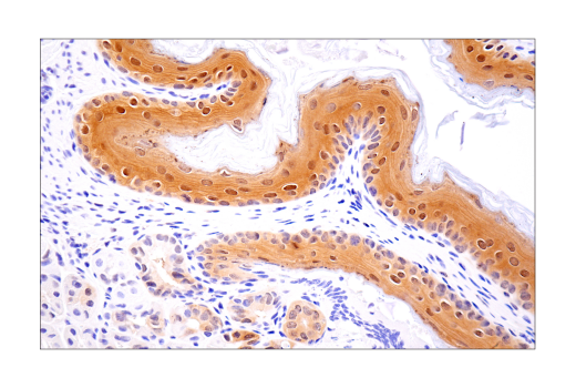
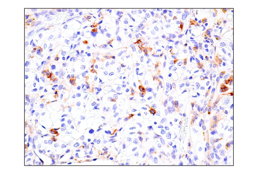
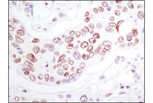
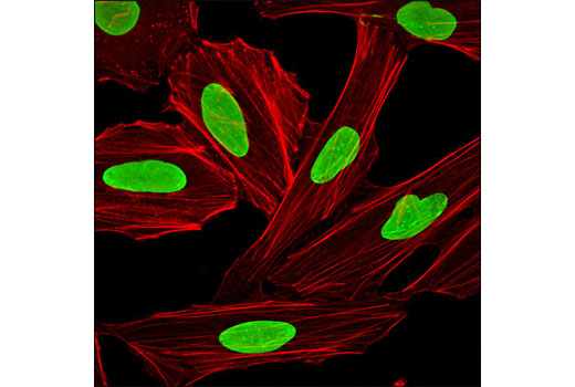
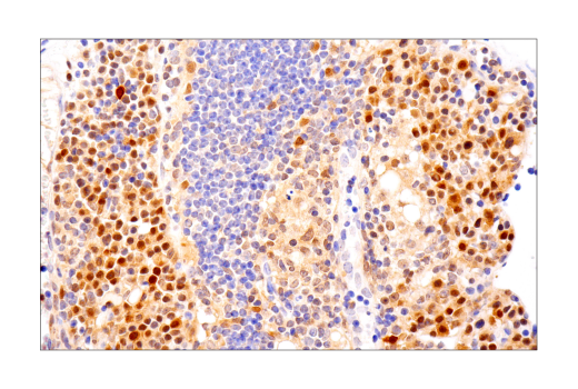
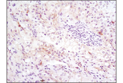
Ki-67 is a nuclear nonhistone protein (5) universally expressed among proliferating cells and absent in quiescent cells (6). Axl is a receptor tyrosine kinase that binds Gas6, stimulating regulatory effects on microglial phagocytic response to inflammatory stimuli (7). Hypoxia inducible factor-1 (HIF-1α) is a transcription factor responsible for adaptation to low oxygen environments whose downstream effects have been shown in a number of neurodegenerative diseases. Under normoxic conditions, HIF-1α is proline hydroxylated leading to ubiquitin mediated degradation (8). Stat2 is critical to the transcriptional responses induced by type I interferons, IFN-alpha/beta (9,10). In response to IFN-alpha/beta, Stat2 is activated by phosphorylation at site Tyr690 through associations with receptor-bound Jak kinases (11). Lamins are nuclear membrane structural components important for maintaining normal cell functions. Lamin A/C is cleaved by caspase-6 and serves as a marker for caspase-6 activation. The cleavage of lamins results in nuclear dysregulation and cell death (12,13). IQGAP1 is ubiquitously expressed and has been found to interact with APC (14) and the CLIP170 complex in response to small GTPases, promoting cell polarization and migration (15).
- Friedman, B.A. et al. (2018) Cell Rep 22, 832-47.
- Zhang, Y. et al. (2014) J Neurosci 34, 11929-47.
- Kitamura, D. et al. (1995) Biochem Biophys Res Commun 208, 1137-46.
- Srinivasula, S.M. et al. (2002) J Biol Chem 277, 21119-22.
- Gerdes, J. et al. (1983) Int J Cancer 31, 13-20.
- Weigel, M.T. and Dowsett, M. (2010) Endocr Relat Cancer 17, R245-62.
- Grommes, C. et al. (2008) J Neuroimmune Pharmacol 3, 130-40.
- Zhang, Z. et al. (2011) Curr Med Chem 18, 4335-43.
- Fu, X.Y. et al. (1992) Proc Natl Acad Sci U S A 89, 7840-3.
- Ihle, J.N. (2001) Curr Opin Cell Biol 13, 211-7.
- Improta, T. et al. (1994) Proc Natl Acad Sci U S A 91, 4776-80.
- Oberhammer, F.A. et al. (1994) J Cell Biol 126, 827-37.
- Rao, L. et al. (1996) J Cell Biol 135, 1441-55.
- Watanabe, T. et al. (2004) Dev Cell 7, 871-83.
- Fukata, M. et al. (2002) Cell 109, 873-85.
Background References
Trademarks and Patents
使用に関する制限
法的な権限を与えられたCSTの担当者が署名した書面によって別途明示的に合意された場合を除き、 CST、その関連会社または代理店が提供する製品には以下の条件が適用されます。お客様が定める条件でここに定められた条件に含まれるものを超えるもの、 または、ここに定められた条件と異なるものは、法的な権限を与えられたCSTの担当者が別途書面にて受諾した場合を除き、拒絶され、 いかなる効力も効果も有しません。
研究専用 (For Research Use Only) またはこれに類似する表示がされた製品は、 いかなる目的についても FDA または外国もしくは国内のその他の規制機関により承認、認可または許可を受けていません。 お客様は製品を診断もしくは治療目的で使用してはならず、また、製品に表示された内容に違反する方法で使用してはなりません。 CST が販売または使用許諾する製品は、エンドユーザーであるお客様に対し、使途を研究および開発のみに限定して提供されるものです。 診断、予防もしくは治療目的で製品を使用することまたは製品を再販売 (単独であるか他の製品等の一部であるかを問いません) もしくはその他の商業的利用の目的で購入することについては、CST から別途許諾を得る必要があります。 お客様は以下の事項を遵守しなければなりません。(a) CST の製品 (単独であるか他の資材と一緒であるかを問いません) を販売、使用許諾、貸与、寄付もしくはその他の態様で第三者に譲渡したり使用させたりしてはなりません。また、商用の製品を製造するために CST の製品を使用してはなりません。(b) 複製、改変、リバースエンジニアリング、逆コンパイル、 分解または他の方法により製品の構造または技術を解明しようとしてはなりません。また、 CST の製品またはサービスと競合する製品またはサービスを開発する目的で CST の製品を使用してはなりません。(c) CST の製品の商標、商号、ロゴ、特許または著作権に関する通知または表示を除去したり改変したりしてはなりません。(d) CST の製品をCST 製品販売条件(CST’s Product Terms of Sale) および該当する書面のみに従って使用しなければなりません。(e) CST の製品に関連してお客様が使用する第三者の製品またはサービスに関する使用許諾条件、 サービス提供条件またはこれに類する合意事項を遵守しなければなりません。