WB
H
Endogenous
20
Rabbit
#P20963
919
Product Information
Product Usage Information
| Application | Dilution |
|---|---|
| Western Blotting | 1:1000 |
Storage
Specificity / Sensitivity
Species Reactivity:
Human
Species predicted to react based on 100% sequence homology
The antigen sequence used to produce this antibody shares
100% sequence homology with the species listed here, but
reactivity has not been tested or confirmed to work by CST.
Use of this product with these species is not covered under
our
Product Performance Guarantee.
Pig
Source / Purification
Polyclonal antibodies are produced by immunizing animals with a synthetic phosphopeptide corresponding to residues surrounding Tyr142 of human CD3ζ protein. Antibodies are purified by peptide affinity chromatography.
Background
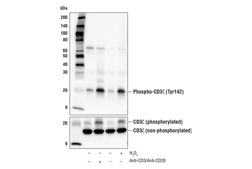
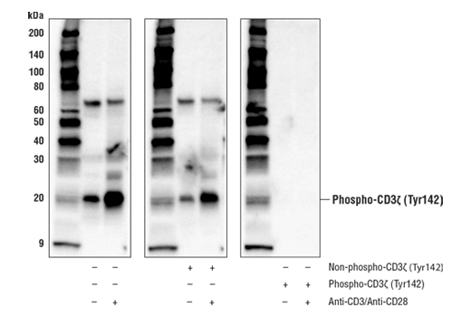
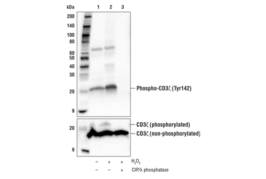
When T cells encounter antigens via the T cell receptor (TCR), information about the quantity and quality of antigens is relayed to the intracellular signal transduction machinery (1). This activation process depends mainly on CD3 (Cluster of Differentiation 3), a multiunit protein complex that directly associates with the TCR. CD3 is composed of four polypeptides: ζ, γ, ε, and δ. Each of these polypeptides contains at least one immunoreceptor tyrosine-based activation motif (ITAM) (2). Engagement of the TCR complex with foreign antigens induces tyrosine phosphorylation in the ITAM motifs and phosphorylated ITAMs function as docking sites for signaling molecules such as ZAP-70 and the p85 subunit of PI-3 kinase (3,4). TCR ligation also induces a conformational change in CD3ε, such that a proline region is exposed and then associates with the adaptor protein Nck (5).
The CD3ζ invariant chain is a type-I transmembrane protein that exists in the TCR signaling complex as a disulfide-linked homodimer (6). The cytoplasmic tail of each CD3ζ monomer contains three distinct ITAM motifs, each containing two tyrosine residues. Phosphorylation of CD3ζ ITAM tyrosine residues, including Y142, is driven by recruitment of the Lck and Fyn tyrosine kinases to the TCR (7). Lck/Fyn-mediated ITAM phosphorylation creates docking sites that promote the SH2 domain-dependent recruitment and activation of Zap-70 (8-10), which drives amplification of signaling events downstream of the TCR that facilitate T cell activation (10). Phosphorylation of a pool of p16 CD3ζ leads to the generation of p21 and p23 species, which differ in the degree of ITAM phosphorylation. It has been proposed that the ratio of p21/p23 contributes to regulating the amplitude of T cell activation (11). CD3ζ plays an important role in the assembly and surface expression of the TCR complex. Indeed, research studies have demonstrated that CD3ζ is degraded in response to Ag-dependent TCR stimulation as a mechanism to tightly control T cell activation (12).
- Kuhns, M.S. et al. (2006) Immunity 24, 133-139.
- Pitcher, L.A. and van Oers, N.S. (2003) Trends Immunol. 24, 554-560.
- Osman, N. et al. (1996) Eur. J. Immunol. 26, 1063-1068.
- Hatada, M.H. et al. (1995) Nature 377, 32-38.
- Gil, D. et al. (2002) Cell 109, 901-912.
- Call, M.E. et al. (2006) Cell 127, 355-68.
- Housden, H.R. et al. (2003) Eur J Biochem 270, 2369-76.
- Hatada, M.H. et al. (1995) Nature 377, 32-8.
- Visco, C. et al. (2000) Biochemistry 39, 2784-91.
- Iwashima, M. et al. (1994) Science 263, 1136-9.
- Pitcher, L.A. et al. (2003) Immunol Rev 191, 47-61.
- Dumont, C. et al. (2002) J Immunol 169, 1705-12.
Species Reactivity
Species reactivity is determined by testing in at least one approved application (e.g., western blot).
Western Blot Buffer
IMPORTANT: For western blots, incubate membrane with diluted primary antibody in 5% w/v BSA, 1X TBS, 0.1% Tween® 20 at 4°C with gentle shaking, overnight.
Applications Key
WB: Western Blotting
Cross-Reactivity Key
H: human M: mouse R: rat Hm: hamster Mk: monkey Vir: virus Mi: mink C: chicken Dm: D. melanogaster X: Xenopus Z: zebrafish B: bovine Dg: dog Pg: pig Sc: S. cerevisiae Ce: C. elegans Hr: horse GP: Guinea Pig Rab: rabbit All: all species expected
Trademarks and Patents
使用に関する制限
法的な権限を与えられたCSTの担当者が署名した書面によって別途明示的に合意された場合を除き、 CST、その関連会社または代理店が提供する製品には以下の条件が適用されます。お客様が定める条件でここに定められた条件に含まれるものを超えるもの、 または、ここに定められた条件と異なるものは、法的な権限を与えられたCSTの担当者が別途書面にて受諾した場合を除き、拒絶され、 いかなる効力も効果も有しません。
研究専用 (For Research Use Only) またはこれに類似する表示がされた製品は、 いかなる目的についても FDA または外国もしくは国内のその他の規制機関により承認、認可または許可を受けていません。 お客様は製品を診断もしくは治療目的で使用してはならず、また、製品に表示された内容に違反する方法で使用してはなりません。 CST が販売または使用許諾する製品は、エンドユーザーであるお客様に対し、使途を研究および開発のみに限定して提供されるものです。 診断、予防もしくは治療目的で製品を使用することまたは製品を再販売 (単独であるか他の製品等の一部であるかを問いません) もしくはその他の商業的利用の目的で購入することについては、CST から別途許諾を得る必要があります。 お客様は以下の事項を遵守しなければなりません。(a) CST の製品 (単独であるか他の資材と一緒であるかを問いません) を販売、使用許諾、貸与、寄付もしくはその他の態様で第三者に譲渡したり使用させたりしてはなりません。また、商用の製品を製造するために CST の製品を使用してはなりません。(b) 複製、改変、リバースエンジニアリング、逆コンパイル、 分解または他の方法により製品の構造または技術を解明しようとしてはなりません。また、 CST の製品またはサービスと競合する製品またはサービスを開発する目的で CST の製品を使用してはなりません。(c) CST の製品の商標、商号、ロゴ、特許または著作権に関する通知または表示を除去したり改変したりしてはなりません。(d) CST の製品をCST 製品販売条件(CST’s Product Terms of Sale) および該当する書面のみに従って使用しなければなりません。(e) CST の製品に関連してお客様が使用する第三者の製品またはサービスに関する使用許諾条件、 サービス提供条件またはこれに類する合意事項を遵守しなければなりません。