WB, W-S, IP, IHC-P
H M
Endogenous
185
Rabbit
#P21860
2065
Product Information
Product Usage Information
| Application | Dilution |
|---|---|
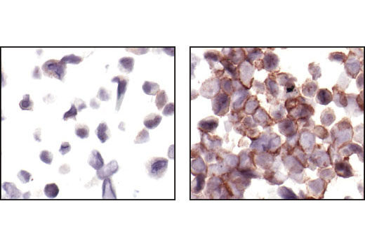
| Western Blotting | 1:1000 |
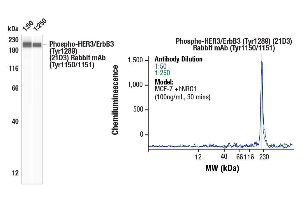
| Simple Western™ | 1:50 - 1:250 |
| Immunoprecipitation | 1:100 |
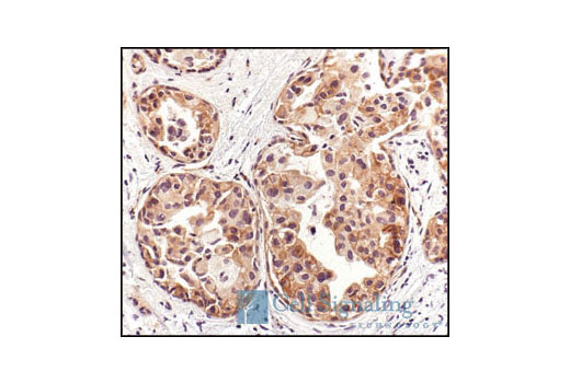
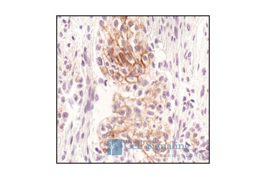
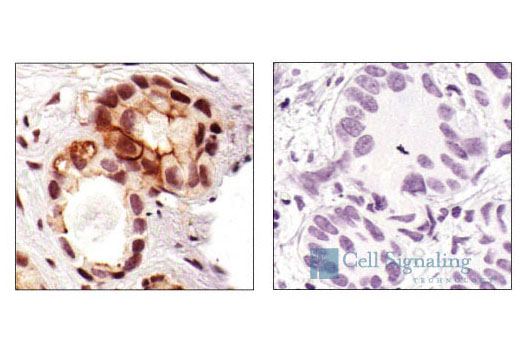
| Immunohistochemistry (Paraffin) | 1:1000 |
Storage
For a carrier free (BSA and azide free) version of this product see product #73367.
Specificity / Sensitivity
Species Reactivity:
Human, Mouse
Species predicted to react based on 100% sequence homology
The antigen sequence used to produce this antibody shares
100% sequence homology with the species listed here, but
reactivity has not been tested or confirmed to work by CST.
Use of this product with these species is not covered under
our
Product Performance Guarantee.
Rat, Dog
Source / Purification
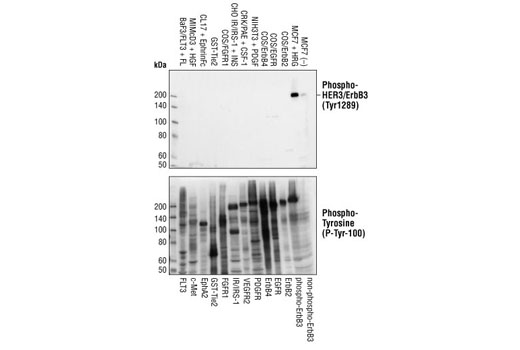
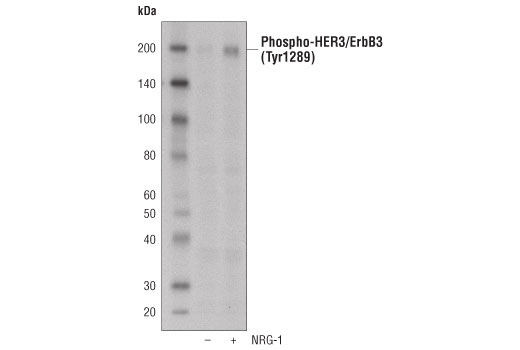
Monoclonal antibody is produced by immunizing animals with a synthetic phosphopeptide corresponding to residues surrounding Tyr1289 of human HER3/ErbB3.
Background
HER3/ErbB3 is a member of the ErbB receptor protein tyrosine kinase family, but it lacks tyrosine kinase activity. Tyrosine phosphorylation of ErbB3 depends on its association with other ErbB tyrosine kinases. Upon ligand binding, heterodimers form between ErbB3 and other ErbB proteins, and ErbB3 is phosphorylated on tyrosine residues by the activated ErbB kinase (1,2). There are at least 9 potential tyrosine phosphorylation sites in the carboxy-terminal tail of ErbB3. These sites serve as consensus binding sites for signal transducing proteins, including Src family members, Grb2, and the p85 subunit of PI3 kinase, which mediate ErbB downstream signaling (3). Both Tyr1222 and Tyr1289 of ErbB3 reside within a YXXM motif and participate in signaling to PI3K (4).
Investigators have found that ErbB3 is highly expressed in many cancer cells (5) and activation of the ErbB3/PI3K pathway is correlated with malignant phenotypes of adenocarcinomas (6). Research studies have demonstrated that in tumor development, ErbB3 may function as an oncogenic unit together with other ErbB members (e.g., ErbB2 requires ErbB3 to drive breast tumor cell proliferation) (7). Thus, investigators view inhibiting interaction between ErbB3 and ErbB tyrosine kinases as a novel strategy for anti-tumor therapy.
- Yarden, Y. and Sliwkowski, M.X. (2001) Nat Rev Mol Cell Biol 2, 127-37.
- Guy, P.M. et al. (1994) Proc Natl Acad Sci U S A 91, 8132-6.
- Songyang, Z. et al. (1993) Cell 72, 767-78.
- Kim, H.H. et al. (1994) J Biol Chem 269, 24747-55.
- Sithanandam, G. et al. (2003) Carcinogenesis 24, 1581-92.
- Kobayashi, M. et al. (2003) Oncogene 22, 1294-301.
- Holbro, T. et al. (2003) Proc Natl Acad Sci U S A 100, 8933-8.
Species Reactivity
Species reactivity is determined by testing in at least one approved application (e.g., western blot).
Western Blot Buffer
IMPORTANT: For western blots, incubate membrane with diluted primary antibody in 5% w/v BSA, 1X TBS, 0.1% Tween® 20 at 4°C with gentle shaking, overnight.
Applications Key
WB: Western Blotting W-S: Simple Western™ IP: Immunoprecipitation IHC-P: Immunohistochemistry (Paraffin)
Cross-Reactivity Key
H: human M: mouse R: rat Hm: hamster Mk: monkey Vir: virus Mi: mink C: chicken Dm: D. melanogaster X: Xenopus Z: zebrafish B: bovine Dg: dog Pg: pig Sc: S. cerevisiae Ce: C. elegans Hr: horse GP: Guinea Pig Rab: rabbit All: all species expected
Trademarks and Patents
使用に関する制限
法的な権限を与えられたCSTの担当者が署名した書面によって別途明示的に合意された場合を除き、 CST、その関連会社または代理店が提供する製品には以下の条件が適用されます。お客様が定める条件でここに定められた条件に含まれるものを超えるもの、 または、ここに定められた条件と異なるものは、法的な権限を与えられたCSTの担当者が別途書面にて受諾した場合を除き、拒絶され、 いかなる効力も効果も有しません。
研究専用 (For Research Use Only) またはこれに類似する表示がされた製品は、 いかなる目的についても FDA または外国もしくは国内のその他の規制機関により承認、認可または許可を受けていません。 お客様は製品を診断もしくは治療目的で使用してはならず、また、製品に表示された内容に違反する方法で使用してはなりません。 CST が販売または使用許諾する製品は、エンドユーザーであるお客様に対し、使途を研究および開発のみに限定して提供されるものです。 診断、予防もしくは治療目的で製品を使用することまたは製品を再販売 (単独であるか他の製品等の一部であるかを問いません) もしくはその他の商業的利用の目的で購入することについては、CST から別途許諾を得る必要があります。 お客様は以下の事項を遵守しなければなりません。(a) CST の製品 (単独であるか他の資材と一緒であるかを問いません) を販売、使用許諾、貸与、寄付もしくはその他の態様で第三者に譲渡したり使用させたりしてはなりません。また、商用の製品を製造するために CST の製品を使用してはなりません。(b) 複製、改変、リバースエンジニアリング、逆コンパイル、 分解または他の方法により製品の構造または技術を解明しようとしてはなりません。また、 CST の製品またはサービスと競合する製品またはサービスを開発する目的で CST の製品を使用してはなりません。(c) CST の製品の商標、商号、ロゴ、特許または著作権に関する通知または表示を除去したり改変したりしてはなりません。(d) CST の製品をCST 製品販売条件(CST’s Product Terms of Sale) および該当する書面のみに従って使用しなければなりません。(e) CST の製品に関連してお客様が使用する第三者の製品またはサービスに関する使用許諾条件、 サービス提供条件またはこれに類する合意事項を遵守しなければなりません。