WB, IP, IF-IC, FC-FP
H M R Hm Mk Dg
Endogenous
125
Rabbit IgG
#P49736
4171
Product Information
Product Usage Information
| Application | Dilution |
|---|---|
| Western Blotting | 1:1000 |
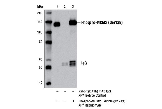
| Immunoprecipitation | 1:50 |
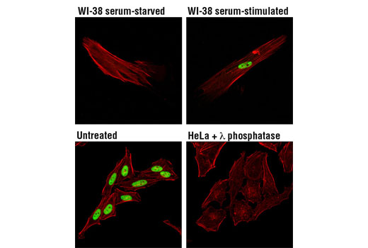
| Immunofluorescence (Immunocytochemistry) | 1:100 |
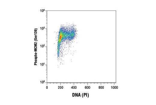
| Flow Cytometry (Fixed/Permeabilized) | 1:200 |
Storage
Specificity / Sensitivity
Species Reactivity:
Human, Mouse, Rat, Hamster, Monkey, Dog
Species predicted to react based on 100% sequence homology
The antigen sequence used to produce this antibody shares
100% sequence homology with the species listed here, but
reactivity has not been tested or confirmed to work by CST.
Use of this product with these species is not covered under
our
Product Performance Guarantee.
Xenopus
Source / Purification
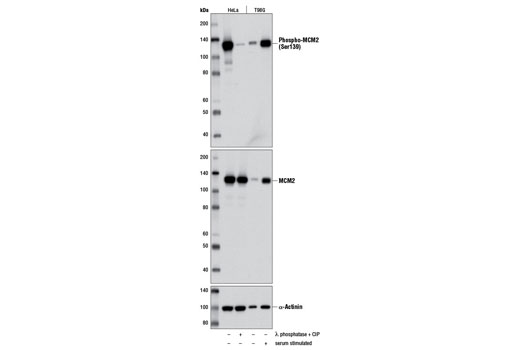
Monoclonal antibody is produced by immunizing animals with a synthetic peptide corresponding to residues surrounding Ser139 of human MCM2 protein.
Background
The minichromosome maintenance (MCM) 2-7 proteins are a family of six related proteins required for initiation and elongation of DNA replication. MCM2-7 bind together to form the heterohexameric MCM complex that is thought to act as a replicative helicase at the DNA replication fork (1-5). This complex is a key component of the pre-replication complex (pre-RC) (reviewed in 1). Cdc6 and CDT1 recruit the MCM complex to the origin recognition complex (ORC) during late mitosis/early G1 phase forming the pre-RC and licensing the DNA for replication (reviewed in 2). Licensing of the chromatin permits the DNA to replicate only once per cell cycle, thereby helping to ensure that genetic alterations and malignant cell growth do not occur (reviewed in 3). Phosphorylation of the MCM2, MCM3, MCM4, and MCM6 subunits appears to regulate MCM complex activity and the initiation of DNA synthesis (6-8). CDK1 phosphorylation of MCM3 at Ser112 during late mitosis/early G1 phase has been shown to initiate complex formation and chromatin loading in vitro (8). Phosphorylation of MCM2 at serine 139 by cdc7/dbf4 coincides with the initiation of DNA replication (9). MCM proteins are removed during DNA replication, causing chromatin to become unlicensed through inhibition of pre-RC reformation. Studies have shown that the MCM complex is involved in checkpoint control by protecting the structure of the replication fork and assisting in restarting replication by recruiting checkpoint proteins after arrest (reviewed in 3,10).
- Lei, M. and Tye, B.K. (2001) J Cell Sci 114, 1447-54.
- Lygerou, Z. and Nurse, P. (2000) Science 290, 2271-3.
- Forsburg, S.L. (2004) Microbiol Mol Biol Rev 68, 109-31.
- Tye, B.K. and Sawyer, S. (2000) J Biol Chem 275, 34833-6.
- Labib, K. et al. (2000) Science 288, 1643-7.
- Charych, D.H. et al. (2008) J Cell Biochem 104, 1075-86.
- Masai, H. et al. (2006) J Biol Chem 281, 39249-61.
- Lin, D.I. et al. (2008) Proc Natl Acad Sci USA 105, 8079-84.
- Tsuji, T. et al. (2006) Mol Biol Cell 17, 4459-72.
- Bailis, J.M. et al. (2008) Mol Cell Biol 28, 1724-38.
Species Reactivity
Species reactivity is determined by testing in at least one approved application (e.g., western blot).
Western Blot Buffer
IMPORTANT: For western blots, incubate membrane with diluted primary antibody in 5% w/v BSA, 1X TBS, 0.1% Tween® 20 at 4°C with gentle shaking, overnight.
Applications Key
WB: Western Blotting IP: Immunoprecipitation IF-IC: Immunofluorescence (Immunocytochemistry) FC-FP: Flow Cytometry (Fixed/Permeabilized)
Cross-Reactivity Key
H: human M: mouse R: rat Hm: hamster Mk: monkey Vir: virus Mi: mink C: chicken Dm: D. melanogaster X: Xenopus Z: zebrafish B: bovine Dg: dog Pg: pig Sc: S. cerevisiae Ce: C. elegans Hr: horse GP: Guinea Pig Rab: rabbit All: all species expected
Trademarks and Patents
使用に関する制限
法的な権限を与えられたCSTの担当者が署名した書面によって別途明示的に合意された場合を除き、 CST、その関連会社または代理店が提供する製品には以下の条件が適用されます。お客様が定める条件でここに定められた条件に含まれるものを超えるもの、 または、ここに定められた条件と異なるものは、法的な権限を与えられたCSTの担当者が別途書面にて受諾した場合を除き、拒絶され、 いかなる効力も効果も有しません。
研究専用 (For Research Use Only) またはこれに類似する表示がされた製品は、 いかなる目的についても FDA または外国もしくは国内のその他の規制機関により承認、認可または許可を受けていません。 お客様は製品を診断もしくは治療目的で使用してはならず、また、製品に表示された内容に違反する方法で使用してはなりません。 CST が販売または使用許諾する製品は、エンドユーザーであるお客様に対し、使途を研究および開発のみに限定して提供されるものです。 診断、予防もしくは治療目的で製品を使用することまたは製品を再販売 (単独であるか他の製品等の一部であるかを問いません) もしくはその他の商業的利用の目的で購入することについては、CST から別途許諾を得る必要があります。 お客様は以下の事項を遵守しなければなりません。(a) CST の製品 (単独であるか他の資材と一緒であるかを問いません) を販売、使用許諾、貸与、寄付もしくはその他の態様で第三者に譲渡したり使用させたりしてはなりません。また、商用の製品を製造するために CST の製品を使用してはなりません。(b) 複製、改変、リバースエンジニアリング、逆コンパイル、 分解または他の方法により製品の構造または技術を解明しようとしてはなりません。また、 CST の製品またはサービスと競合する製品またはサービスを開発する目的で CST の製品を使用してはなりません。(c) CST の製品の商標、商号、ロゴ、特許または著作権に関する通知または表示を除去したり改変したりしてはなりません。(d) CST の製品をCST 製品販売条件(CST’s Product Terms of Sale) および該当する書面のみに従って使用しなければなりません。(e) CST の製品に関連してお客様が使用する第三者の製品またはサービスに関する使用許諾条件、 サービス提供条件またはこれに類する合意事項を遵守しなければなりません。