WB, W-S, IP, IHC-P, IF-IC, FC-FP
H M Mk
Endogenous
150
Rabbit IgG
#P19174
5335
Product Information
Product Usage Information
| Application | Dilution |
|---|---|
| Western Blotting | 1:1000 |
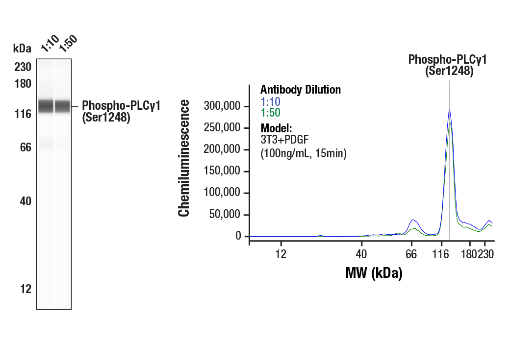
| Simple Western™ | 1:10 - 1:50 |
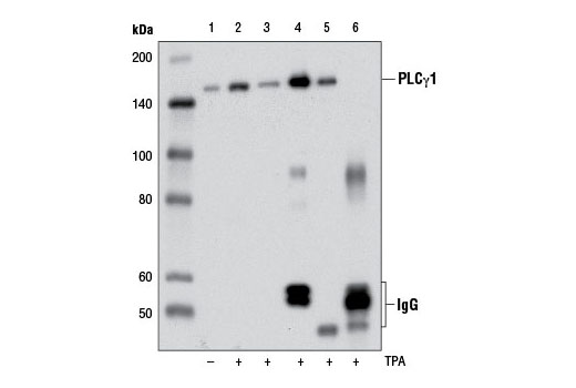
| Immunoprecipitation | 1:50 |
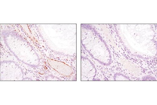
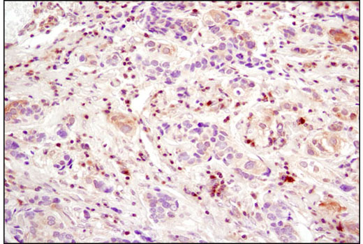
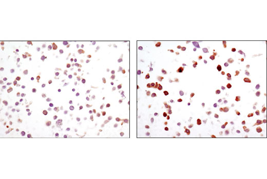
| Immunohistochemistry (Paraffin) | 1:100 - 1:400 |
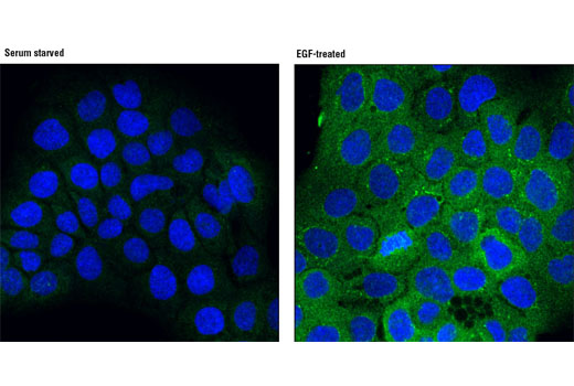
| Immunofluorescence (Immunocytochemistry) | 1:100 - 1:200 |
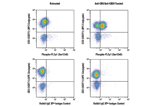
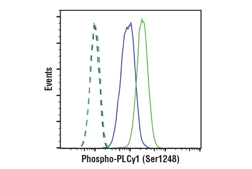
| Flow Cytometry (Fixed/Permeabilized) | 1:800 |
Storage
Specificity / Sensitivity
Species Reactivity:
Human, Mouse, Monkey
Species predicted to react based on 100% sequence homology
The antigen sequence used to produce this antibody shares
100% sequence homology with the species listed here, but
reactivity has not been tested or confirmed to work by CST.
Use of this product with these species is not covered under
our
Product Performance Guarantee.
Rat
Source / Purification
Monoclonal antibody is produced by immunizing animals with a synthetic peptide corresponding to residues surrounding Ser1248 of human PLCγ1 protein.
Background
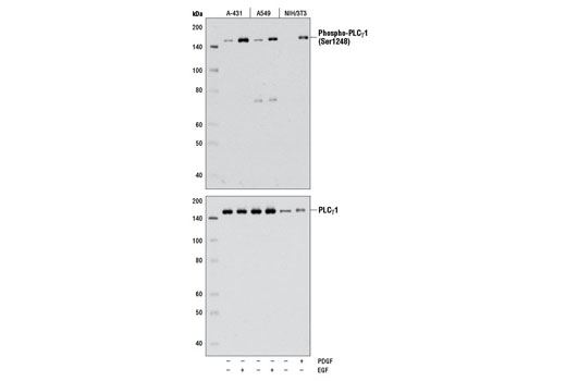
Phosphoinositide-specific phospholipase C (PLC) plays a significant role in transmembrane signaling. In response to extracellular stimuli, such as hormones, growth factors, and neurotransmitters, PLC hydrolyzes phosphatidylinositol 4,5-bisphosphate (PIP2) to generate two secondary messengers: inositol 1,4,5-triphosphate (IP3) and diacylglycerol (DAG) (1). At least four families of PLCs have been identified: PLCβ, PLCγ, PLCδ, and PLCε. Phosphorylation is one of the key mechanisms that regulate the activity of PLC. PLCγ is activated by both receptor and non-receptor tyrosine kinases (2). PLCγ forms a complex with EGF and PDGF receptors, which leads to the phosphorylation of PLCγ at Tyr771, 783, and 1248 (3). Phosphorylation by Syk at Tyr783 activates the enzymatic activity of PLCγ1 (4). PLCγ2 is engaged in antigen-dependent signaling in B cells and collagen-dependent signaling in platelets. Phosphorylation by Btk or Lck at Tyr753, 759, 1197, and 1217 is correlated with PLCγ2 activity (5,6).
Two mammalian PLCγ isoforms (γ1 and γ2) have been cloned and characterized (7,8). Like other PLC-family members, PLCγ1 and PLCγ2 contain calcium-binding (EF-hand, C2) and lipid-interacting (PH, EF-hand) domains necessary for their enzymatic activity and substrate recognition. Uniquely, PLCγ isoforms have additional, conserved SH2 and SH3 domains critical for their functions as signaling molecules and scaffolding proteins. Upon growth factor stimulation, PLCγ1 is recruited (via SH2 domains) to phosphotyrosine residues within the cytoplasmic tail of many RTKs where it serves as a substrate for the RTK and provides docking sites for additional proteins involved in RTK signaling (4-6,9-12). PLCγ1 and γ2 can also be activated downstream of receptors lacking intrinsic tyrosine kinase activity. This has been reported downstream of multiple G protein-coupled receptors and the T cell receptor in which tyrosine kinases of the Src, Syk, and Tec families serve to bind, phosphorylate, and activate PLCγ (reviewed in 13-15). Phosphorylation at tyrosine residues by both receptor and non-receptor tyrosine kinases results in robust activation of PLCγ1 activity, leading to generation of second messengers. In response to agonists, PLCγ1 is phosphorylated on Tyr783, Tyr711, and Tyr1253 (Tyr753, Tyr759, and Tyr1217 in PLCγ2) resulting in robust PI-4,5-P2 hydrolysis (4-6,9-12). Interestingly recent evidence suggests a role for tyrosine kinase-independent regulation of PLCγ in some systems. For example, in response to EGF, proline-rich regions of Akt interact with the SH3 domain of PLCγ1 resulting in association of the two enzymes, phosphorylation of PLCγ1 at Ser1248, and enhanced cellular motility (16). This finding demonstrates that PLCγ1 can function as a "scaffold" between RTKs and Akt, thereby establishing a mechanism by which the Akt signaling pathway cross-talks with tyrosine kinases. However, the mechanism and functional significance of phosphorylation at Ser1248 remains to be fully clarified, as it has also been shown that PKA-mediated phosphorylation at this site is inhibitory to PLCγ1 tyrosine phosphorylation and phospholipase activity in CD3-treated Jurkat cells (17), suggesting that Ser1248 may be an allosteric regulator of PLCγ1 activity.
- Singer, W.D. et al. (1997) Annu Rev Biochem 66, 475-509.
- Margolis, B. et al. (1989) Cell 57, 1101-7.
- Kim, H.K. et al. (1991) Cell 65, 435-41.
- Wang, Z. et al. (1998) Mol Cell Biol 18, 590-7.
- Watanabe, D. et al. (2001) J Biol Chem 276, 38595-601.
- Ozdener, F. et al. (2002) Mol Pharmacol 62, 672-9.
- Burgess, W.H. et al. (1990) Mol Cell Biol 10, 4770-7.
- Ohta, S. et al. (1988) FEBS Lett 242, 31-5.
- Rodriguez, R. et al. (2001) J Biol Chem 276, 47982-92.
- Humphries, L.A. et al. (2004) J Biol Chem 279, 37651-61.
- Kim, Y.J. et al. (2004) Mol Cell Biol 24, 9986-99.
- Sekiya, F. et al. (2004) J Biol Chem 279, 32181-90.
- Carpenter, G. and Ji, Q. (1999) Exp Cell Res 253, 15-24.
- Rebecchi, M.J. and Pentyala, S.N. (2000) Physiol Rev 80, 1291-335.
- Rhee, S.G. (2001) Annu Rev Biochem 70, 281-312.
- Wang, Y. et al. (2006) Mol Biol Cell 17, 2267-77.
- Park, D.J. et al. (1992) J Biol Chem 267, 1496-501.
Species Reactivity
Species reactivity is determined by testing in at least one approved application (e.g., western blot).
Western Blot Buffer
IMPORTANT: For western blots, incubate membrane with diluted primary antibody in 5% w/v BSA, 1X TBS, 0.1% Tween® 20 at 4°C with gentle shaking, overnight.
Applications Key
WB: Western Blotting W-S: Simple Western™ IP: Immunoprecipitation IHC-P: Immunohistochemistry (Paraffin) IF-IC: Immunofluorescence (Immunocytochemistry) FC-FP: Flow Cytometry (Fixed/Permeabilized)
Cross-Reactivity Key
H: human M: mouse R: rat Hm: hamster Mk: monkey Vir: virus Mi: mink C: chicken Dm: D. melanogaster X: Xenopus Z: zebrafish B: bovine Dg: dog Pg: pig Sc: S. cerevisiae Ce: C. elegans Hr: horse GP: Guinea Pig Rab: rabbit All: all species expected
Trademarks and Patents
使用に関する制限
法的な権限を与えられたCSTの担当者が署名した書面によって別途明示的に合意された場合を除き、 CST、その関連会社または代理店が提供する製品には以下の条件が適用されます。お客様が定める条件でここに定められた条件に含まれるものを超えるもの、 または、ここに定められた条件と異なるものは、法的な権限を与えられたCSTの担当者が別途書面にて受諾した場合を除き、拒絶され、 いかなる効力も効果も有しません。
研究専用 (For Research Use Only) またはこれに類似する表示がされた製品は、 いかなる目的についても FDA または外国もしくは国内のその他の規制機関により承認、認可または許可を受けていません。 お客様は製品を診断もしくは治療目的で使用してはならず、また、製品に表示された内容に違反する方法で使用してはなりません。 CST が販売または使用許諾する製品は、エンドユーザーであるお客様に対し、使途を研究および開発のみに限定して提供されるものです。 診断、予防もしくは治療目的で製品を使用することまたは製品を再販売 (単独であるか他の製品等の一部であるかを問いません) もしくはその他の商業的利用の目的で購入することについては、CST から別途許諾を得る必要があります。 お客様は以下の事項を遵守しなければなりません。(a) CST の製品 (単独であるか他の資材と一緒であるかを問いません) を販売、使用許諾、貸与、寄付もしくはその他の態様で第三者に譲渡したり使用させたりしてはなりません。また、商用の製品を製造するために CST の製品を使用してはなりません。(b) 複製、改変、リバースエンジニアリング、逆コンパイル、 分解または他の方法により製品の構造または技術を解明しようとしてはなりません。また、 CST の製品またはサービスと競合する製品またはサービスを開発する目的で CST の製品を使用してはなりません。(c) CST の製品の商標、商号、ロゴ、特許または著作権に関する通知または表示を除去したり改変したりしてはなりません。(d) CST の製品をCST 製品販売条件(CST’s Product Terms of Sale) および該当する書面のみに従って使用しなければなりません。(e) CST の製品に関連してお客様が使用する第三者の製品またはサービスに関する使用許諾条件、 サービス提供条件またはこれに類する合意事項を遵守しなければなりません。