WB, IF-IC, FC-FP
H
Endogenous
57
Rabbit IgG
#P08670
7431
Product Information
Product Usage Information
| Application | Dilution |
|---|---|
| Western Blotting | 1:1000 |
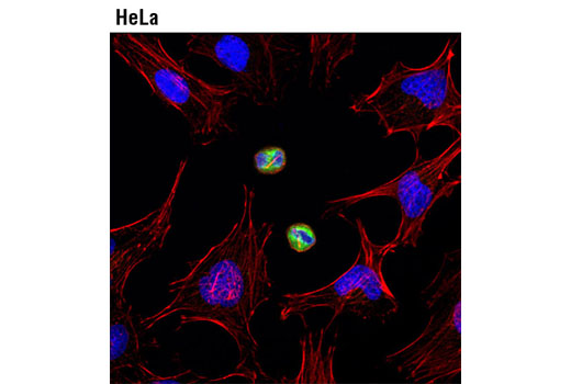
| Immunofluorescence (Immunocytochemistry) | 1:100 |
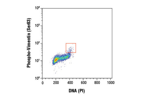
| Flow Cytometry (Fixed/Permeabilized) | 1:200 |
Storage
Specificity / Sensitivity
Species Reactivity:
Human
Species predicted to react based on 100% sequence homology
The antigen sequence used to produce this antibody shares
100% sequence homology with the species listed here, but
reactivity has not been tested or confirmed to work by CST.
Use of this product with these species is not covered under
our
Product Performance Guarantee.
Mouse, Rat
Source / Purification
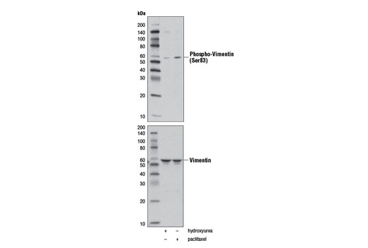
Monoclonal antibody is produced by immunizing animals with a synthetic phosphopeptide corresponding to residues surrounding Ser83 of human vimentin protein.
Background
The cytoskeleton consists of three types of cytosolic fibers: microfilaments (actin filaments), intermediate filaments, and microtubules. Major types of intermediate filaments are distinguished by their cell-specific expression: cytokeratins (epithelial cells), glial fibrillary acidic protein (GFAP) (glial cells), desmin (skeletal, visceral, and certain vascular smooth muscle cells), vimentin (mesenchyme origin), and neurofilaments (neurons). GFAP and vimentin form intermediate filaments in astroglial cells and modulate their motility and shape (1). In particular, vimentin filaments are present at early developmental stages, while GFAP filaments are characteristic of differentiated and mature brain astrocytes. Thus, GFAP is commonly used as a marker for intracranial and intraspinal tumors arising from astrocytes (2). Research studies have shown that vimentin is present in sarcomas, but not carcinomas, and its expression is examined in conjunction with that of other markers to distinguish between the two (3). Vimentin's dynamic structural changes and spatial re-organization in response to extracellular stimuli help to coordinate various signaling pathways (4). Phosphorylation of vimentin at Ser56 in smooth muscle cells regulates the structural arrangement of vimentin filaments in response to serotonin (5,6). Remodeling of vimentin and other intermediate filaments is important during lymphocyte adhesion and migration through the endothelium (7).
During mitosis, CDK1 phosphorylates vimentin at Ser56. This phosphorylation provides a PLK binding site for vimentin-PLK interaction. PLK further phosphorylates vimentin at Ser83, which might serve as memory phosphorylation site and play a regulatory role in vimentin filament disassembly (8,9). Additionally, studies using various soft-tissue sarcoma cells have shown that phosphorylation of vimentin at Ser39 by Akt1 enhances cell migration and survival, suggesting that vimentin could be a potential target for soft-tissue sarcoma targeted therapy (10,11).
CDK1 phosphorylates vimentin at Ser56 during mitosis, providing a PLK binding site for vimentin-PLK interaction. PLK further phosphorylates vimentin at Ser83, which might serve as a memory phosphorylation site and play a regulatory role in vimentin filament disassembly (8,9).
- Eng, L.F. et al. (2000) Neurochem Res 25, 1439-51.
- Goebel, H.H. et al. (1987) Acta Histochem Suppl 34, 81-93.
- Leader, M. et al. (1987) Histopathology 11, 63-72.
- Helfand, B.T. et al. (2004) J Cell Sci 117, 133-41.
- Tang, D.D. et al. (2005) Biochem J 388, 773-83.
- Fomina, I.G. et al. (1990) Klin Med (Mosk) 68, 125-7.
- Nieminen, M. et al. (2006) Nat Cell Biol 8, 156-62.
- Yamaguchi, T. et al. (2005) J Cell Biol 171, 431-6.
- Oguri, T. et al. (2006) Genes Cells 11, 531-40.
- Zhu, Q.S. et al. (2011) Oncogene 30, 457-70.
- Xue, G. and Hemmings, B.A. (2013) J Natl Cancer Inst 105, 393-404.
- Yamaguchi, T. et al. (2005) J Cell Biol 171, 431-6.
- Oguri, T. et al. (2006) Genes Cells 11, 531-40.
Species Reactivity
Species reactivity is determined by testing in at least one approved application (e.g., western blot).
Western Blot Buffer
IMPORTANT: For western blots, incubate membrane with diluted primary antibody in 5% w/v BSA, 1X TBS, 0.1% Tween® 20 at 4°C with gentle shaking, overnight.
Applications Key
WB: Western Blotting IF-IC: Immunofluorescence (Immunocytochemistry) FC-FP: Flow Cytometry (Fixed/Permeabilized)
Cross-Reactivity Key
H: human M: mouse R: rat Hm: hamster Mk: monkey Vir: virus Mi: mink C: chicken Dm: D. melanogaster X: Xenopus Z: zebrafish B: bovine Dg: dog Pg: pig Sc: S. cerevisiae Ce: C. elegans Hr: horse GP: Guinea Pig Rab: rabbit All: all species expected
Trademarks and Patents
使用に関する制限
法的な権限を与えられたCSTの担当者が署名した書面によって別途明示的に合意された場合を除き、 CST、その関連会社または代理店が提供する製品には以下の条件が適用されます。お客様が定める条件でここに定められた条件に含まれるものを超えるもの、 または、ここに定められた条件と異なるものは、法的な権限を与えられたCSTの担当者が別途書面にて受諾した場合を除き、拒絶され、 いかなる効力も効果も有しません。
研究専用 (For Research Use Only) またはこれに類似する表示がされた製品は、 いかなる目的についても FDA または外国もしくは国内のその他の規制機関により承認、認可または許可を受けていません。 お客様は製品を診断もしくは治療目的で使用してはならず、また、製品に表示された内容に違反する方法で使用してはなりません。 CST が販売または使用許諾する製品は、エンドユーザーであるお客様に対し、使途を研究および開発のみに限定して提供されるものです。 診断、予防もしくは治療目的で製品を使用することまたは製品を再販売 (単独であるか他の製品等の一部であるかを問いません) もしくはその他の商業的利用の目的で購入することについては、CST から別途許諾を得る必要があります。 お客様は以下の事項を遵守しなければなりません。(a) CST の製品 (単独であるか他の資材と一緒であるかを問いません) を販売、使用許諾、貸与、寄付もしくはその他の態様で第三者に譲渡したり使用させたりしてはなりません。また、商用の製品を製造するために CST の製品を使用してはなりません。(b) 複製、改変、リバースエンジニアリング、逆コンパイル、 分解または他の方法により製品の構造または技術を解明しようとしてはなりません。また、 CST の製品またはサービスと競合する製品またはサービスを開発する目的で CST の製品を使用してはなりません。(c) CST の製品の商標、商号、ロゴ、特許または著作権に関する通知または表示を除去したり改変したりしてはなりません。(d) CST の製品をCST 製品販売条件(CST’s Product Terms of Sale) および該当する書面のみに従って使用しなければなりません。(e) CST の製品に関連してお客様が使用する第三者の製品またはサービスに関する使用許諾条件、 サービス提供条件またはこれに類する合意事項を遵守しなければなりません。