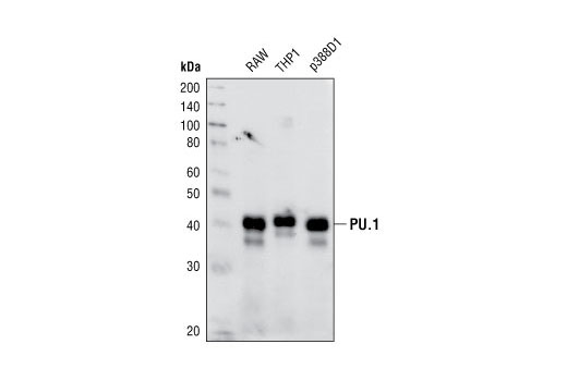
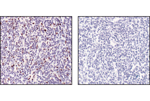
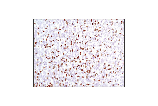
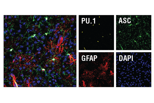
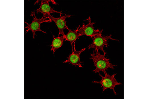
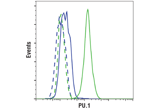
WB, IHC-P, IF-F, IF-IC, FC-FP
H M
Endogenous
42
Rabbit IgG
#P17947
6688
Product Information
Product Usage Information
This formulation is ideal for use with technologies requiring specialized or custom antibody labeling, including fluorophores, metals, lanthanides, and oligonucleotides. It is not recommended for ChIP, ChIP-seq, CUT&RUN or CUT&Tag assays. If you require a carrier free formulation for chromatin profiling, please contact us. Optimal dilutions/concentrations should be determined by the end user.
Formulation
Storage
Specificity / Sensitivity
Species Reactivity:
Human, Mouse
Species predicted to react based on 100% sequence homology
The antigen sequence used to produce this antibody shares
100% sequence homology with the species listed here, but
reactivity has not been tested or confirmed to work by CST.
Use of this product with these species is not covered under
our
Product Performance Guarantee.
Pig
Source / Purification
Monoclonal antibody is produced by immunizing animals with a synthetic peptide corresponding to the sequence of human PU.1 protein.
Background
PU.1 is a member of the Ets family of transcription factors and activates target genes through the purine-rich PU-box (1). PU.1 plays a pivotal role in the differentiation of myeloid cells and lymphocytes and is expressed in several hematopoietic cells including B lymphocytes, macrophages, neutrophils, mast cells, early erythroid cells, and megakaryocytes (1,2). The concentration of PU.1 is critical for both the determination of hematopoietic cell lineage and the regulation of differentiation versus stem cell proliferation (3,4). In addition, PU.1 activity is influenced by phosphorylation and interactions with other hematopoietic transcription factors. Phosphorylation of PU.1 at Ser146 by CK2 promotes binding to IRF4 and synergistic activation through the immunoglobulin κ 3' enhancer (5). Treatment of pro-B cells with IL-3 leads to phosphorylation of PU.1 at Ser140, resulting in increased PU.1 activity and activation of the anti-apoptotic gene MCL-1 (6). GATA1 binding blocks PU.1 activity during erythroid cell development (7). Overexpression of PU.1 resulting from proviral insertion during Friend virus infection can induce erythroleukemia, while reduced expression has been associated with acute myeloid leukemia (8).
- Lloberas, J. et al. (1999) Immunol Today 20, 184-9.
- Klemsz, M.J. et al. (1990) Cell 61, 113-24.
- Dahl, R. and Simon, M.C. (2003) Blood Cells Mol Dis 31, 229-33.
- DeKoter, R.P. and Singh, H. (2000) Science 288, 1439-41.
- Pongubala, J.M. et al. (1993) Science 259, 1622-5.
- Wang, J.M. et al. (2003) Mol Cell Biol 23, 1896-909.
- Zhang, P. et al. (1999) Proc Natl Acad Sci U S A 96, 8705-10.
- Moreau-Gachelin, F. et al. (1988) Nature 331, 277-80.
Species Reactivity
Species reactivity is determined by testing in at least one approved application (e.g., western blot).
Applications Key
WB: Western Blotting IHC-P: Immunohistochemistry (Paraffin) IF-F: Immunofluorescence (Frozen) IF-IC: Immunofluorescence (Immunocytochemistry) FC-FP: Flow Cytometry (Fixed/Permeabilized)
Cross-Reactivity Key
H: human M: mouse R: rat Hm: hamster Mk: monkey Vir: virus Mi: mink C: chicken Dm: D. melanogaster X: Xenopus Z: zebrafish B: bovine Dg: dog Pg: pig Sc: S. cerevisiae Ce: C. elegans Hr: horse GP: Guinea Pig Rab: rabbit All: all species expected
Trademarks and Patents
使用に関する制限
法的な権限を与えられたCSTの担当者が署名した書面によって別途明示的に合意された場合を除き、 CST、その関連会社または代理店が提供する製品には以下の条件が適用されます。お客様が定める条件でここに定められた条件に含まれるものを超えるもの、 または、ここに定められた条件と異なるものは、法的な権限を与えられたCSTの担当者が別途書面にて受諾した場合を除き、拒絶され、 いかなる効力も効果も有しません。
研究専用 (For Research Use Only) またはこれに類似する表示がされた製品は、 いかなる目的についても FDA または外国もしくは国内のその他の規制機関により承認、認可または許可を受けていません。 お客様は製品を診断もしくは治療目的で使用してはならず、また、製品に表示された内容に違反する方法で使用してはなりません。 CST が販売または使用許諾する製品は、エンドユーザーであるお客様に対し、使途を研究および開発のみに限定して提供されるものです。 診断、予防もしくは治療目的で製品を使用することまたは製品を再販売 (単独であるか他の製品等の一部であるかを問いません) もしくはその他の商業的利用の目的で購入することについては、CST から別途許諾を得る必要があります。 お客様は以下の事項を遵守しなければなりません。(a) CST の製品 (単独であるか他の資材と一緒であるかを問いません) を販売、使用許諾、貸与、寄付もしくはその他の態様で第三者に譲渡したり使用させたりしてはなりません。また、商用の製品を製造するために CST の製品を使用してはなりません。(b) 複製、改変、リバースエンジニアリング、逆コンパイル、 分解または他の方法により製品の構造または技術を解明しようとしてはなりません。また、 CST の製品またはサービスと競合する製品またはサービスを開発する目的で CST の製品を使用してはなりません。(c) CST の製品の商標、商号、ロゴ、特許または著作権に関する通知または表示を除去したり改変したりしてはなりません。(d) CST の製品をCST 製品販売条件(CST’s Product Terms of Sale) および該当する書面のみに従って使用しなければなりません。(e) CST の製品に関連してお客様が使用する第三者の製品またはサービスに関する使用許諾条件、 サービス提供条件またはこれに類する合意事項を遵守しなければなりません。