| Product Includes | Product # | Quantity | Mol. Wt | Isotype/Source |
|---|---|---|---|---|
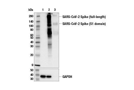
| SARS-CoV-2 Spike Protein (S1) (E5S3V) Rabbit mAb | 99423 | 20 µl | 110, 220 kDa | Rabbit IgG |
| SARS-CoV-2 Spike Protein (RBD) (E7B3E) Rabbit mAb | 63847 | 20 µl | 110, 220 kDa | Rabbit IgG |
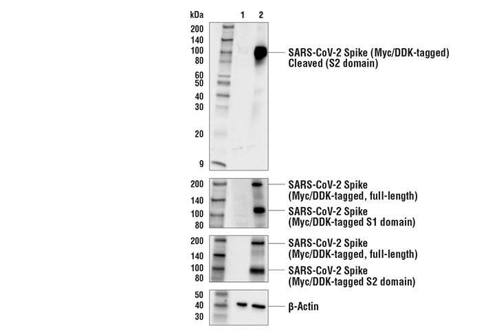
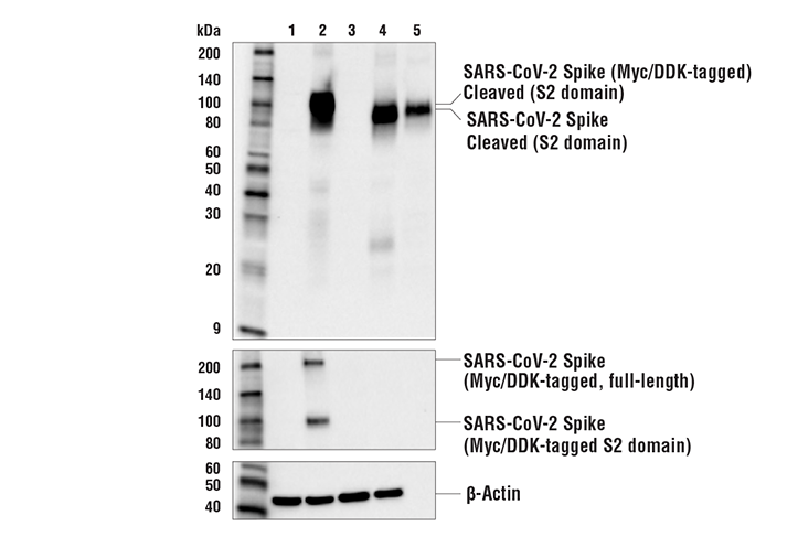
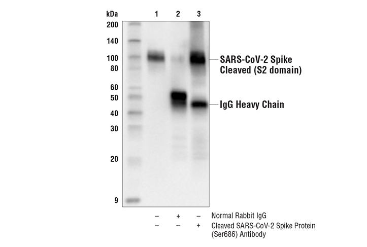
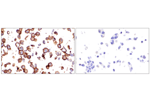
| Cleaved SARS-CoV-2 Spike Protein (Ser686) Antibody | 84534 | 20 µl | 100 kDa | Rabbit |
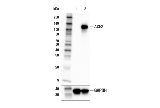
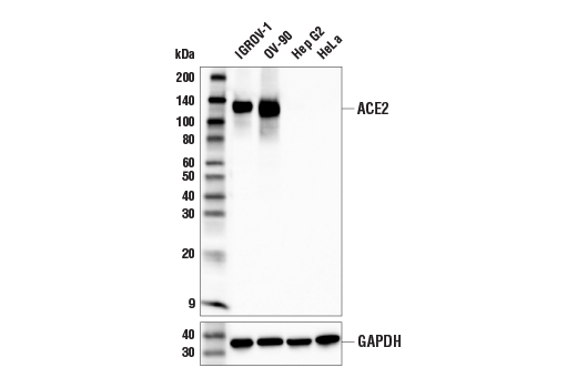
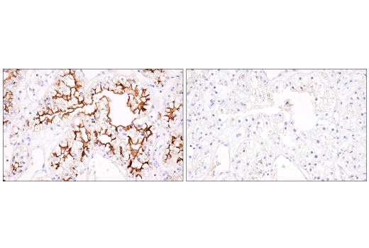
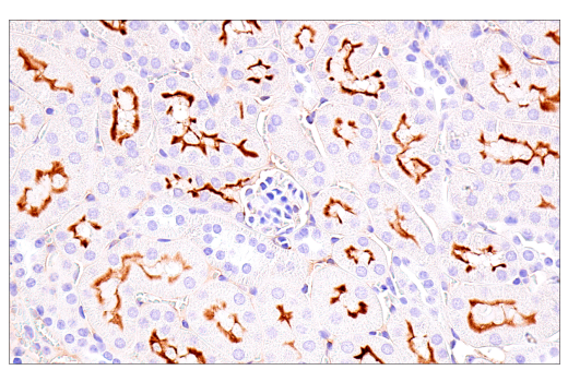
| ACE2 (E5O6J) XP® Rabbit mAb | 92485 | 20 µl | 120-135 kDa | Rabbit IgG |
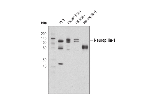
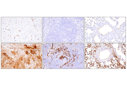
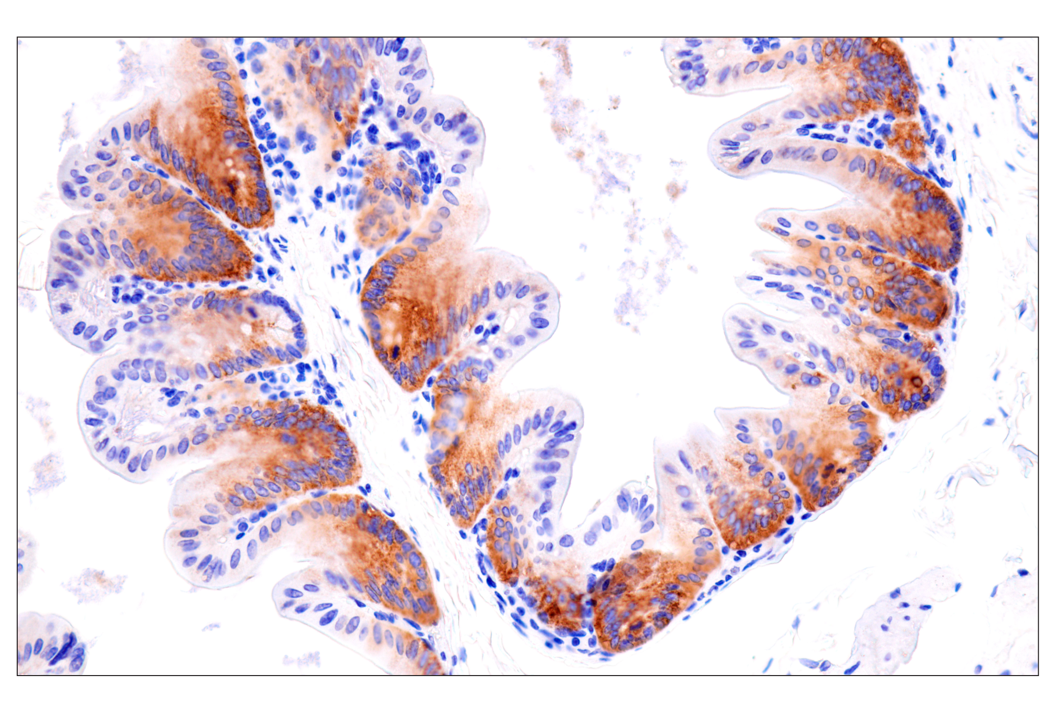
| Neuropilin-1 (D62C6) Rabbit mAb | 3725 | 20 µl | 120-140 kDa | Rabbit IgG |
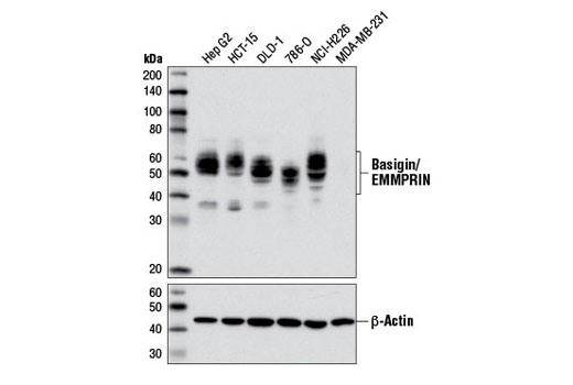
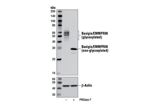
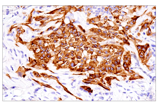
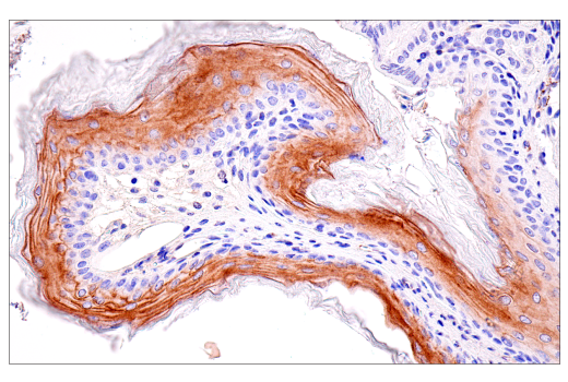
| Basigin/EMMPRIN (E1S1V) Rabbit mAb | 13287 | 20 µl | 38-58 kDa | Rabbit IgG |
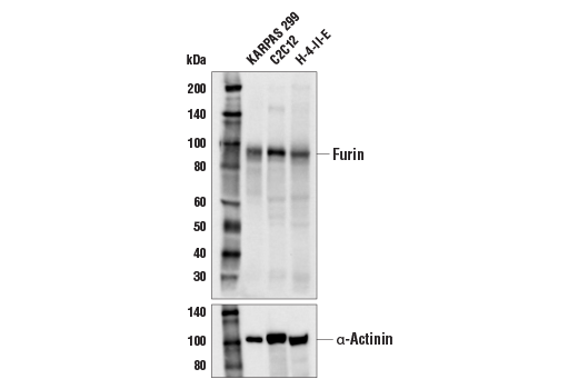
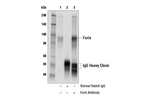
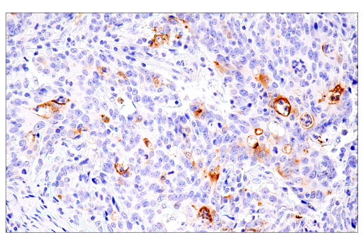
| Furin Antibody | 43996 | 20 µl | 90 kDa | Rabbit |
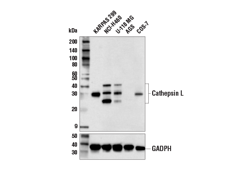
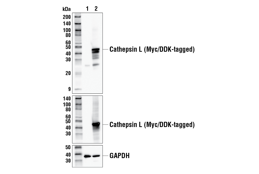
| Cathepsin L Antibody | 71298 | 20 µl | 25-42 kDa | Rabbit |
| Anti-rabbit IgG, HRP-linked Antibody | 7074 | 100 µl | Goat |
Please visit cellsignal.com for individual component applications, species cross-reactivity, dilutions, protocols, and additional product information.
Description
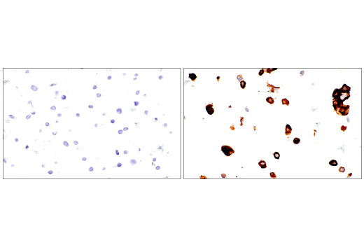
The SARS-CoV-2 Virus-Host Interaction Antibody Sampler Kit provides an economical means of detecting key viral and host proteins involved in SARS-CoV-2 infection of human host cells. The kit includes enough antibodies to perform two western blot experiments with each primary antibody.
Storage
Background
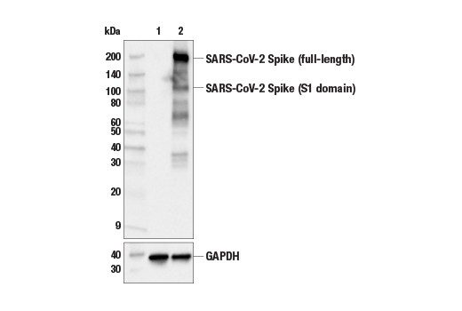
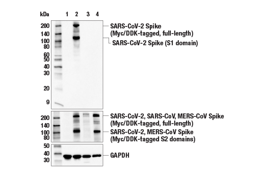
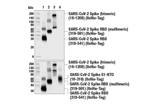
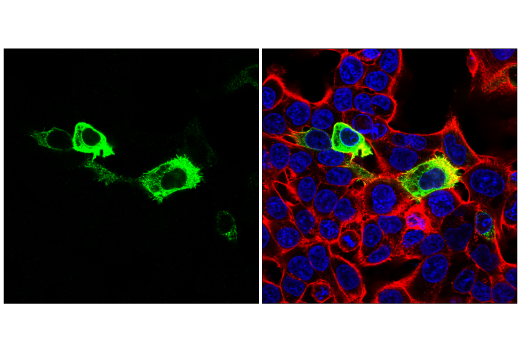
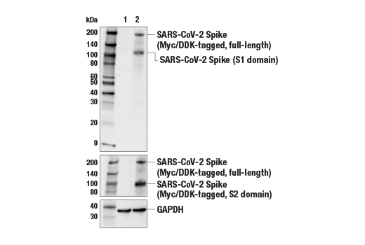
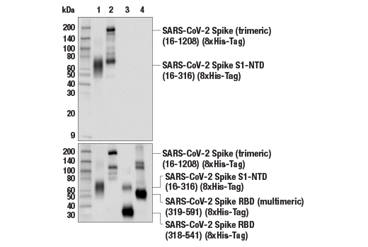
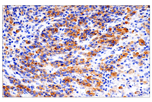
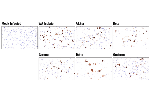
The cause of the COVID-19 pandemic is a novel and highly pathogenic coronavirus, termed SARS-CoV-2 (severe acute respiratory syndrome coronavirus-2). SARS-CoV-2 is a member of the Coronaviridae family of viruses (1). The SARS-CoV-2 virion is comprised of four key structural proteins: spike (S), envelope (E), membrane (M), and nucleocapsid (N) (2). Coronavirus spike proteins are class I fusion proteins and harbor an ectodomain, a transmembrane domain, and an intracellular tail (3,4). The highly glycosylated ectodomain projects from the viral envelope surface and facilitates attachment and fusion with the host cell membrane. The ectodomain can be further subdivided into the receptor-binding domain (RBD) S1 and membrane-fusion (S2) subunits, which are produced upon proteolysis by host proteases. S1 and S2 subunits are reassociated after cleavage, assembling into crown-like homotrimers (2,4).
The SARS-CoV-2 spike protein contains a novel tetrabasic "furin cleavage site" (FCS) at the S1/S2 junction. Research studies suggest this site is cleaved by proprotein convertases (e.g., furin) or lysosomal proteases (e.g., cathepsin L) (5,6). S1/S2 cleavage elicits a confirmational change in the spike protein that positions elements of the trimeric RBD in an exposed "up" position, priming it for interaction with host receptor proteins. Cleavage can occur at multiple steps of the viral lifecycle, including during viral packaging, or upon contact of the intact virion with the host cell surface. This novel cleavage event has been suggested to contribute to the high infectivity rate of the SARS-CoV-2 virus (7).
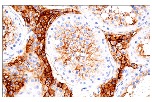
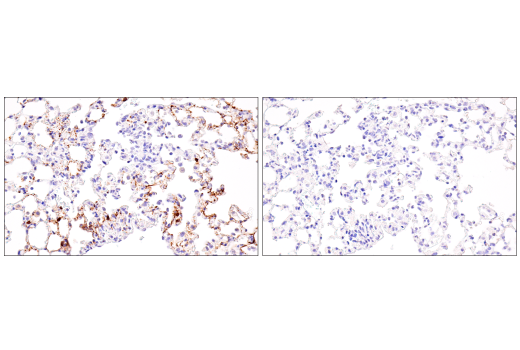
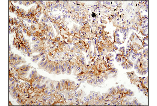
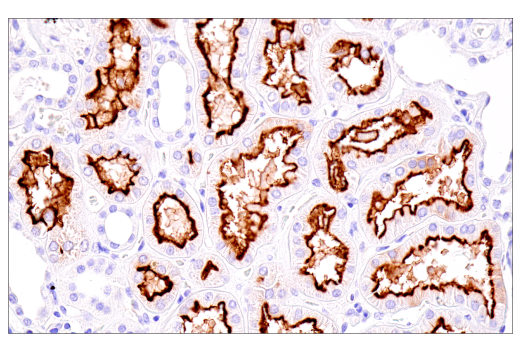
The SARS-CoV-2 virus has been shown to utilize the angiotensin-converting enzyme 2 (ACE2) protein as its primary receptor for cellular entry (8). However, research studies have suggested that other cell surface proteins may serve as receptors or co-receptors for SARS-CoV-2. These include neuropilin-1 (NPN1), a single-pass transmembrane receptor that can function as part of a semaphorin receptor complex, and as a vascular endothelial growth factor (VEGF) receptor (9), and Basigin/EMMPRIN (CD147), a type I integral membrane receptor belonging to the immunoglobulin superfamily (10).
- Zhou, P. et al. (2020) Nature 579, 270-273.
- Tortorici, M.A. and Veesler, D. (2019) Adv Virus Res 105, 93-116.
- Li, F. et al. (2006) J Virol 80, 6794-800.
- Li, F. (2016) Annu Rev Virol 3, 237-261.
- Coutard, B. et al. (2020) Antiviral Res 176, 104742.
- Jaimes, J.A. et al. (2020) iScience 23, 101212.
- Hasan, A. et al. (2021) J Biomol Struct Dyn 39, 3025-3033.
- Shang, J. et al. (2020) Nature 581, 221-224.
- Cantuti-Castelvetri, L. et al. (2020) Science 370, 856-860.
- Wang, K. et al. (2020) Signal Transduct Target Ther 5, 283.
Background References
Trademarks and Patents
使用に関する制限
法的な権限を与えられたCSTの担当者が署名した書面によって別途明示的に合意された場合を除き、 CST、その関連会社または代理店が提供する製品には以下の条件が適用されます。お客様が定める条件でここに定められた条件に含まれるものを超えるもの、 または、ここに定められた条件と異なるものは、法的な権限を与えられたCSTの担当者が別途書面にて受諾した場合を除き、拒絶され、 いかなる効力も効果も有しません。
研究専用 (For Research Use Only) またはこれに類似する表示がされた製品は、 いかなる目的についても FDA または外国もしくは国内のその他の規制機関により承認、認可または許可を受けていません。 お客様は製品を診断もしくは治療目的で使用してはならず、また、製品に表示された内容に違反する方法で使用してはなりません。 CST が販売または使用許諾する製品は、エンドユーザーであるお客様に対し、使途を研究および開発のみに限定して提供されるものです。 診断、予防もしくは治療目的で製品を使用することまたは製品を再販売 (単独であるか他の製品等の一部であるかを問いません) もしくはその他の商業的利用の目的で購入することについては、CST から別途許諾を得る必要があります。 お客様は以下の事項を遵守しなければなりません。(a) CST の製品 (単独であるか他の資材と一緒であるかを問いません) を販売、使用許諾、貸与、寄付もしくはその他の態様で第三者に譲渡したり使用させたりしてはなりません。また、商用の製品を製造するために CST の製品を使用してはなりません。(b) 複製、改変、リバースエンジニアリング、逆コンパイル、 分解または他の方法により製品の構造または技術を解明しようとしてはなりません。また、 CST の製品またはサービスと競合する製品またはサービスを開発する目的で CST の製品を使用してはなりません。(c) CST の製品の商標、商号、ロゴ、特許または著作権に関する通知または表示を除去したり改変したりしてはなりません。(d) CST の製品をCST 製品販売条件(CST’s Product Terms of Sale) および該当する書面のみに従って使用しなければなりません。(e) CST の製品に関連してお客様が使用する第三者の製品またはサービスに関する使用許諾条件、 サービス提供条件またはこれに類する合意事項を遵守しなければなりません。