View in English?
View in English?
View in English?

| Cat. # | Size | Qty. | Price | Inventory |
|---|---|---|---|---|
| 45394T | 1 Kit (8 x 20 microliters) |
|
| Product Includes | Quantity | Applications | Reactivity | MW(kDa) | Isotype |
|---|---|---|---|---|---|
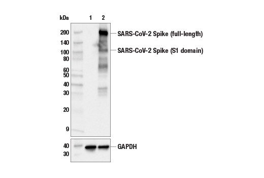
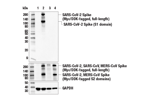
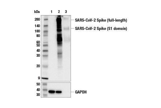
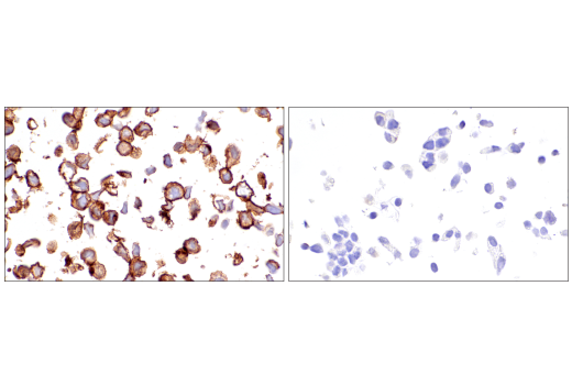
| SARS-CoV-2 Spike Protein (S1) (E5S3V) Rabbit mAb 99423 | 20 µl |
|
Vir | 110, 220 | Rabbit IgG |
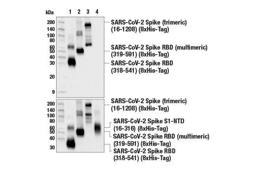
| SARS-CoV-2 Spike Protein (RBD) (E7B3E) Rabbit mAb 63847 | 20 µl |
|
Vir | 110, 220 | Rabbit IgG |
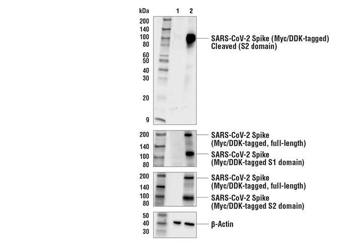
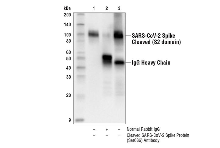
| Cleaved SARS-CoV-2 Spike Protein (Ser686) Antibody 84534 | 20 µl |
|
Vir | 100 | Rabbit |
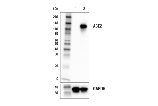
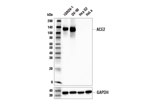
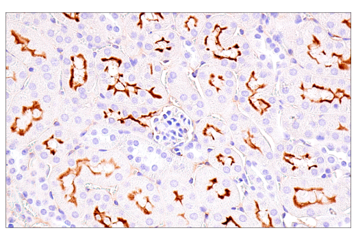
| ACE2 (E5O6J) XP® Rabbit mAb 92485 | 20 µl |
|
H M Hm | 120-135 | Rabbit IgG |
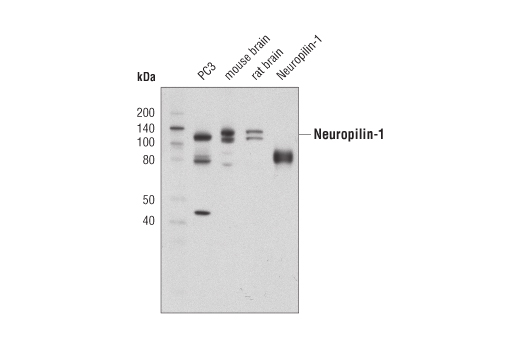
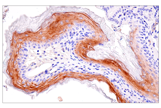
| Neuropilin-1 (D62C6) Rabbit mAb 3725 | 20 µl |
|
H M R | 120-140 | Rabbit IgG |
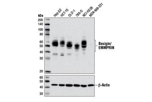
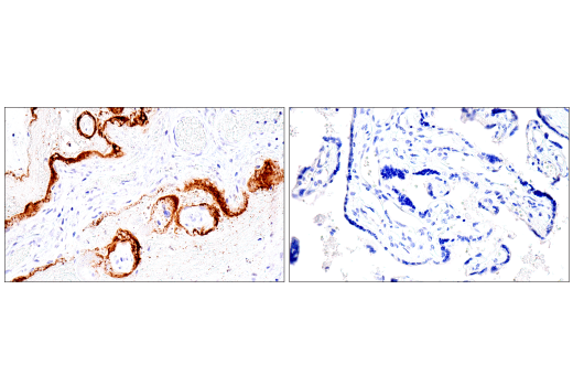
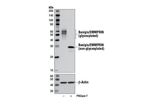
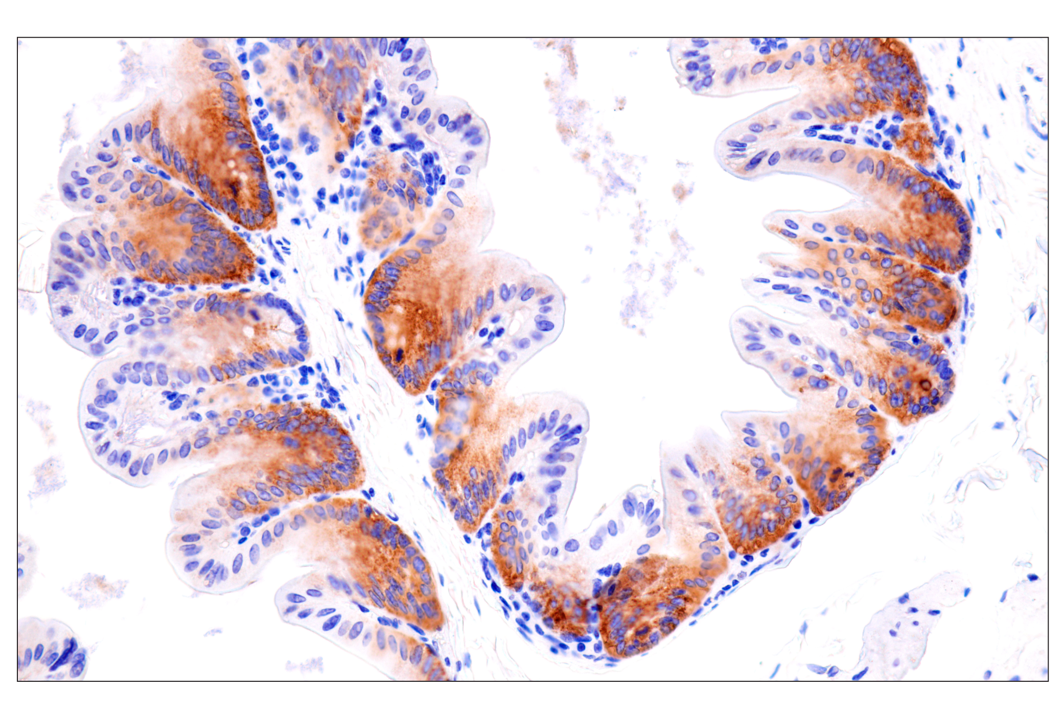
| Basigin/EMMPRIN (E1S1V) Rabbit mAb 13287 | 20 µl |
|
H | 38-58 | Rabbit IgG |
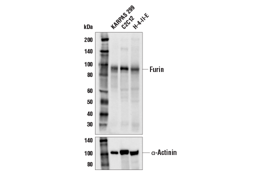
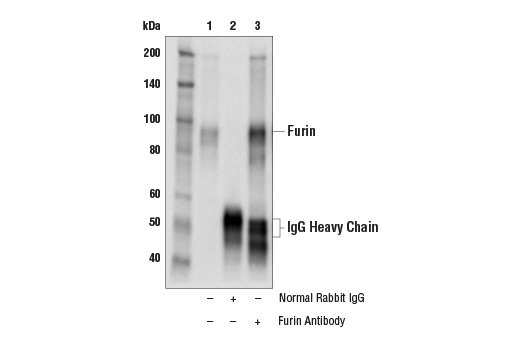
| Furin Antibody 43996 | 20 µl |
|
H M R | 90 | Rabbit |
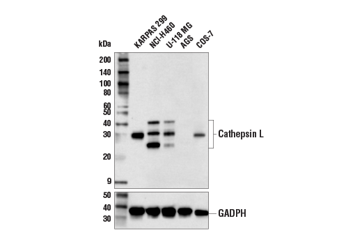
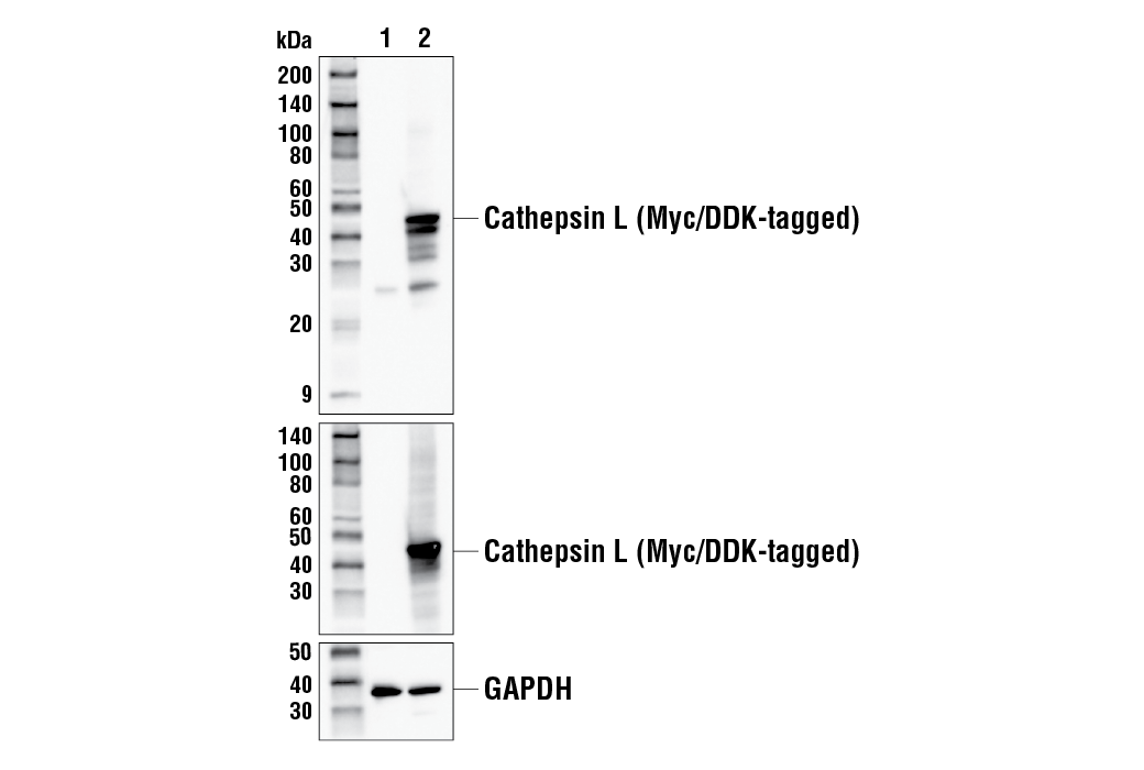
| Cathepsin L Antibody 71298 | 20 µl |
|
H Mk | 25-42 | Rabbit |
| Anti-rabbit IgG, HRP-linked Antibody 7074 | 100 µl |
|
Goat |
Product Information
Monoclonal antibodies are produced by immunizing animals with synthetic peptides corresponding to residues surrounding Ala254 of human EMMPRIN protein, Asp201 of human ACE2 protein, and Ser459 of SARS-CoV-2 spike protein, with a recombinant protein corresponding to the S1 domain of the SARS-CoV-2 spike protein, and with a GST-fusion protein corresponding to residues of mouse neuropilin-1 protein. Polyclonal antibodies are produced by immunizing animals with synthetic peptides corresponding to the amino terminus (Ser668) of the SARS-CoV-2 spike protein S2 domain, Pro229 of human cathepsin L, and Ala237 of human furin protein.
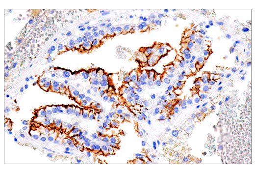
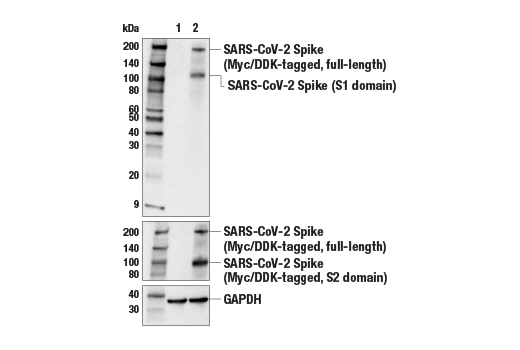
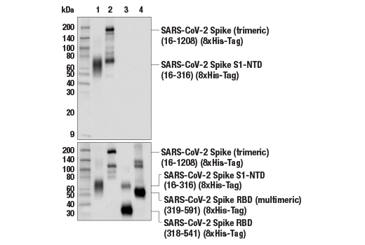
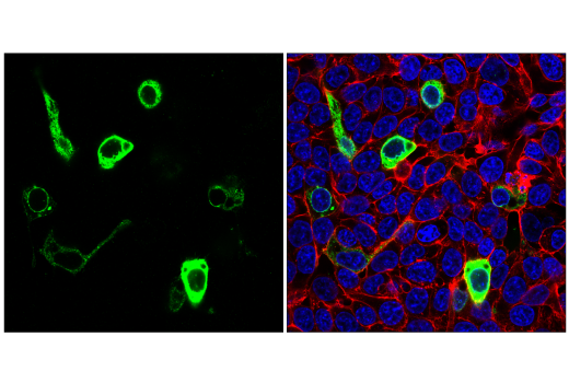
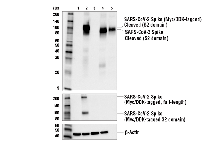
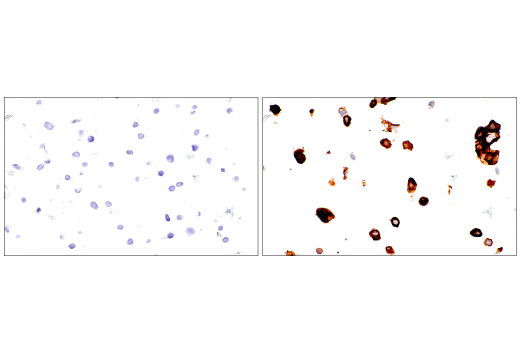
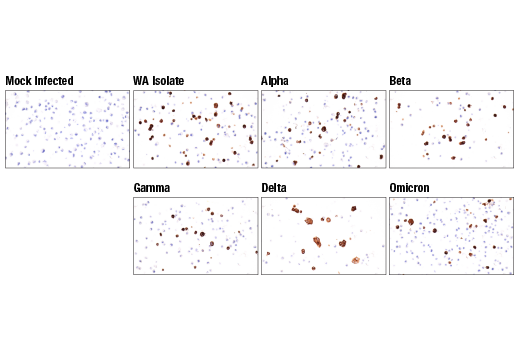
The cause of the COVID-19 pandemic is a novel and highly pathogenic coronavirus, termed SARS-CoV-2 (severe acute respiratory syndrome coronavirus-2). SARS-CoV-2 is a member of the Coronaviridae family of viruses (1). The SARS-CoV-2 virion is comprised of four key structural proteins: spike (S), envelope (E), membrane (M), and nucleocapsid (N) (2). Coronavirus spike proteins are class I fusion proteins and harbor an ectodomain, a transmembrane domain, and an intracellular tail (3,4). The highly glycosylated ectodomain projects from the viral envelope surface and facilitates attachment and fusion with the host cell membrane. The ectodomain can be further subdivided into the receptor-binding domain (RBD) S1 and membrane-fusion (S2) subunits, which are produced upon proteolysis by host proteases. S1 and S2 subunits are reassociated after cleavage, assembling into crown-like homotrimers (2,4).
The SARS-CoV-2 spike protein contains a novel tetrabasic "furin cleavage site" (FCS) at the S1/S2 junction. Research studies suggest this site is cleaved by proprotein convertases (e.g., furin) or lysosomal proteases (e.g., cathepsin L) (5,6). S1/S2 cleavage elicits a confirmational change in the spike protein that positions elements of the trimeric RBD in an exposed "up" position, priming it for interaction with host receptor proteins. Cleavage can occur at multiple steps of the viral lifecycle, including during viral packaging, or upon contact of the intact virion with the host cell surface. This novel cleavage event has been suggested to contribute to the high infectivity rate of the SARS-CoV-2 virus (7).
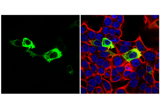
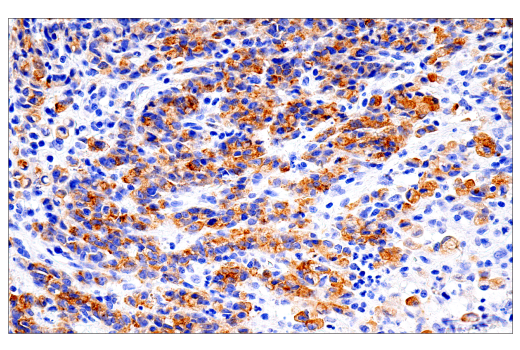
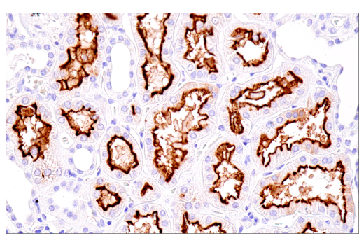
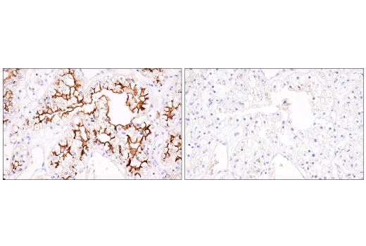
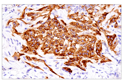
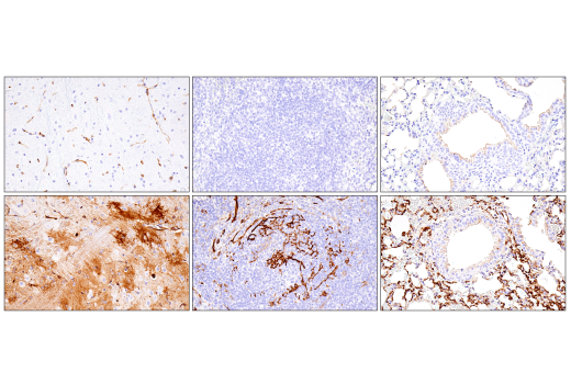
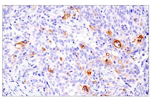
The SARS-CoV-2 virus has been shown to utilize the angiotensin-converting enzyme 2 (ACE2) protein as its primary receptor for cellular entry (8). However, research studies have suggested that other cell surface proteins may serve as receptors or co-receptors for SARS-CoV-2. These include neuropilin-1 (NPN1), a single-pass transmembrane receptor that can function as part of a semaphorin receptor complex, and as a vascular endothelial growth factor (VEGF) receptor (9), and Basigin/EMMPRIN (CD147), a type I integral membrane receptor belonging to the immunoglobulin superfamily (10).
Explore pathways related to this product.
STRING - Known and Predicted Protein-Protein Interactions.
Except as otherwise expressly agreed in a writing signed by a legally authorized representative of CST, the following terms apply to Products provided by CST, its affiliates or its distributors. Any Customer's terms and conditions that are in addition to, or different from, those contained herein, unless separately accepted in writing by a legally authorized representative of CST, are rejected and are of no force or effect.
Products are labeled with For Research Use Only or a similar labeling statement and have not been approved, cleared, or licensed by the FDA or other regulatory foreign or domestic entity, for any purpose. Customer shall not use any Product for any diagnostic or therapeutic purpose, or otherwise in any manner that conflicts with its labeling statement. Products sold or licensed by CST are provided for Customer as the end-user and solely for research and development uses. Any use of Product for diagnostic, prophylactic or therapeutic purposes, or any purchase of Product for resale (alone or as a component) or other commercial purpose, requires a separate license from CST. Customer shall (a) not sell, license, loan, donate or otherwise transfer or make available any Product to any third party, whether alone or in combination with other materials, or use the Products to manufacture any commercial products, (b) not copy, modify, reverse engineer, decompile, disassemble or otherwise attempt to discover the underlying structure or technology of the Products, or use the Products for the purpose of developing any products or services that would compete with CST products or services, (c) not alter or remove from the Products any trademarks, trade names, logos, patent or copyright notices or markings, (d) use the Products solely in accordance with CST Product Terms of Sale and any applicable documentation, and (e) comply with any license, terms of service or similar agreement with respect to any third party products or services used by Customer in connection with the Products.
View in English?