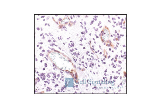
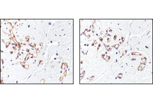
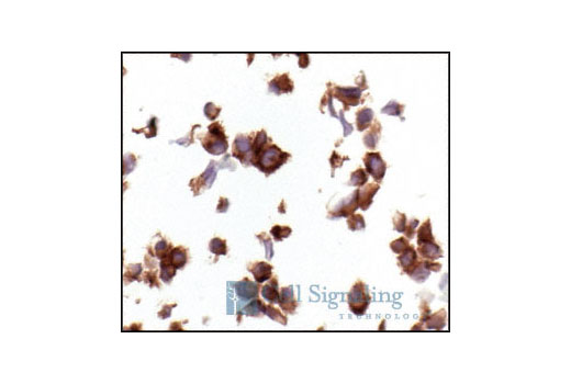
WB, IP, IHC-P, IF-F, IF-IC
H M
Endogenous
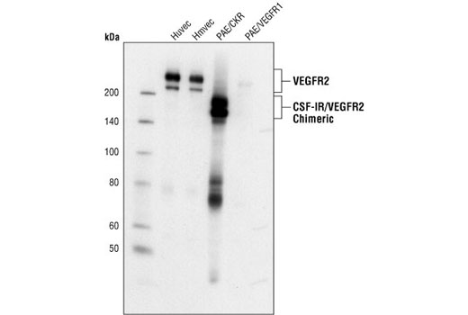
210, 230
Rabbit IgG
#P35968
3791
Product Information
Product Usage Information
| Application | Dilution |
|---|---|
| Western Blotting | 1:1000 |
| Immunoprecipitation | 1:100 |
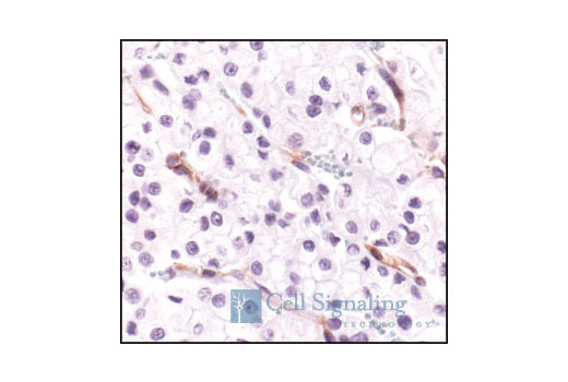
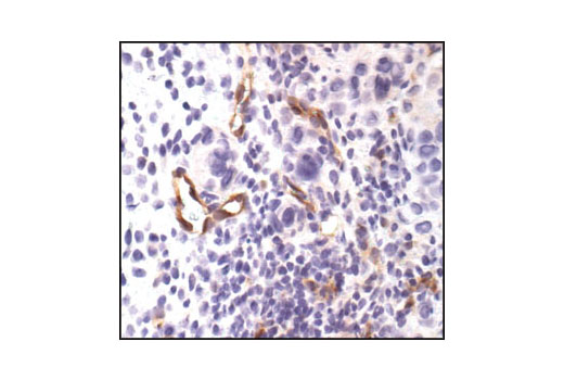
| Immunohistochemistry (Paraffin) | 1:4000 - 1:16000 |
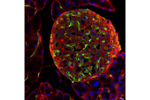
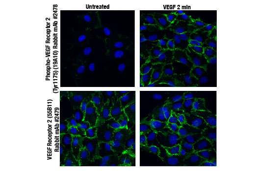
| Immunofluorescence (Frozen) | 1:200 - 1:800 |
| Immunofluorescence (Immunocytochemistry) | 1:200 - 1:800 |
Storage
For a carrier free (BSA and azide free) version of this product see product #28521.
Specificity / Sensitivity
Species Reactivity:
Human, Mouse
Species predicted to react based on 100% sequence homology
The antigen sequence used to produce this antibody shares
100% sequence homology with the species listed here, but
reactivity has not been tested or confirmed to work by CST.
Use of this product with these species is not covered under
our
Product Performance Guarantee.
Bovine
Source / Purification
Monoclonal antibody is produced by immunizing animals with a recombinant protein containing the carboxy-terminal 150 amino acid residues of human VEGF receptor 2.
Background
Vascular endothelial growth factor receptor 2 (VEGFR2, KDR, Flk-1) is a major receptor for VEGF-induced signaling in endothelial cells. Upon ligand binding, VEGFR2 undergoes autophosphorylation and becomes activated (1). Major autophosphorylation sites of VEGFR2 are located in the kinase insert domain (Tyr951/996) and in the tyrosine kinase catalytic domain (Tyr1054/1059) (2). Activation of the receptor leads to rapid recruitment of adaptor proteins, including Shc, GRB2, PI3 kinase, NCK, and the protein tyrosine phosphatases SHP-1 and SHP-2 (3). Phosphorylation at Tyr1212 provides a docking site for GRB2 binding and phospho-Tyr1175 binds the p85 subunit of PI3 kinase and PLCγ, as well as Shb (1,4,5). Signaling from VEGFR2 is necessary for the execution of VEGF-stimulated proliferation, chemotaxis and sprouting, as well as survival of cultured endothelial cells in vitro and angiogenesis in vivo (6-8).
- Meyer, M. et al. (1999) EMBO J 18, 363-74.
- Dougher-Vermazen, M. et al. (1994) Biochem Biophys Res Commun 205, 728-38.
- Kroll, J. and Waltenberger, J. (1997) J Biol Chem 272, 32521-7.
- Takahashi, T. et al. (2001) EMBO J 20, 2768-78.
- Holmqvist, K. et al. (2004) J Biol Chem 279, 22267-75.
- Karkkainen, M.J. and Petrova, T.V. (2000) Oncogene 19, 5598-605.
- Rahimi, N. et al. (2000) J Biol Chem 275, 16986-92.
- Claesson-Welsh, L. (2003) Biochem Soc Trans 31, 20-4.
Species Reactivity
Species reactivity is determined by testing in at least one approved application (e.g., western blot).
Western Blot Buffer
IMPORTANT: For western blots, incubate membrane with diluted primary antibody in 5% w/v BSA, 1X TBS, 0.1% Tween® 20 at 4°C with gentle shaking, overnight.
Applications Key
WB: Western Blotting IP: Immunoprecipitation IHC-P: Immunohistochemistry (Paraffin) IF-F: Immunofluorescence (Frozen) IF-IC: Immunofluorescence (Immunocytochemistry)
Cross-Reactivity Key
H: human M: mouse R: rat Hm: hamster Mk: monkey Vir: virus Mi: mink C: chicken Dm: D. melanogaster X: Xenopus Z: zebrafish B: bovine Dg: dog Pg: pig Sc: S. cerevisiae Ce: C. elegans Hr: horse GP: Guinea Pig Rab: rabbit All: all species expected
Trademarks and Patents
使用に関する制限
法的な権限を与えられたCSTの担当者が署名した書面によって別途明示的に合意された場合を除き、 CST、その関連会社または代理店が提供する製品には以下の条件が適用されます。お客様が定める条件でここに定められた条件に含まれるものを超えるもの、 または、ここに定められた条件と異なるものは、法的な権限を与えられたCSTの担当者が別途書面にて受諾した場合を除き、拒絶され、 いかなる効力も効果も有しません。
研究専用 (For Research Use Only) またはこれに類似する表示がされた製品は、 いかなる目的についても FDA または外国もしくは国内のその他の規制機関により承認、認可または許可を受けていません。 お客様は製品を診断もしくは治療目的で使用してはならず、また、製品に表示された内容に違反する方法で使用してはなりません。 CST が販売または使用許諾する製品は、エンドユーザーであるお客様に対し、使途を研究および開発のみに限定して提供されるものです。 診断、予防もしくは治療目的で製品を使用することまたは製品を再販売 (単独であるか他の製品等の一部であるかを問いません) もしくはその他の商業的利用の目的で購入することについては、CST から別途許諾を得る必要があります。 お客様は以下の事項を遵守しなければなりません。(a) CST の製品 (単独であるか他の資材と一緒であるかを問いません) を販売、使用許諾、貸与、寄付もしくはその他の態様で第三者に譲渡したり使用させたりしてはなりません。また、商用の製品を製造するために CST の製品を使用してはなりません。(b) 複製、改変、リバースエンジニアリング、逆コンパイル、 分解または他の方法により製品の構造または技術を解明しようとしてはなりません。また、 CST の製品またはサービスと競合する製品またはサービスを開発する目的で CST の製品を使用してはなりません。(c) CST の製品の商標、商号、ロゴ、特許または著作権に関する通知または表示を除去したり改変したりしてはなりません。(d) CST の製品をCST 製品販売条件(CST’s Product Terms of Sale) および該当する書面のみに従って使用しなければなりません。(e) CST の製品に関連してお客様が使用する第三者の製品またはサービスに関する使用許諾条件、 サービス提供条件またはこれに類する合意事項を遵守しなければなりません。