View in English?
View in English?
View in English?

| Cat. # | Size | Qty. | Price | Inventory |
|---|---|---|---|---|
| 85314T | 1 Kit (7 x 20 microliters) |
|
| Product Includes | Quantity | Applications | Reactivity | MW(kDa) | Isotype |
|---|---|---|---|---|---|
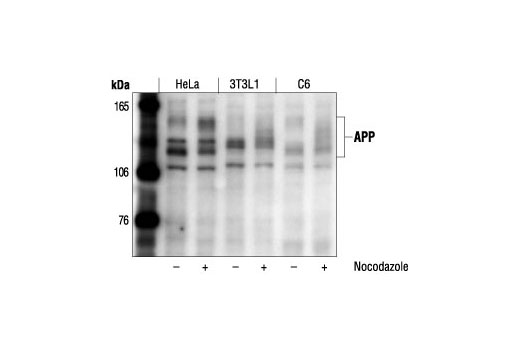
| APP Antibody 2452 | 20 µl |
|
H M R Mk | 100 to 140 | Rabbit |
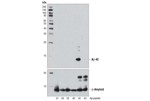
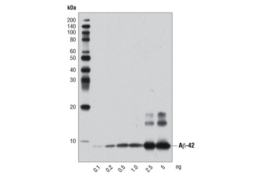
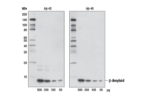
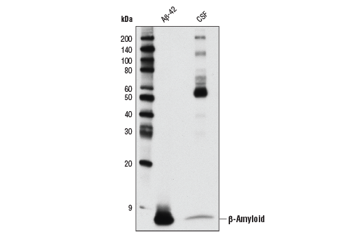
| β-Amyloid (1-42) (D3E10) Rabbit mAb 12843 | 20 µl |
|
H | 4 | Rabbit |
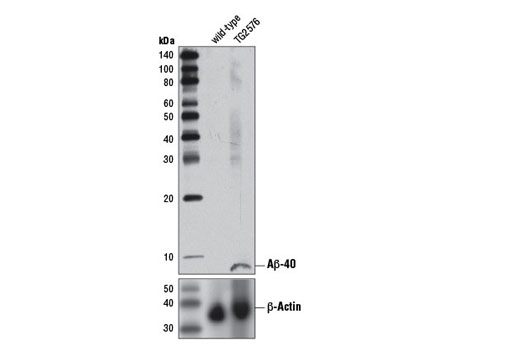
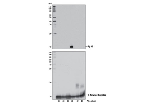
| β-Amyloid (1-40) (D8Q7I) Rabbit mAb 12990 | 20 µl |
|
H | 4 | Rabbit IgG |
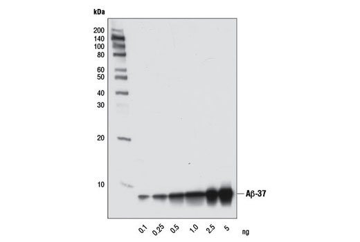
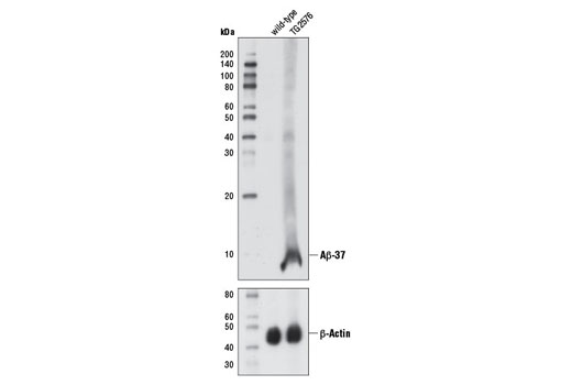
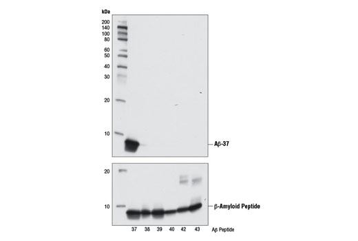
| β-Amyloid (1-37) (D2A6H) Rabbit mAb 12467 | 20 µl |
|
H | 4 | Rabbit IgG |
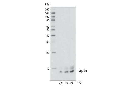
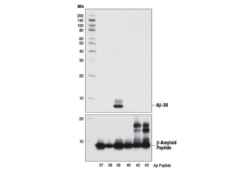
| β-Amyloid (1-39) (D5Y9L) Rabbit mAb 12077 | 20 µl |
|
H | 4 | Rabbit IgG |
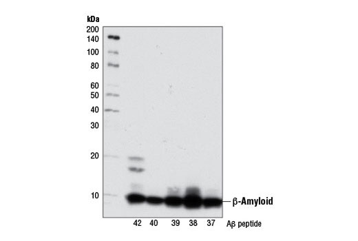
| β-Amyloid (D54D2) XP® Rabbit mAb 8243 | 20 µl |
|
H | 5 | Rabbit IgG |
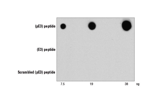
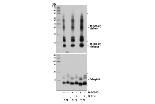
| β-Amyloid (pE3 Peptide) (D5N5H) Rabbit mAb 14975 | 20 µl |
|
H | 4 | Rabbit IgG |
| Anti-rabbit IgG, HRP-linked Antibody 7074 | 100 µl |
|
Rab | Goat |
Product Information
APP Antibody is a polyclonal antibody produced by immunizing animals with a synthetic peptide corresponding to residues surrounding Thr668 of human APP695 protein. Antibodies are purified by protein A and peptide affinity chromatography. β-Amyloid fragment-specific monoclonal antibodies are produced by immunizing rabbits with synthetic peptides corresponding to residues at the carboxy terminus of each indicated human β-Amyloid fragment. β-Amyloid (D54D2) XP® Rabbit mAb is produced by immunizing animals with a synthetic peptide corresponding to residues near the amino terminus of human β-amyloid peptide (Aβ). β-Amyloid (pE3 Peptide) (D5N5H) Rabbit mAb is produced by immunizing animals with a synthetic peptide corresponding to residues near the amino terminus of human β-amyloid (pE3) peptide.
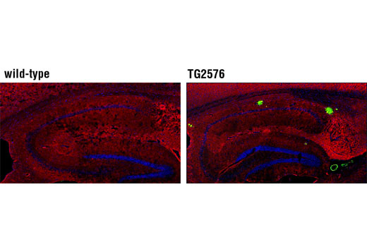
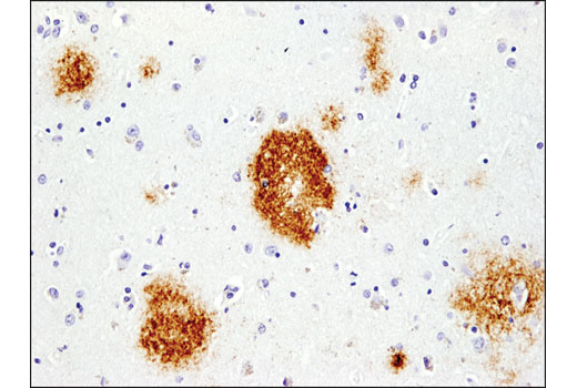
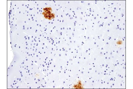
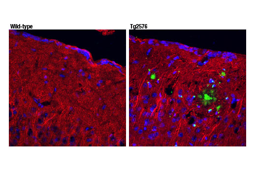
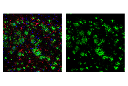
Amyloid β (Aβ) precursor protein (APP) is a 100-140 kDa transmembrane glycoprotein that exists as several isoforms (1). The amino acid sequence of APP contains an amyloid domain, which can be processed and released by two-step proteolytic cleavage (1). The extracellular deposition and accumulation of the released Aβ fragments form the main components of amyloid plaques in Alzheimer's disease (1). Several fragments corresponding to progressive APP processing at alternative cleavage sites have been identified (2). These include Aβ (1-37), Aβ (1-39), Aβ (1-40), and Aβ (1-42) (2). These fragments can also be N-terminally modified to generate pyroglutamate-3 Aβ (pE3-peptide) (3). Fragment-specific and pan-Aβ antibodies are used to detect and examine relative levels of individual Aβ fragments.
Explore pathways related to this product.
STRING - Known and Predicted Protein-Protein Interactions.
Except as otherwise expressly agreed in a writing signed by a legally authorized representative of CST, the following terms apply to Products provided by CST, its affiliates or its distributors. Any Customer's terms and conditions that are in addition to, or different from, those contained herein, unless separately accepted in writing by a legally authorized representative of CST, are rejected and are of no force or effect.
Products are labeled with For Research Use Only or a similar labeling statement and have not been approved, cleared, or licensed by the FDA or other regulatory foreign or domestic entity, for any purpose. Customer shall not use any Product for any diagnostic or therapeutic purpose, or otherwise in any manner that conflicts with its labeling statement. Products sold or licensed by CST are provided for Customer as the end-user and solely for research and development uses. Any use of Product for diagnostic, prophylactic or therapeutic purposes, or any purchase of Product for resale (alone or as a component) or other commercial purpose, requires a separate license from CST. Customer shall (a) not sell, license, loan, donate or otherwise transfer or make available any Product to any third party, whether alone or in combination with other materials, or use the Products to manufacture any commercial products, (b) not copy, modify, reverse engineer, decompile, disassemble or otherwise attempt to discover the underlying structure or technology of the Products, or use the Products for the purpose of developing any products or services that would compete with CST products or services, (c) not alter or remove from the Products any trademarks, trade names, logos, patent or copyright notices or markings, (d) use the Products solely in accordance with CST Product Terms of Sale and any applicable documentation, and (e) comply with any license, terms of service or similar agreement with respect to any third party products or services used by Customer in connection with the Products.
View in English?