View in English?
View in English?
View in English?

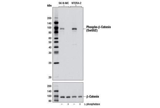
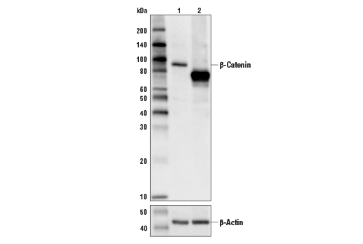
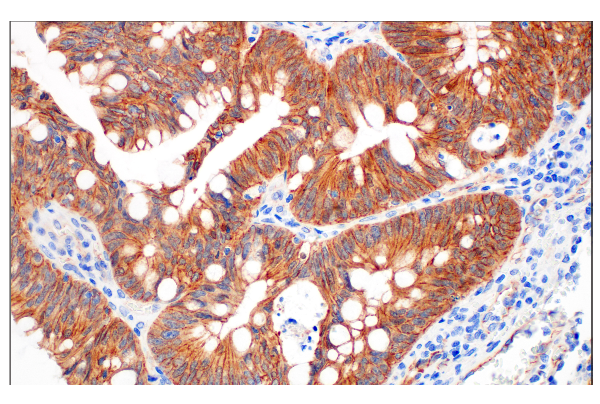
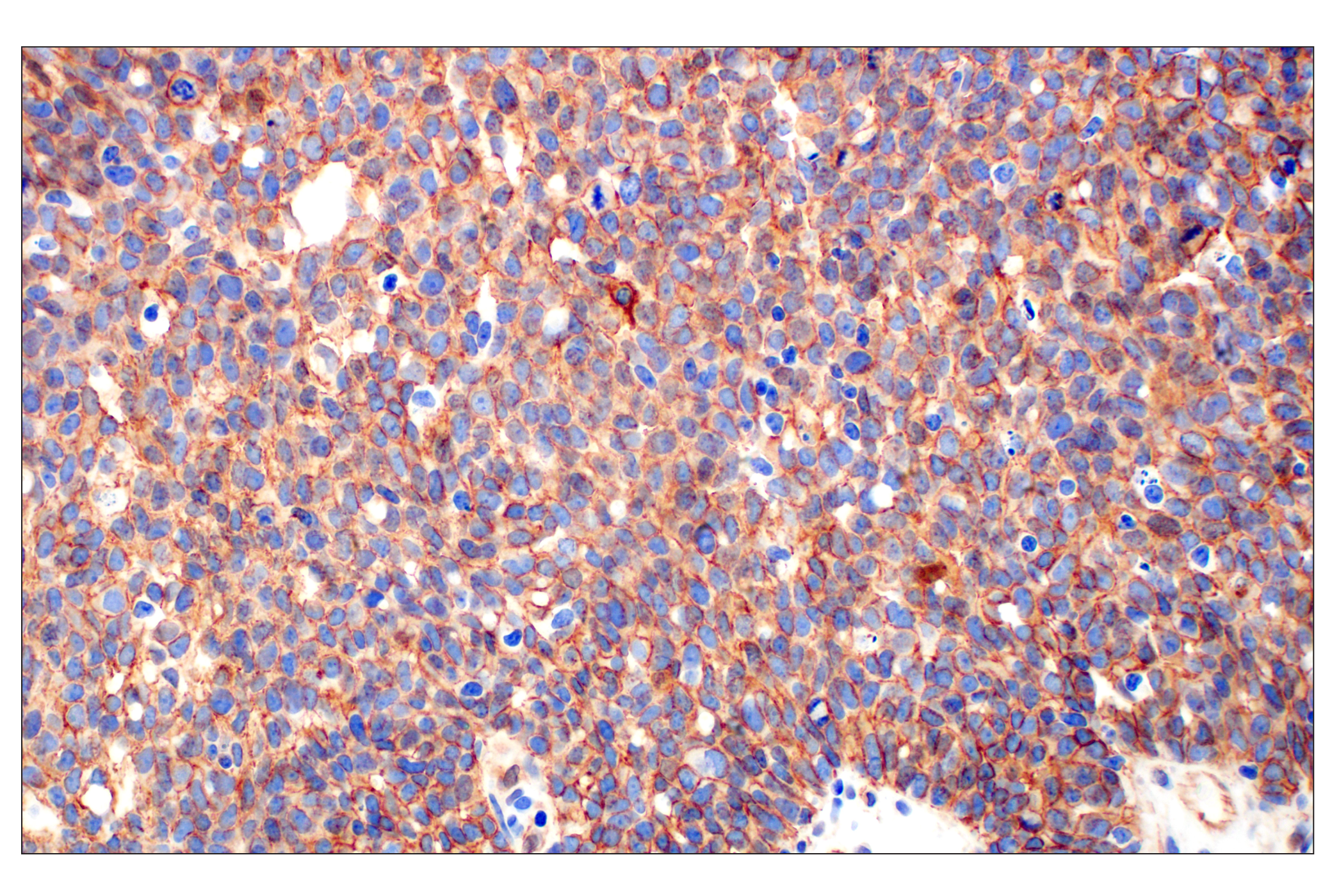
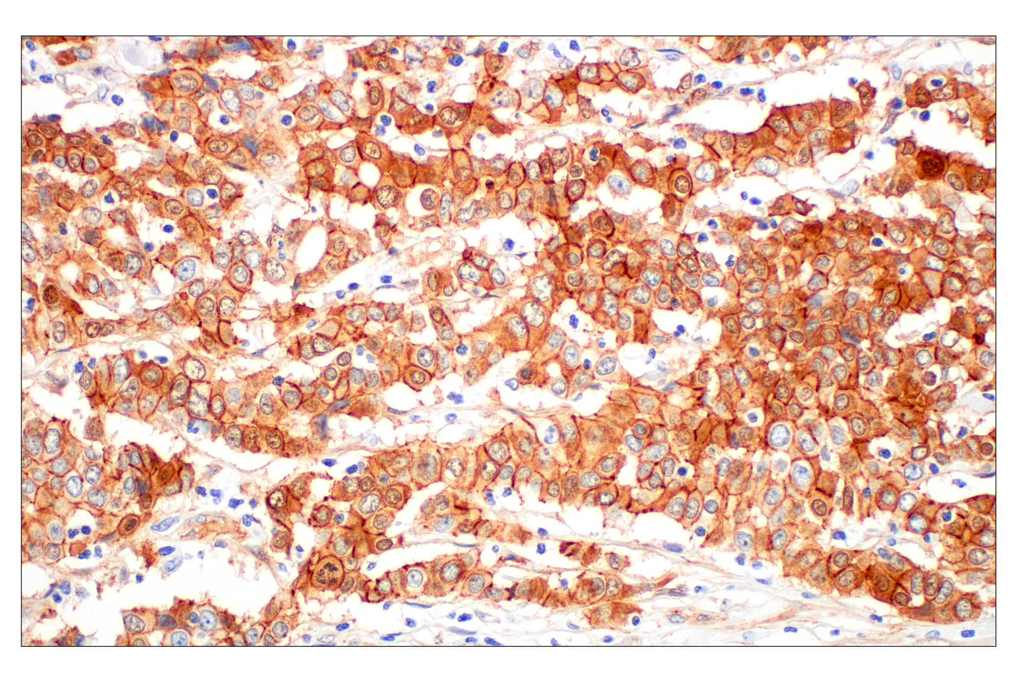
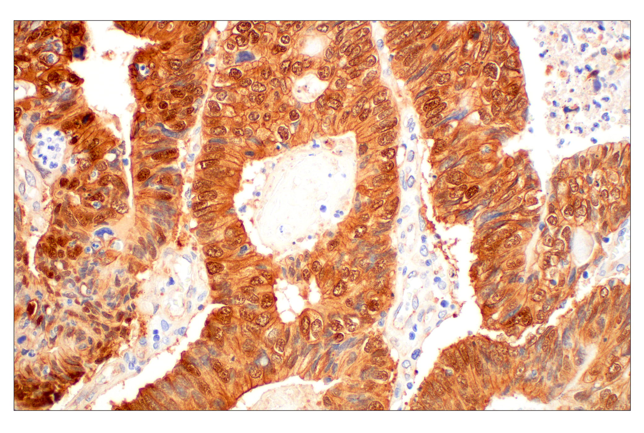
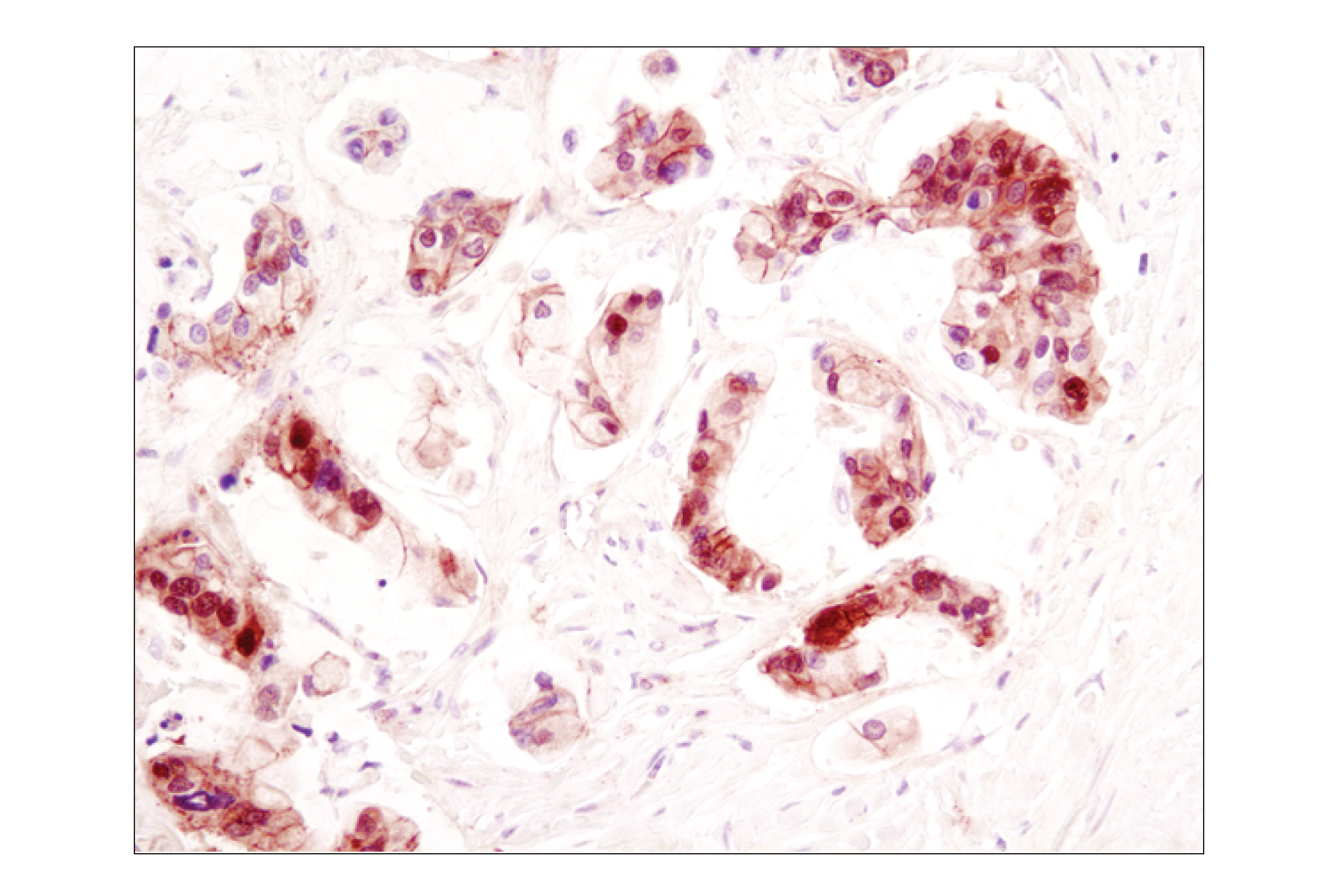
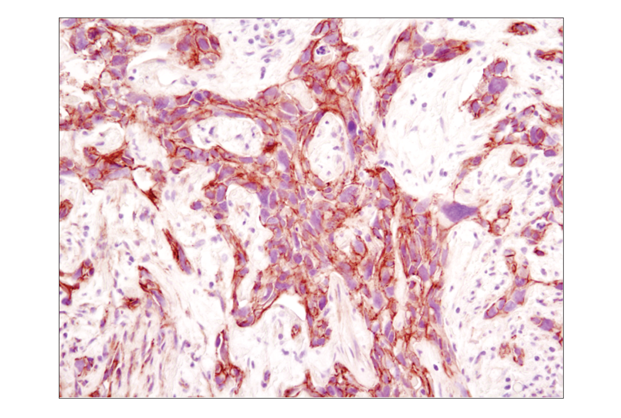
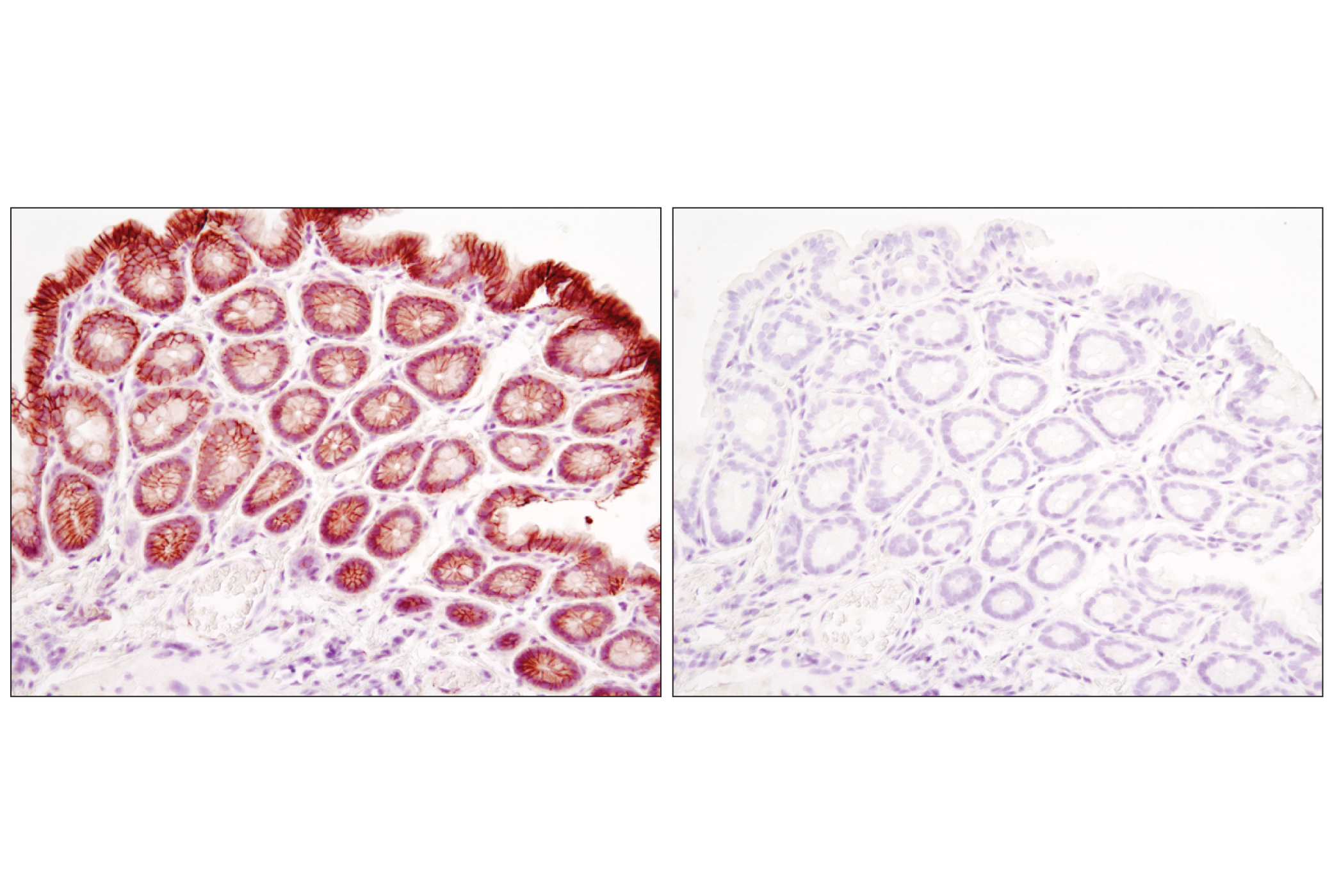
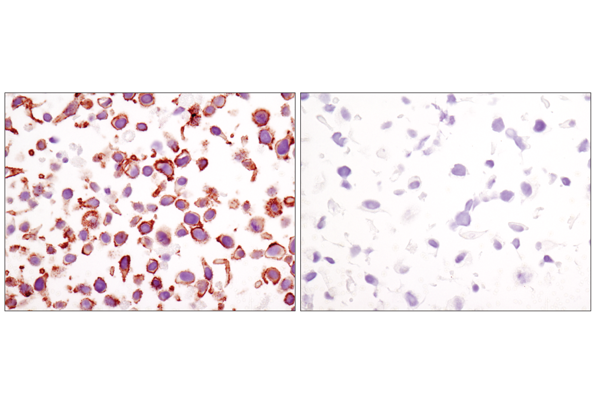
Phospho-β-Catenin (Ser552) (D8E11) Rabbit mAb (upper) or β-Catenin (6B3) Rabbit mAb #9582 (lower).
| Cat. # | Size | Qty. | Price | Inventory |
|---|---|---|---|---|
| 2951T | 1 Kit (5 x 20 microliters) |
|
| Product Includes | Quantity | Applications | Reactivity | MW(kDa) | Isotype |
|---|---|---|---|---|---|
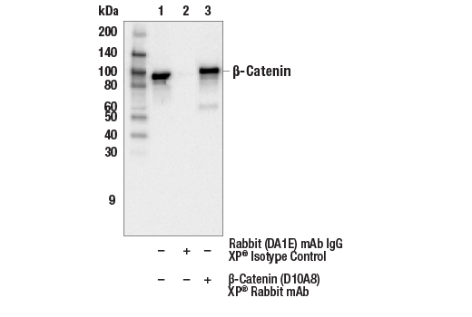
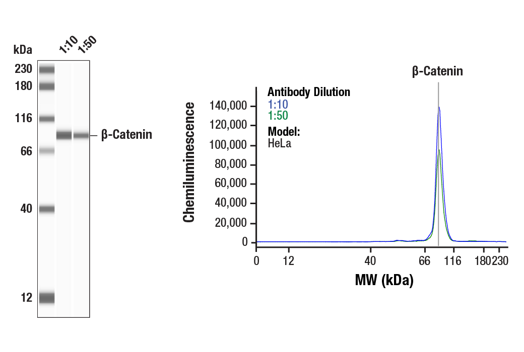
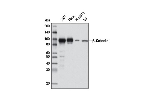
| β-Catenin (D10A8) XP® Rabbit mAb 8480 | 20 µl |
|
H M R Mk | 92 | Rabbit IgG |
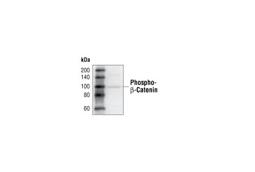
| Phospho-β-Catenin (Ser33/37/Thr41) Antibody 9561 | 20 µl |
|
H M R Mk | 92 | Rabbit |
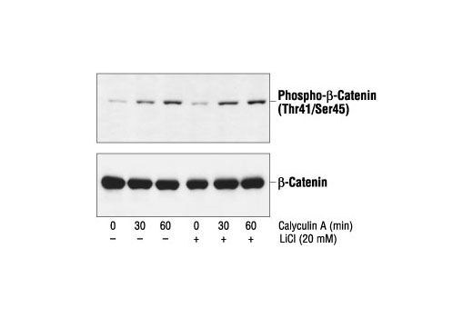
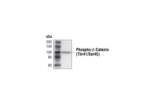
| Phospho-β-Catenin (Thr41/Ser45) Antibody 9565 | 20 µl |
|
H M Mk | 92 | Rabbit |
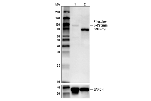
| Phospho-β-Catenin (Ser552) (D8E11) Rabbit mAb 5651 | 20 µl |
|
H M | 92 | Rabbit IgG |
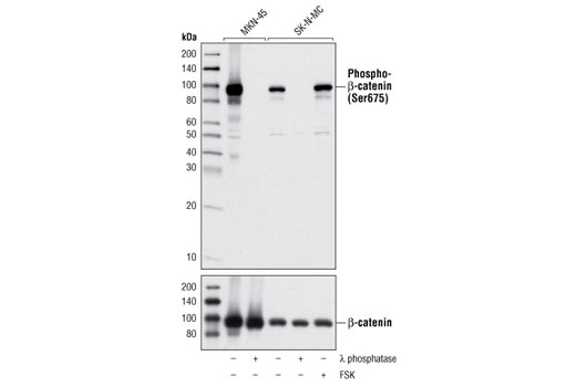
| Phospho-β-Catenin (Ser675) (D2F1) XP® Rabbit mAb 4176 | 20 µl |
|
H M R | 92 | Rabbit IgG |
| Anti-rabbit IgG, HRP-linked Antibody 7074 | 100 µl |
|
Goat |
Product Information
Polyclonal antibodies are produced by immunizing animals with a synthetic phosphopeptide corresponding to residues surrounding Ser33, Ser37 and Thr41 of human β-catenin or residues surrounding Thr41 and Ser45 of human β-catenin. Polyclonal antibodies are purified by protein A and peptide affinity chromatography. Monoclonal antibodies are produced by immunizing animals with a synthetic peptide corresponding to residues surrounding Pro714 of human β-catenin protein, a synthetic phosphopeptide corresponding to residues surrounding Ser552 of human β-catenin protein, or a synthetic phosphopeptide corresponding to residues surrounding Ser675 of human β-catenin.
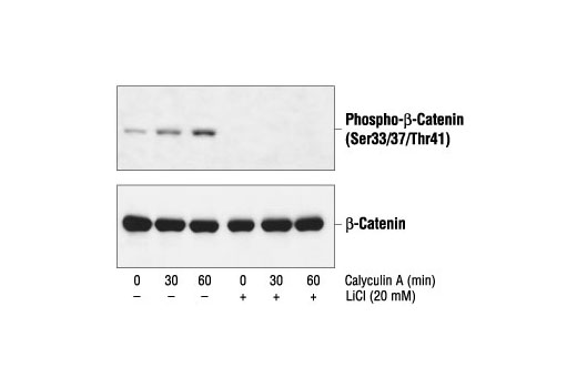
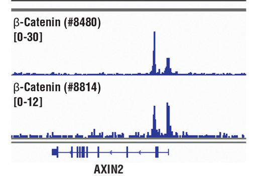
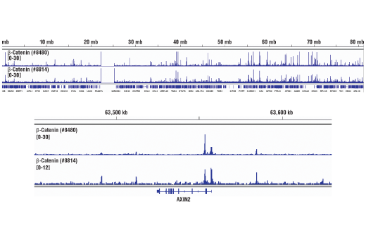
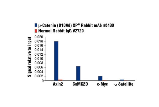
β-catenin is a key downstream effector in the Wnt signaling pathway (1). It is implicated in two major biological processes in vertebrates: early embryonic development (2) and tumorigenesis (3). CK1 phosphorylates β-catenin at Ser45. This phosphorylation event primes β-catenin for subsequent phosphorylation by GSK-3β (4-6). GSK-3β destabilizes β-catenin by phosphorylating it at Ser33, Ser37, and Thr41 (7). Mutations at these sites result in the stabilization of β-catenin protein levels and have been found in many tumor cell lines (8).
Both Akt and PKA were shown to phosphorylate β-catenin at Ser552 and Ser675. Phosphorylation at Ser552 and Ser675 induces β-catenin accumulation in the nucleus and increases its transcriptional activity (9-12).
Explore pathways related to this product.
STRING - Known and Predicted Protein-Protein Interactions.
Except as otherwise expressly agreed in a writing signed by a legally authorized representative of CST, the following terms apply to Products provided by CST, its affiliates or its distributors. Any Customer's terms and conditions that are in addition to, or different from, those contained herein, unless separately accepted in writing by a legally authorized representative of CST, are rejected and are of no force or effect.
Products are labeled with For Research Use Only or a similar labeling statement and have not been approved, cleared, or licensed by the FDA or other regulatory foreign or domestic entity, for any purpose. Customer shall not use any Product for any diagnostic or therapeutic purpose, or otherwise in any manner that conflicts with its labeling statement. Products sold or licensed by CST are provided for Customer as the end-user and solely for research and development uses. Any use of Product for diagnostic, prophylactic or therapeutic purposes, or any purchase of Product for resale (alone or as a component) or other commercial purpose, requires a separate license from CST. Customer shall (a) not sell, license, loan, donate or otherwise transfer or make available any Product to any third party, whether alone or in combination with other materials, or use the Products to manufacture any commercial products, (b) not copy, modify, reverse engineer, decompile, disassemble or otherwise attempt to discover the underlying structure or technology of the Products, or use the Products for the purpose of developing any products or services that would compete with CST products or services, (c) not alter or remove from the Products any trademarks, trade names, logos, patent or copyright notices or markings, (d) use the Products solely in accordance with CST Product Terms of Sale and any applicable documentation, and (e) comply with any license, terms of service or similar agreement with respect to any third party products or services used by Customer in connection with the Products.
View in English?