View in English?
View in English?
View in English?

| Cat. # | Size | Qty. | Price | Inventory |
|---|---|---|---|---|
| 11843T | 1 Kit (4 x 20 microliters) |
|
| Product Includes | Quantity | Applications | Reactivity | MW(kDa) | Isotype |
|---|---|---|---|---|---|
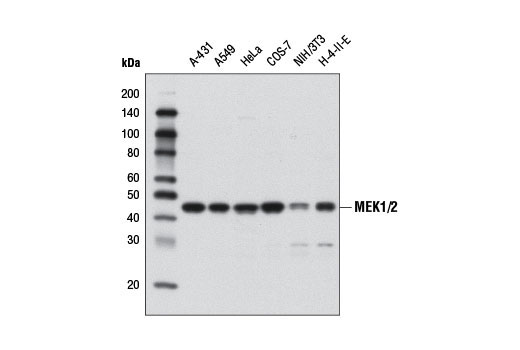
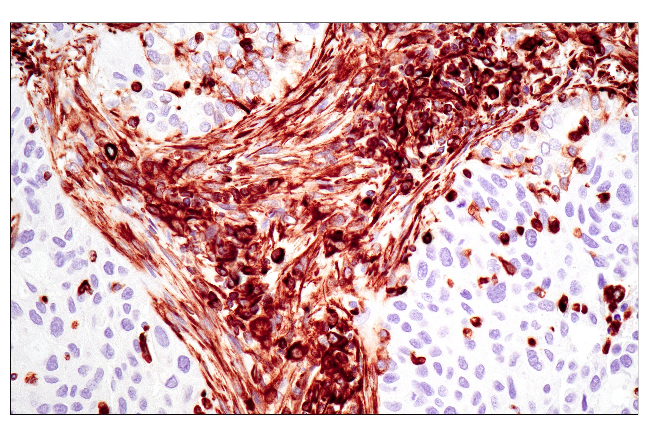
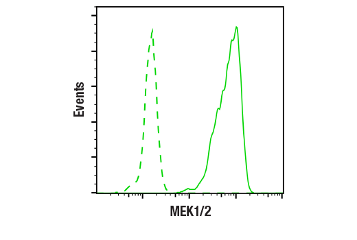
| MEK1/2 (D1A5) Rabbit mAb 8727 | 20 µl |
|
H M R Mk Dm | 45 | Rabbit IgG |
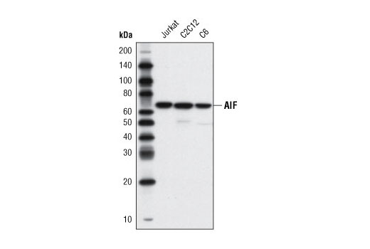
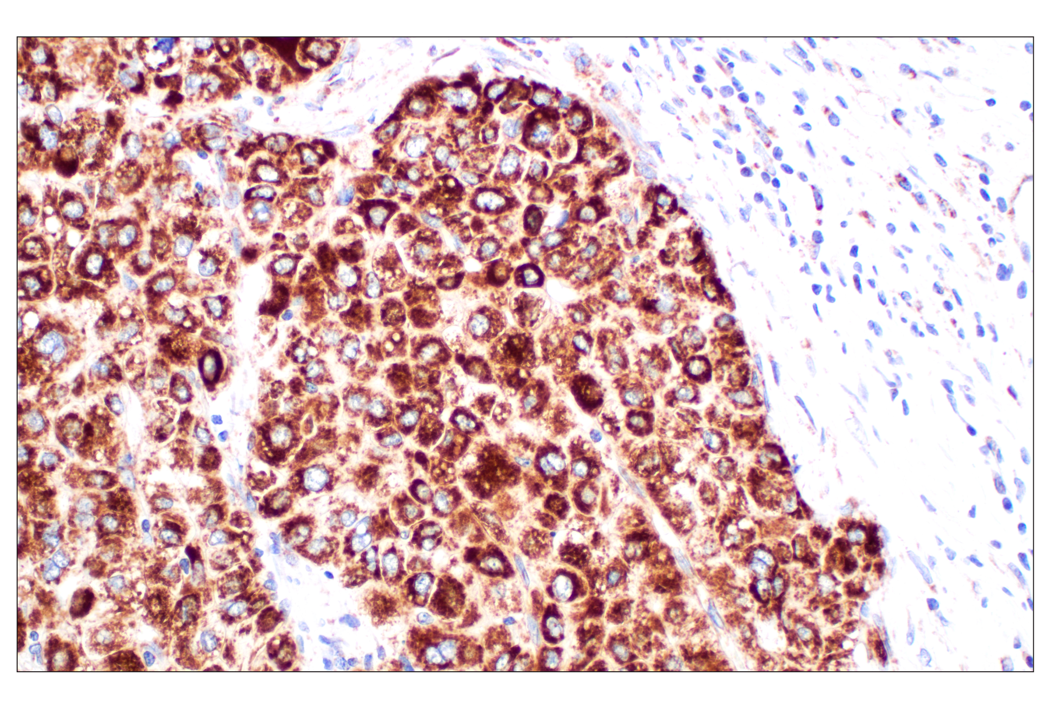
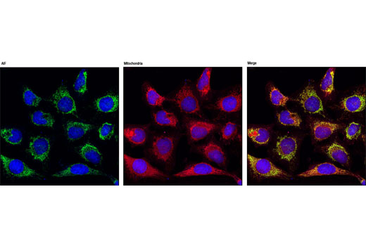
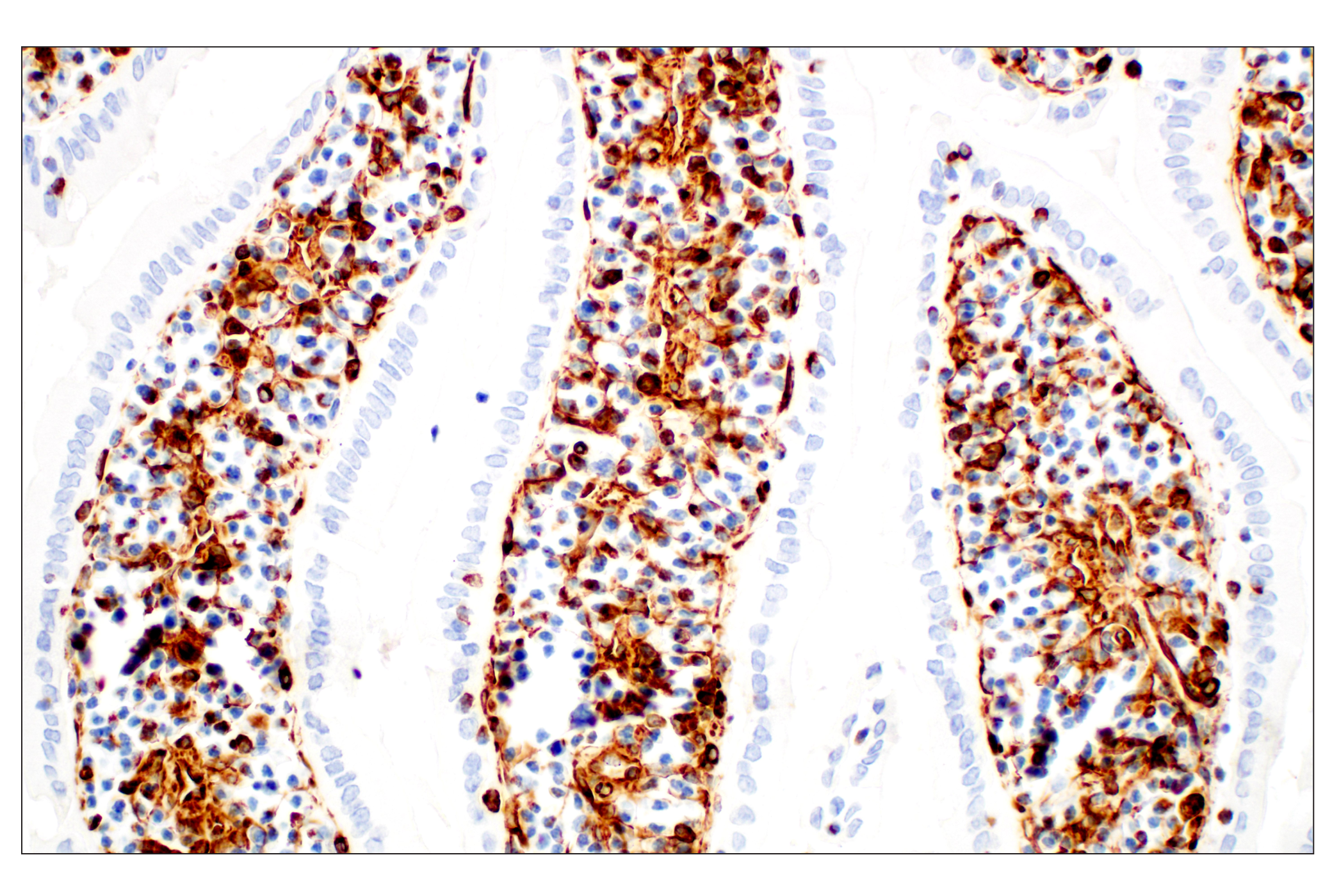
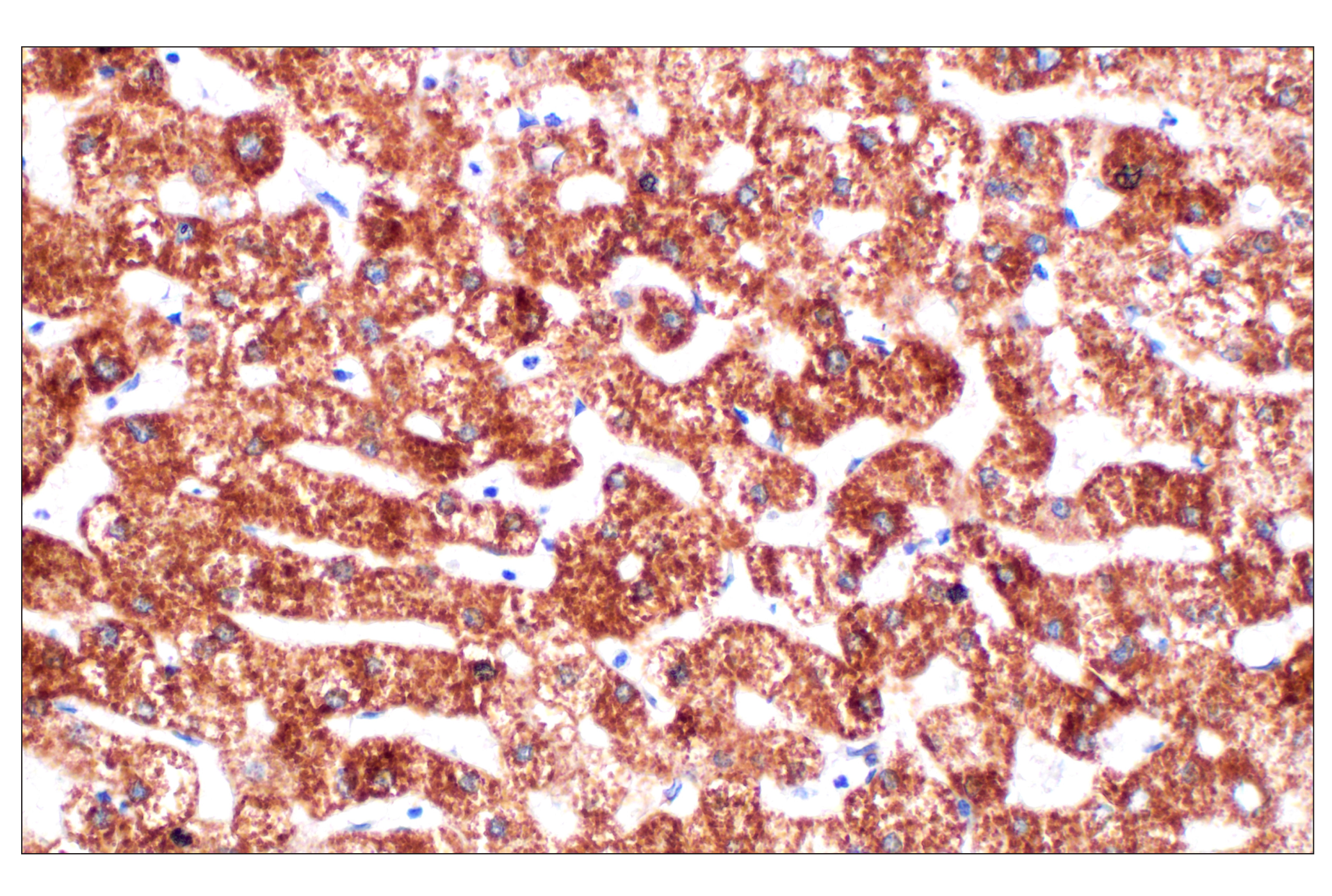
| AIF (D39D2) XP® Rabbit mAb 5318 | 20 µl |
|
H M R Mk | 67 | Rabbit IgG |
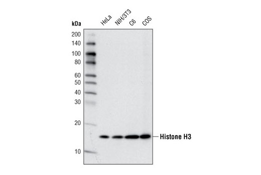
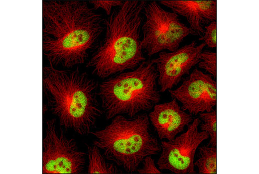
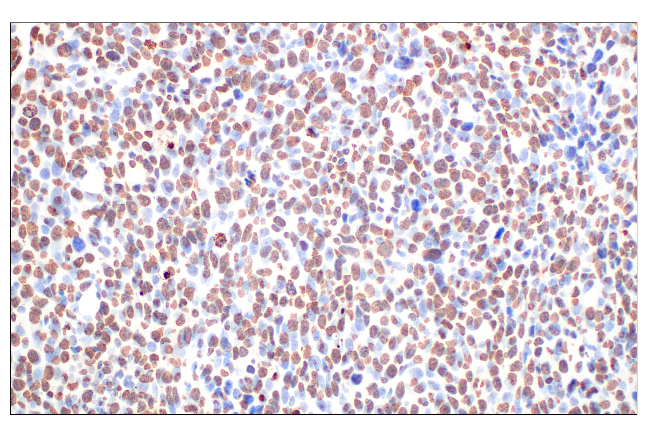
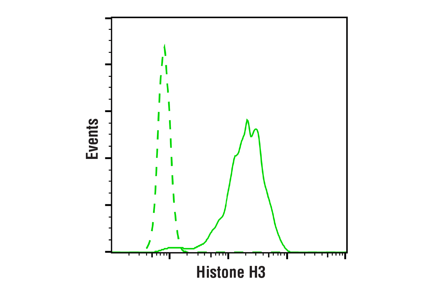
| Histone H3 (D1H2) XP® Rabbit mAb 4499 | 20 µl |
|
H M R Mk | 17 | Rabbit IgG |
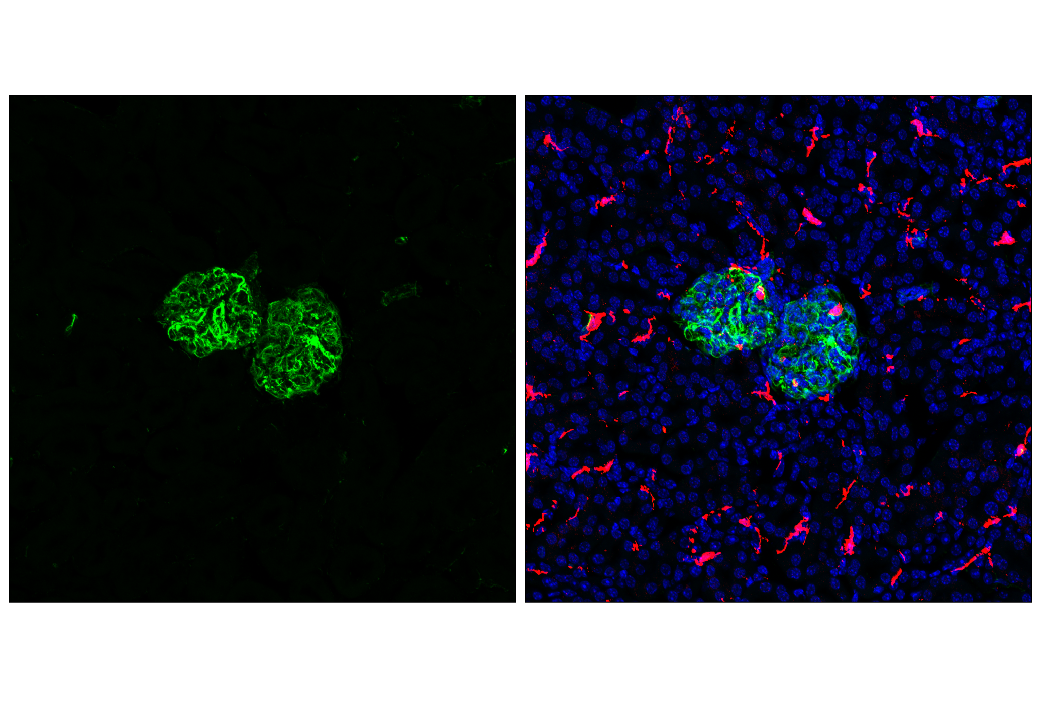
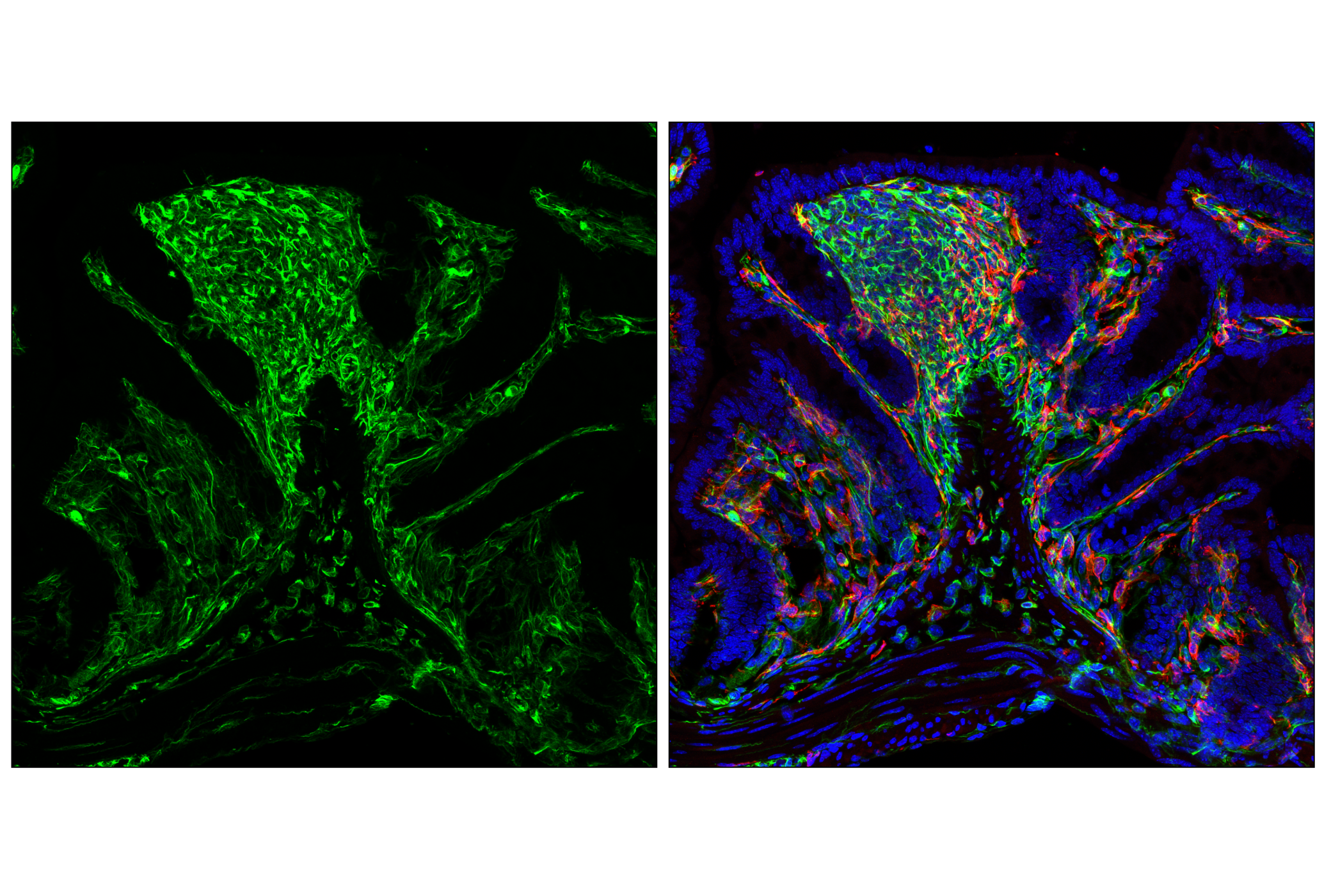
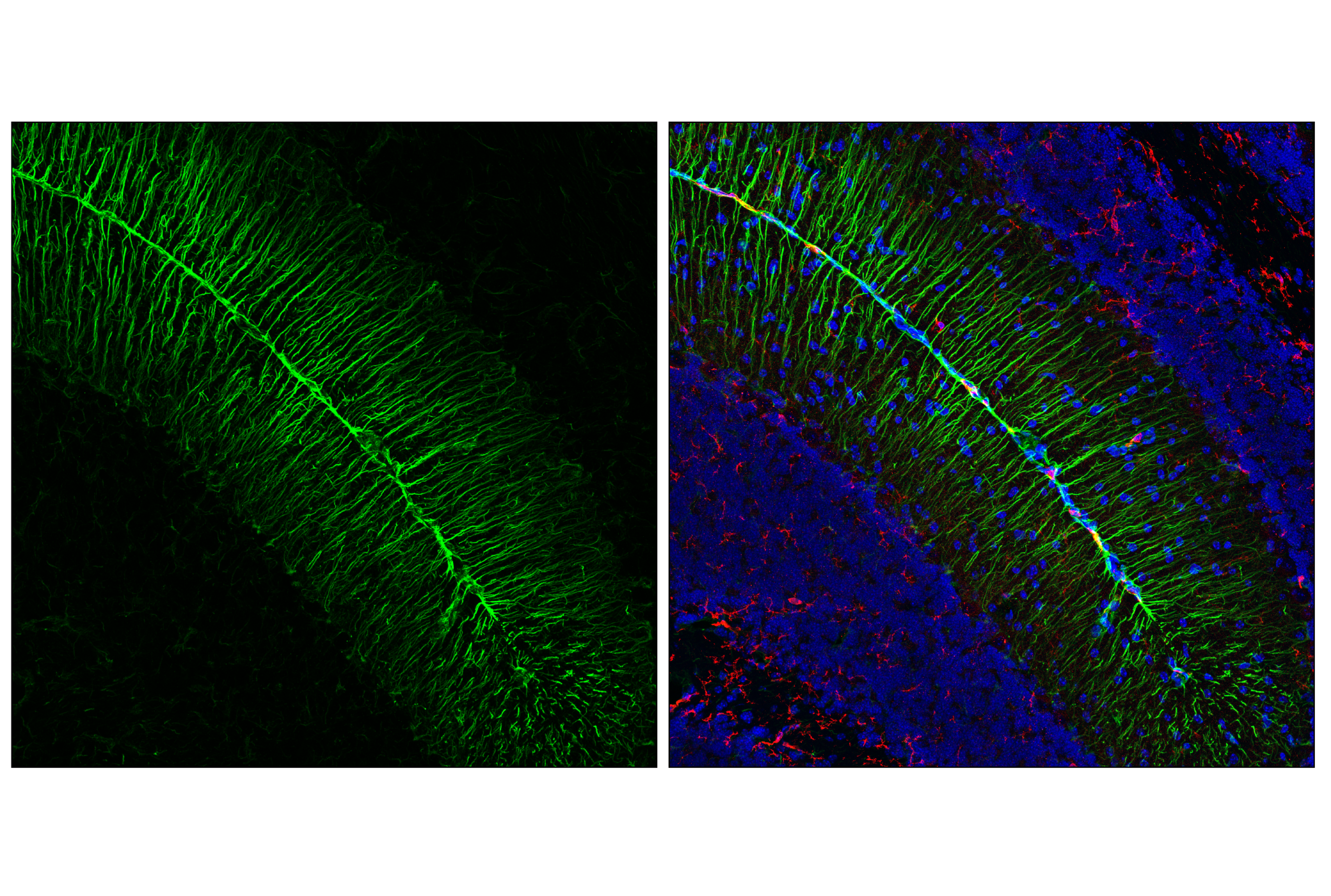
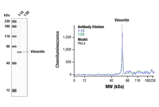
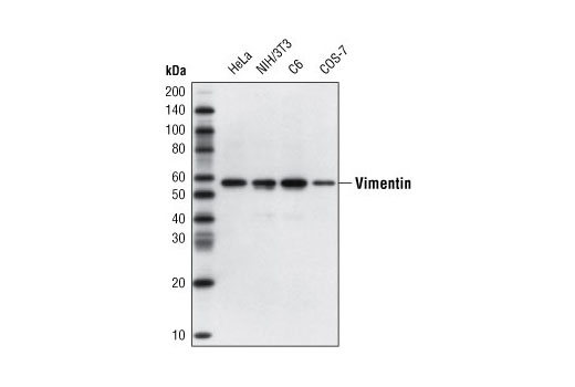
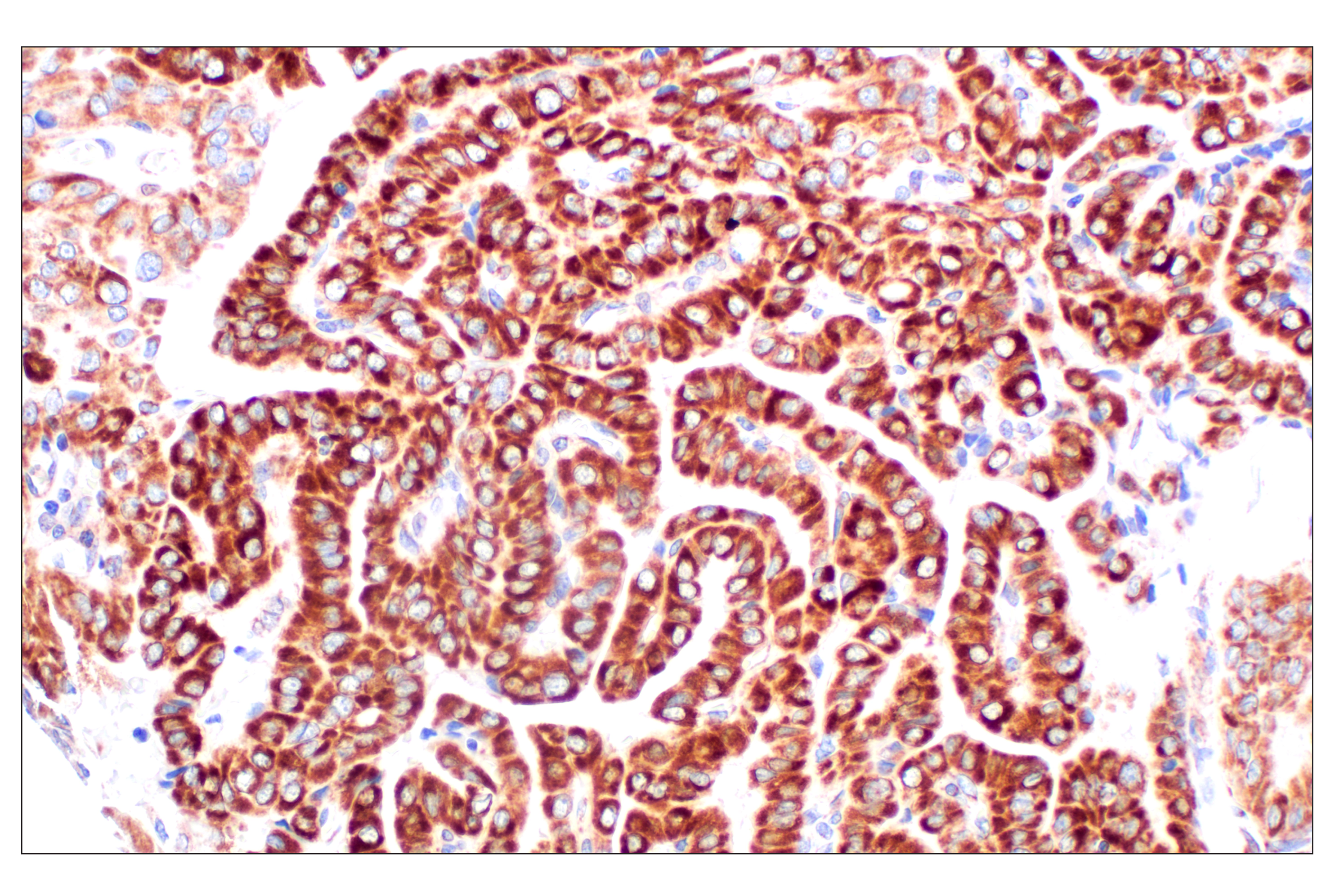
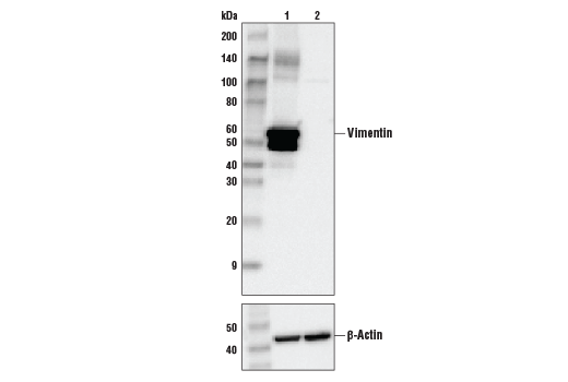
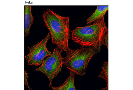
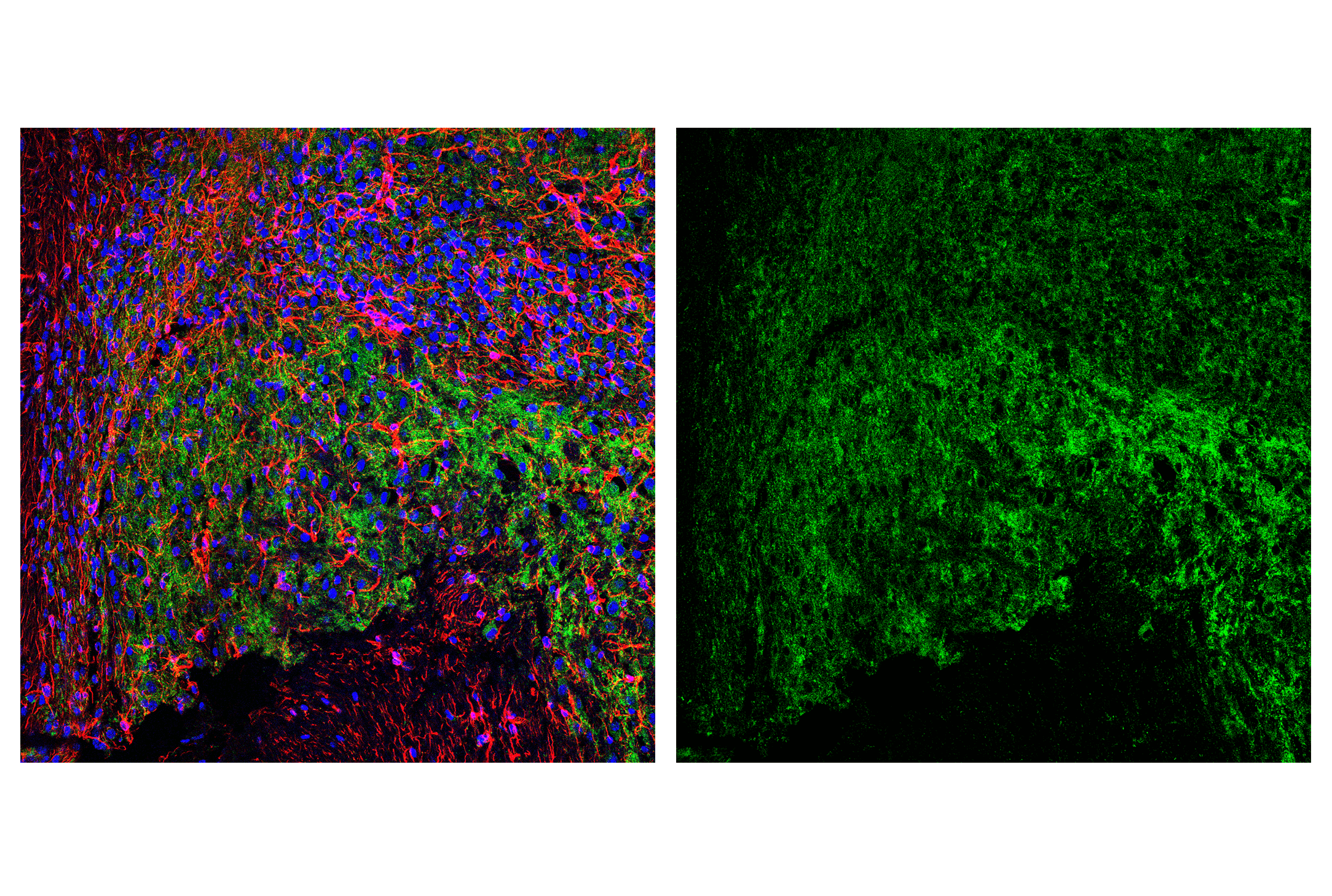
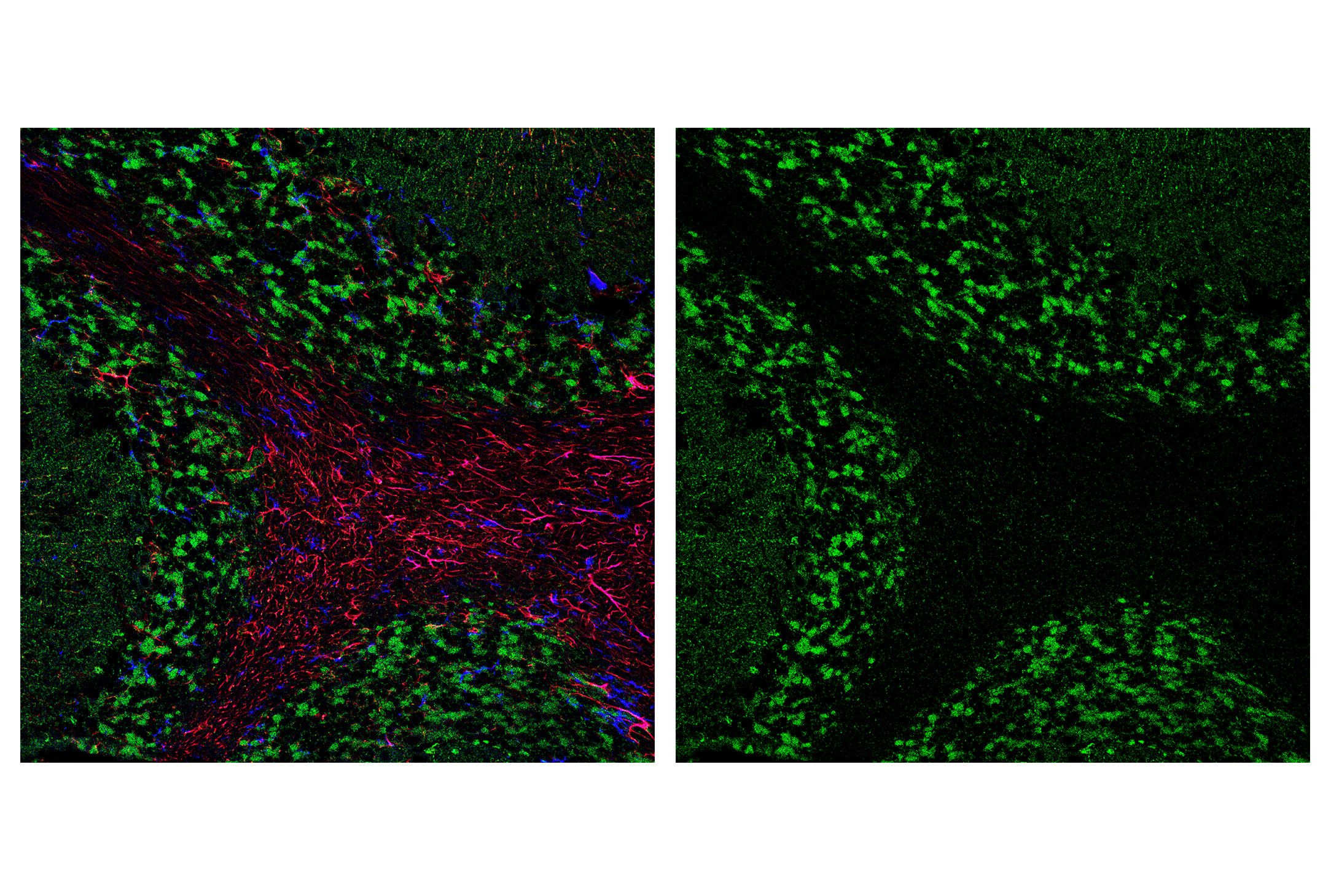
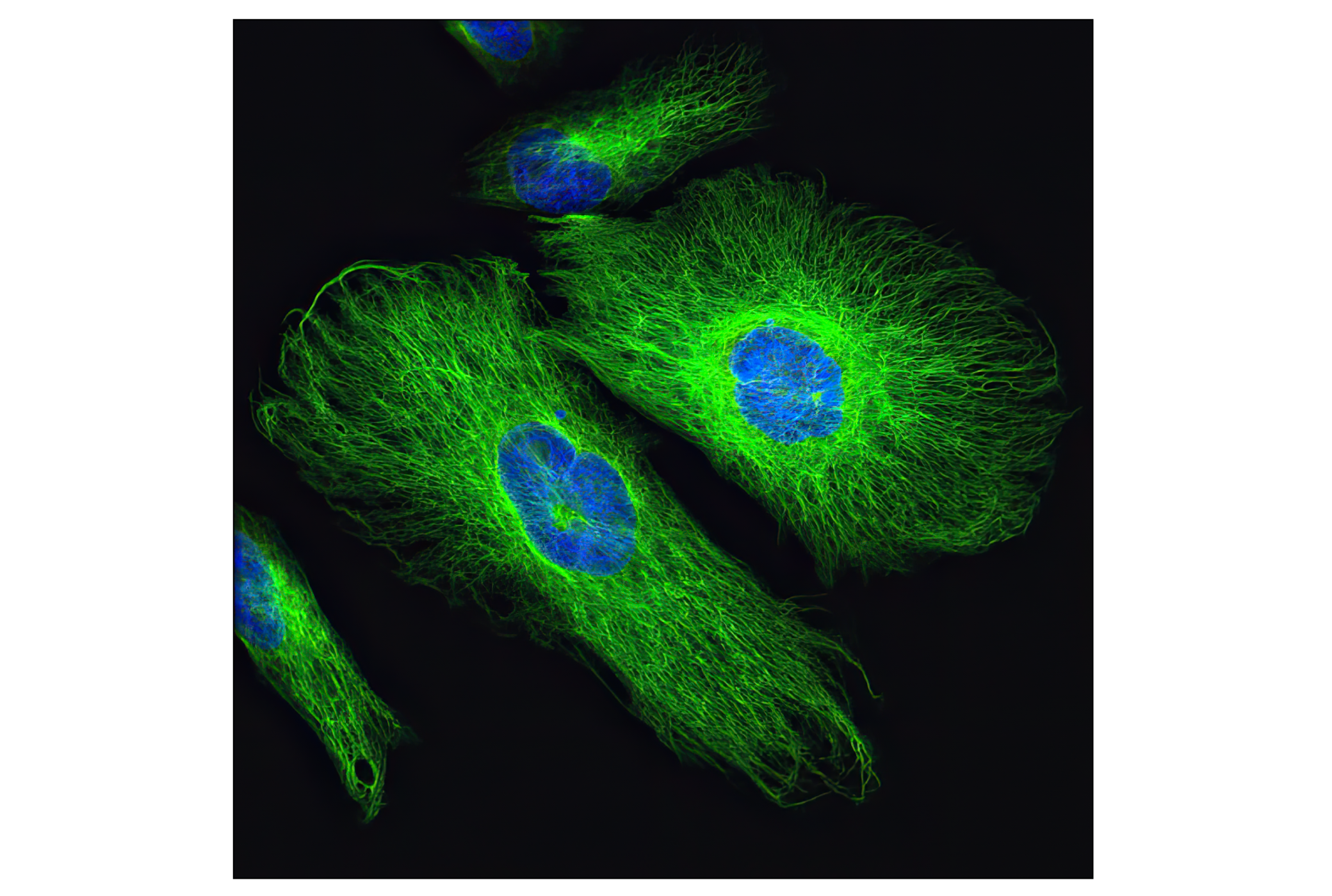
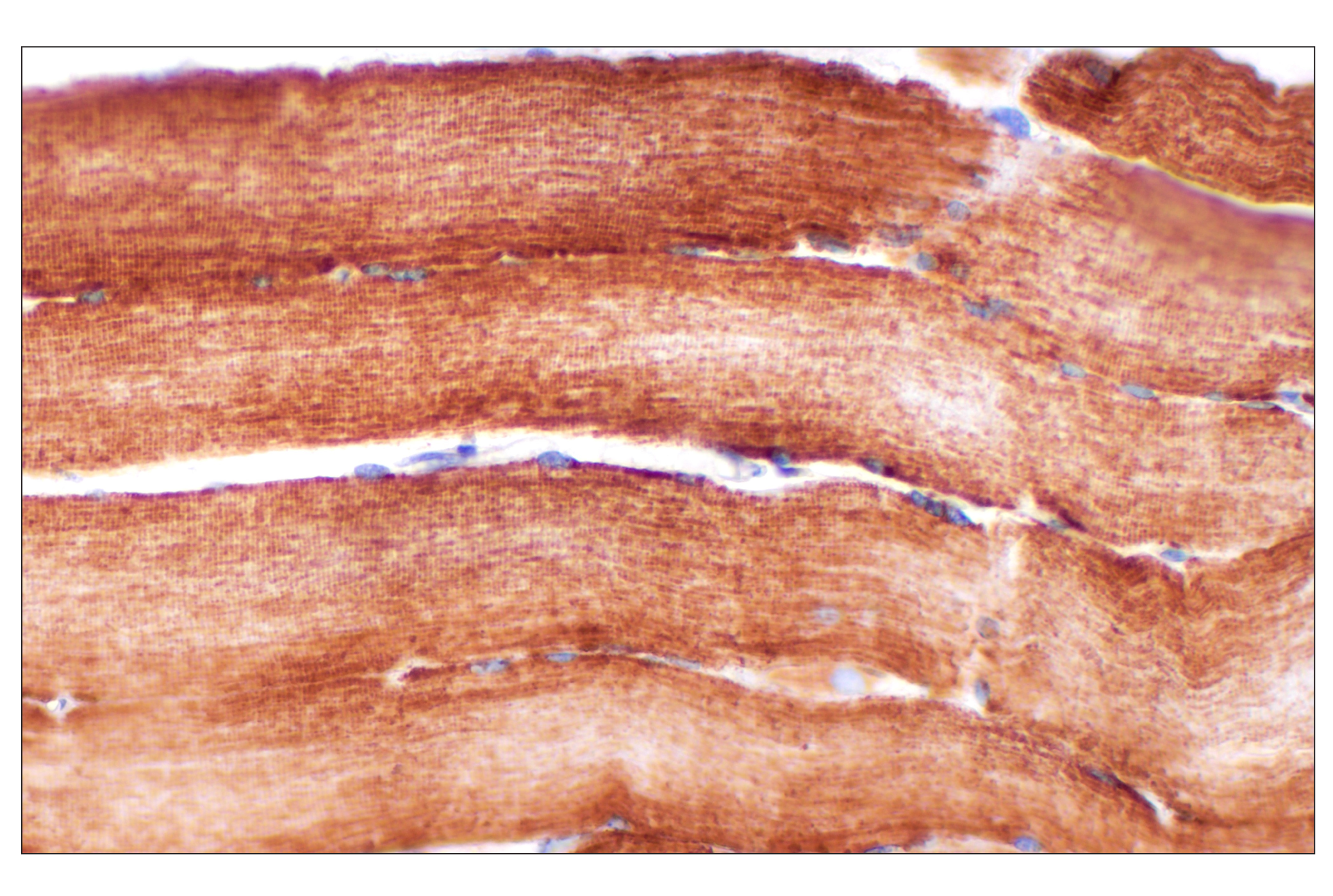
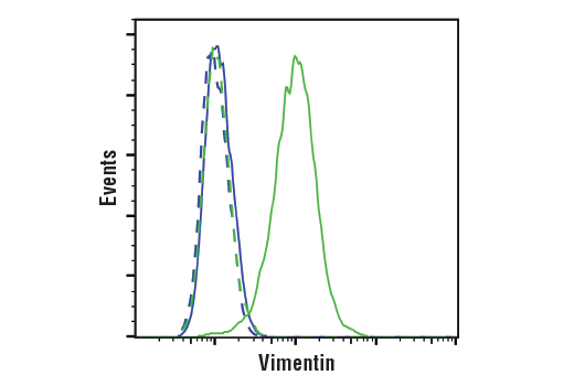
| Vimentin (D21H3) XP® Rabbit mAb 5741 | 20 µl |
|
H M R Mk | 57 | Rabbit IgG |
| Anti-rabbit IgG, HRP-linked Antibody 7074 | 100 µl |
|
Goat |
Product Information
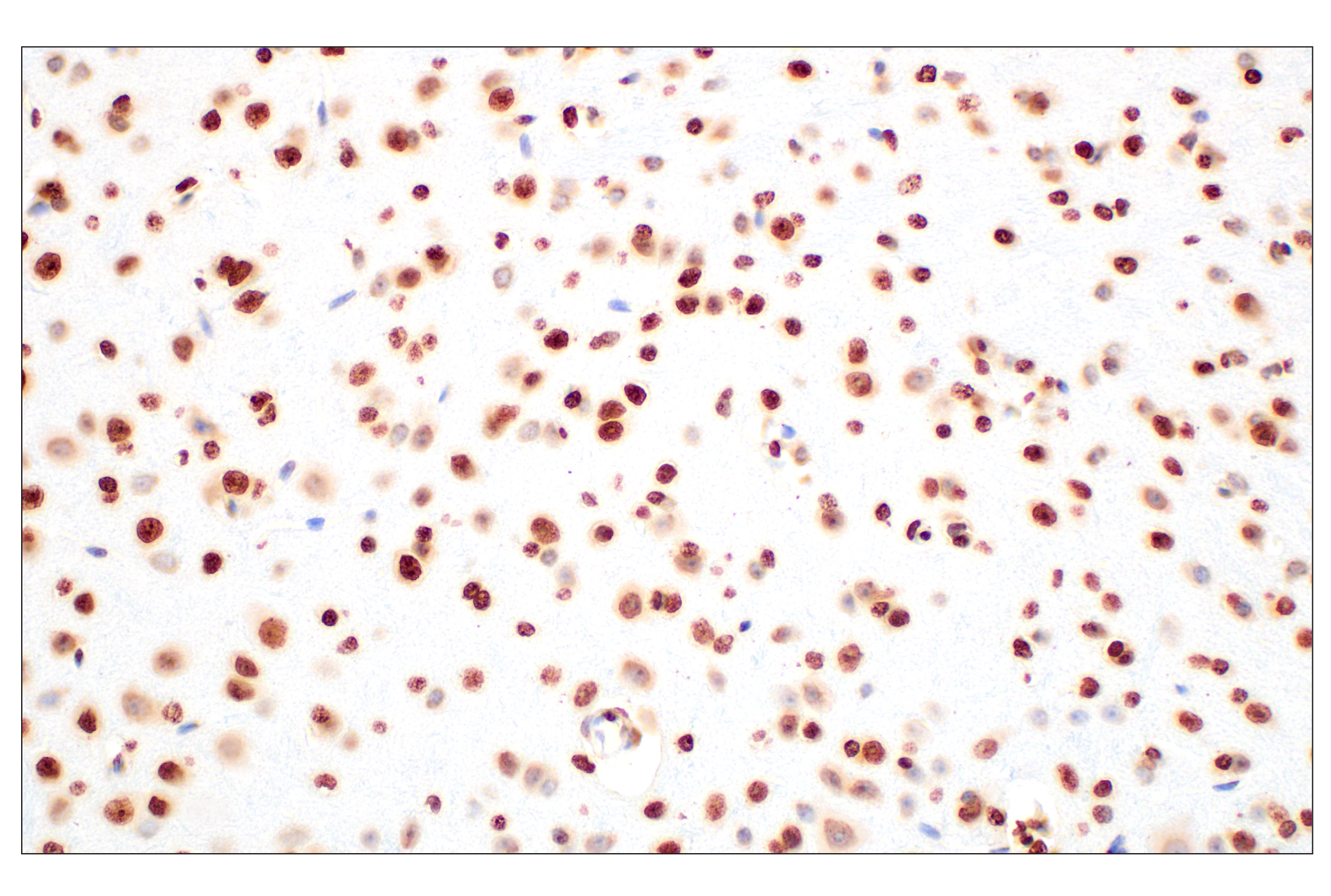
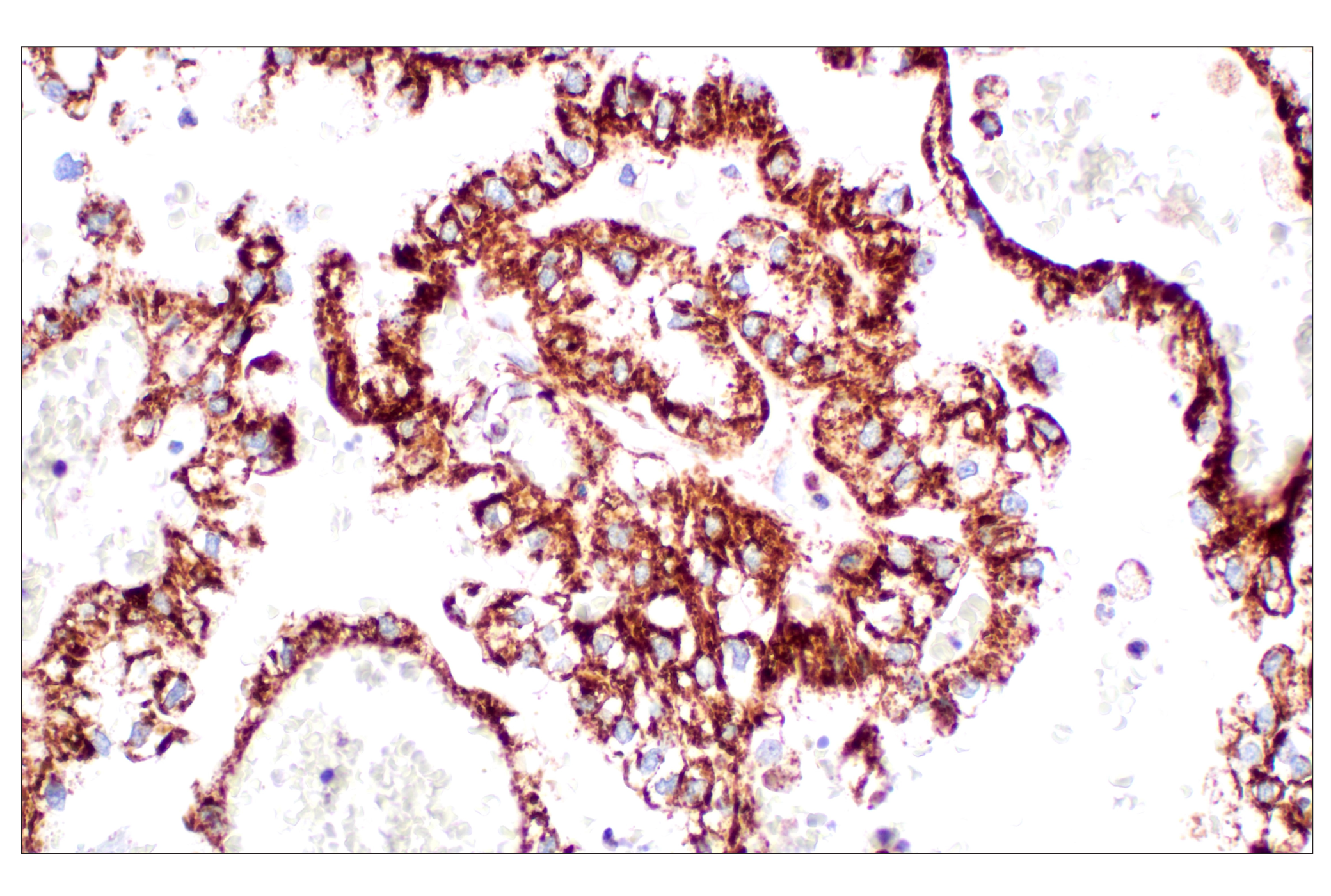
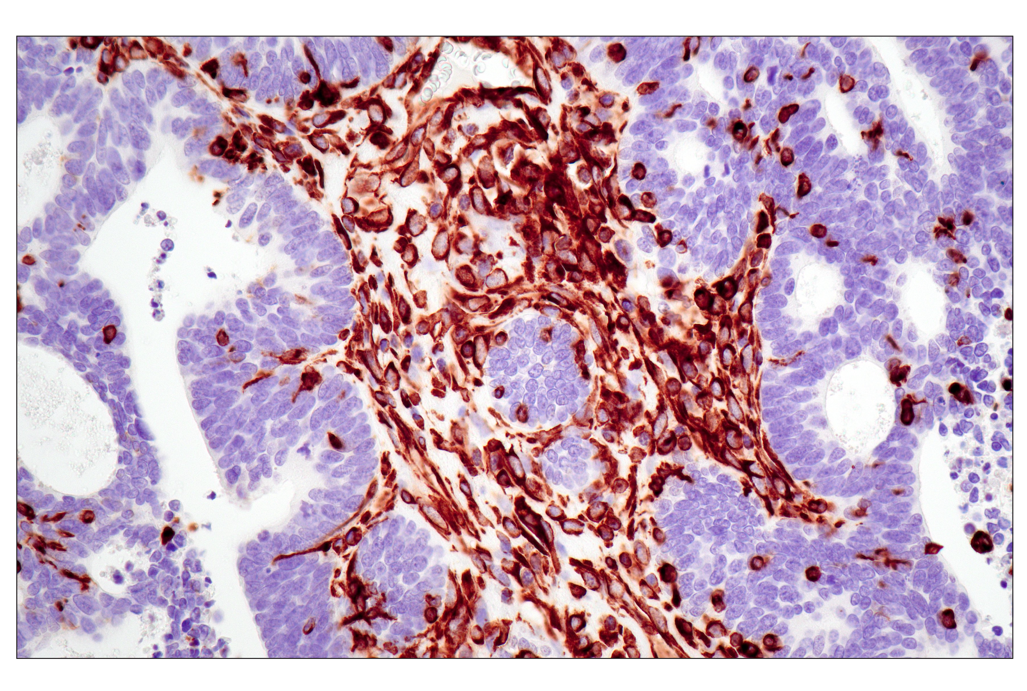
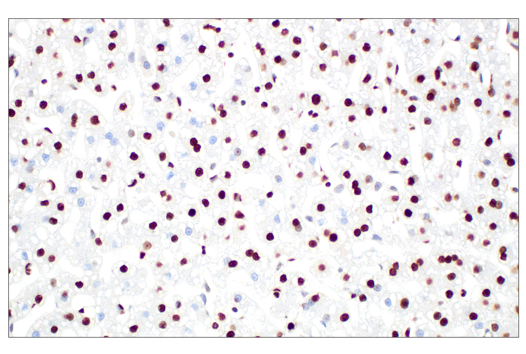
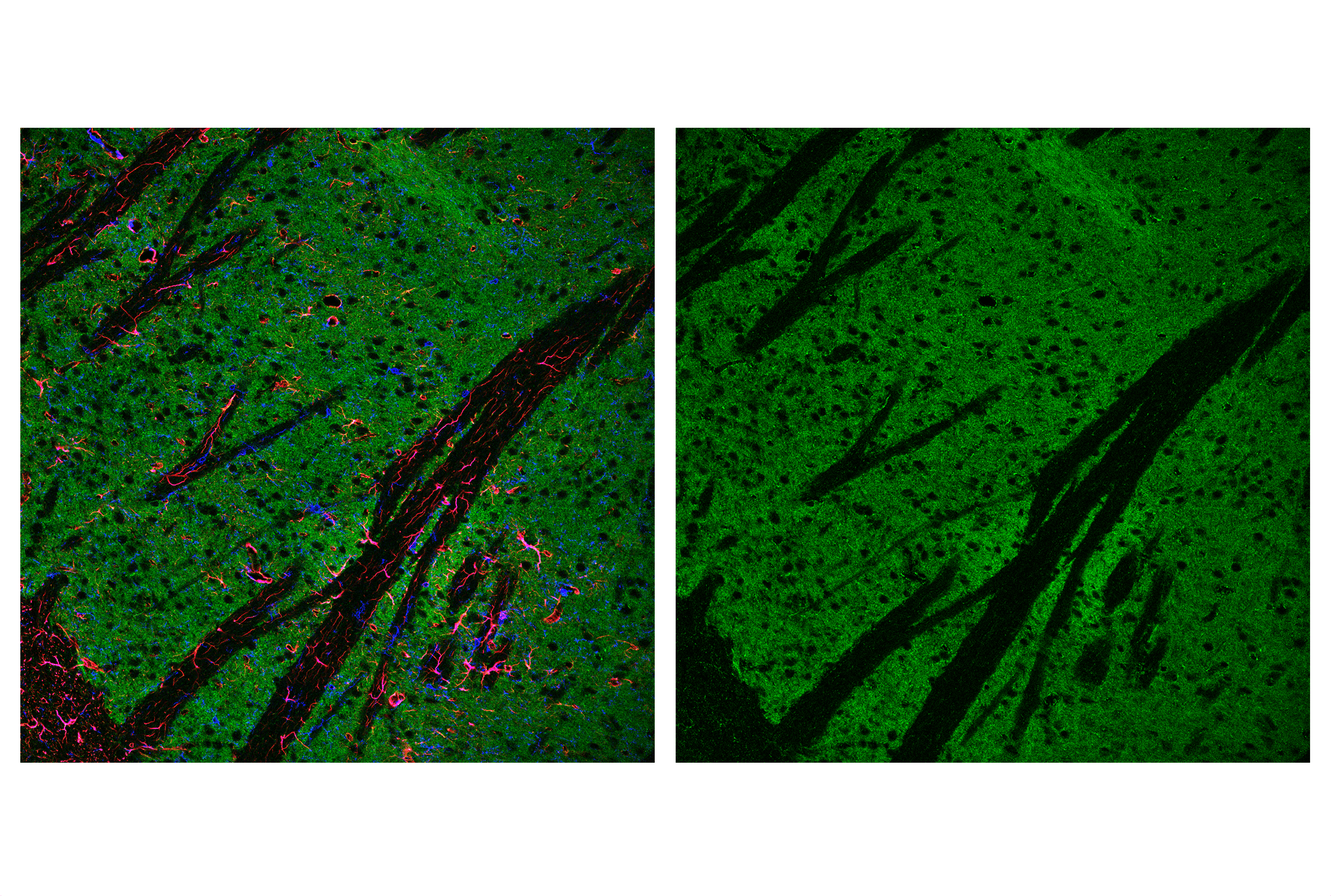
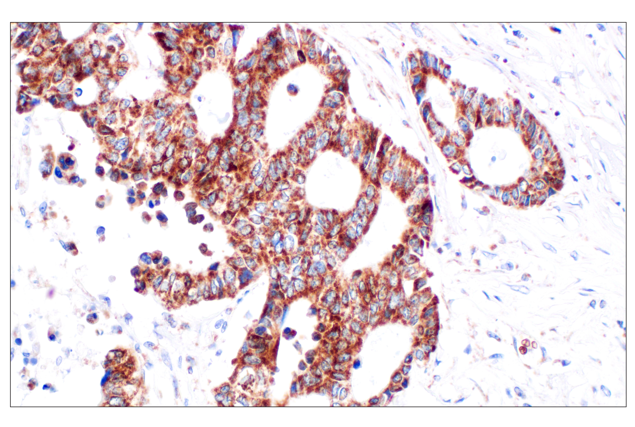
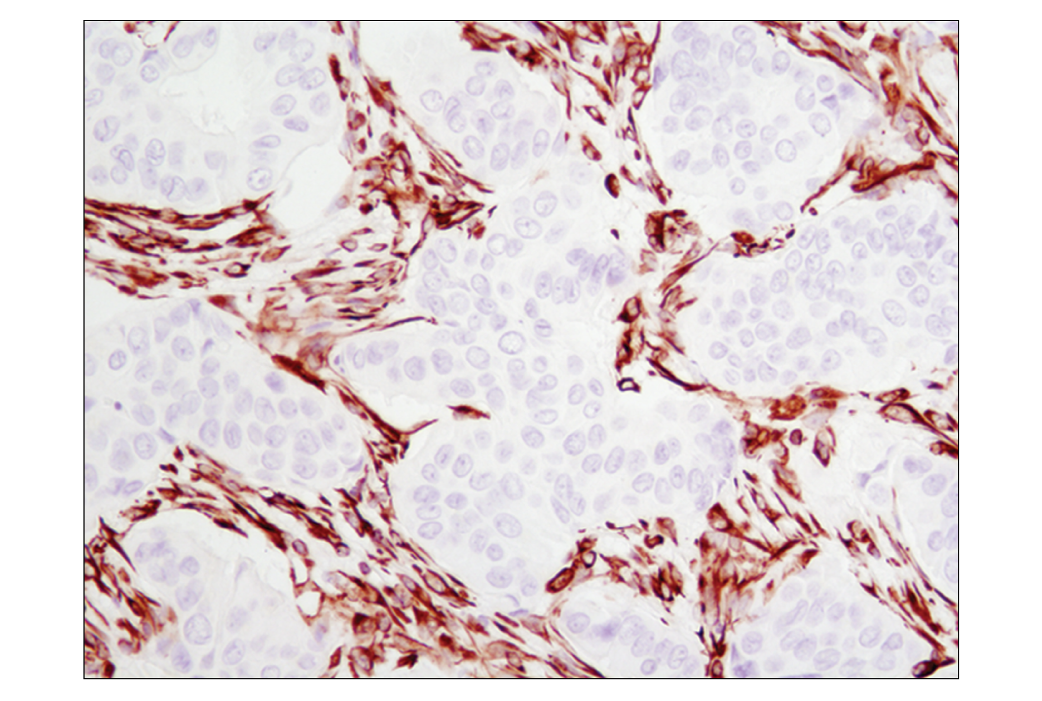
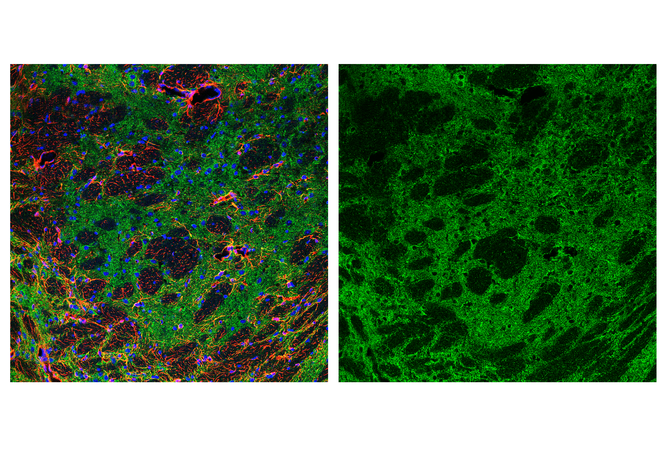
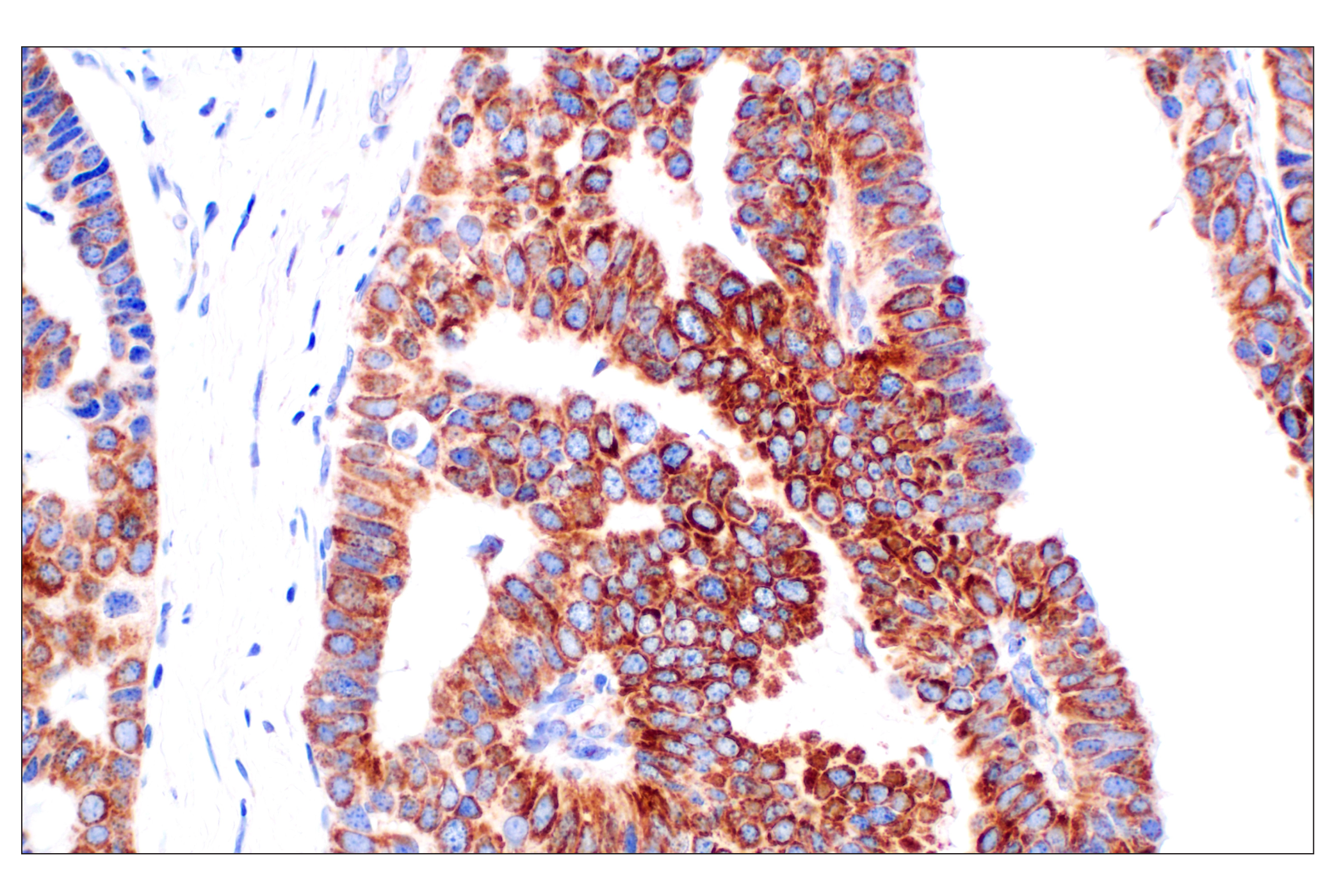
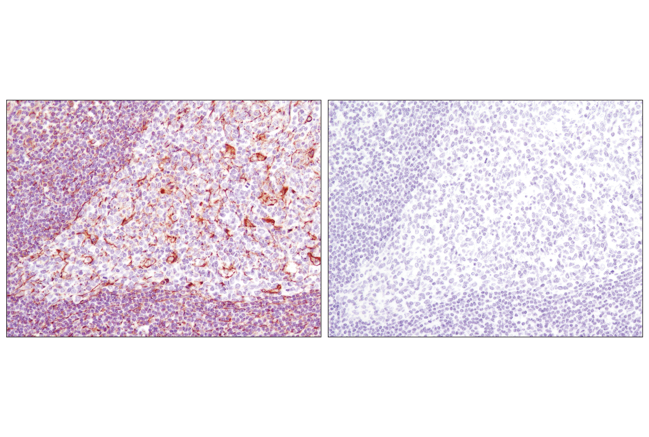
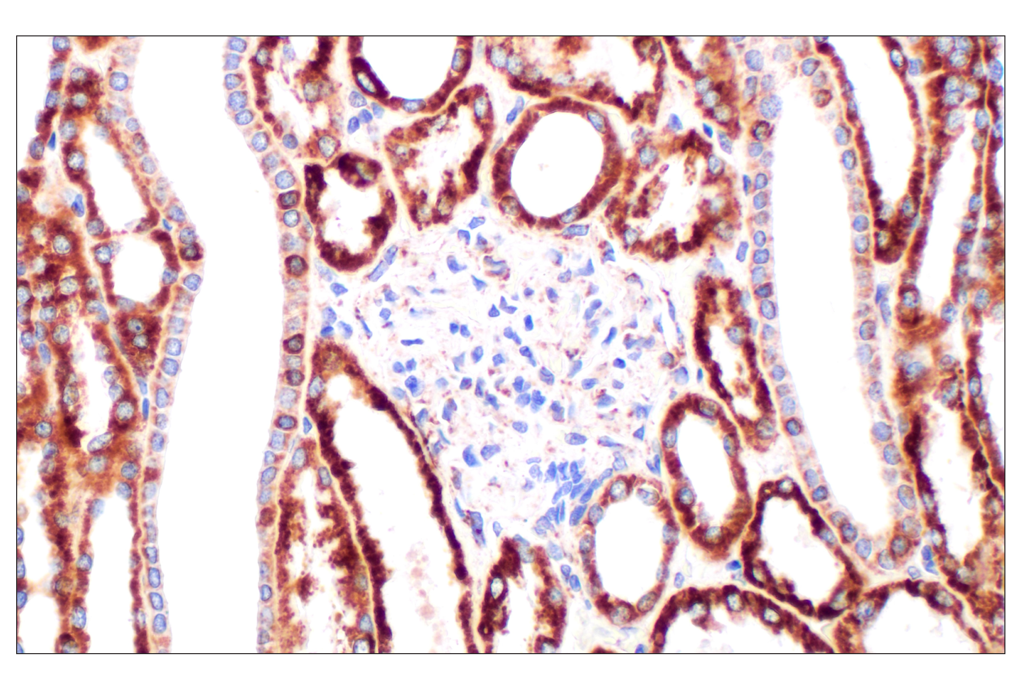
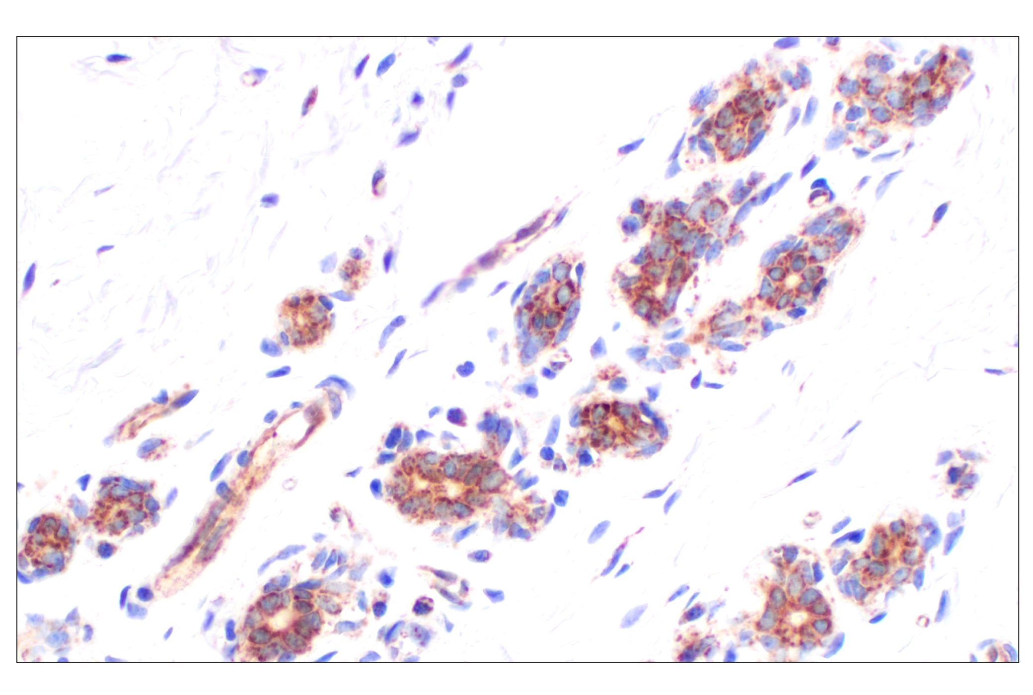
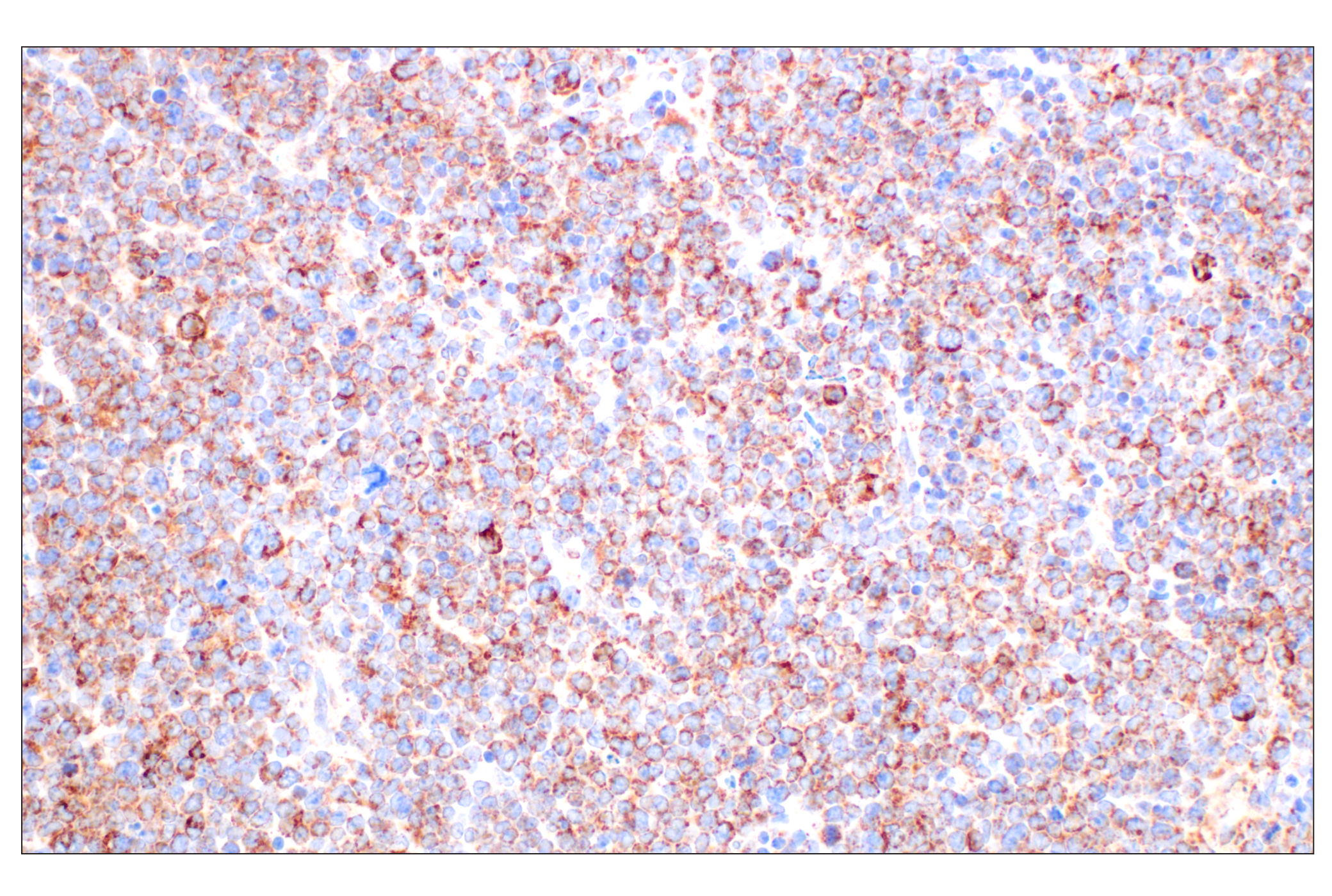
Rabbit monoclonal antibodies are produced by immunizing animals with a synthetic peptide corresponding to residues surrounding Ala520 of human AIF protein, the carboxy terminus of human histone H3 protein, residues surrounding Ala220 of human MEK1 protein, and residues surrounding Arg45 of human vimentin protein.
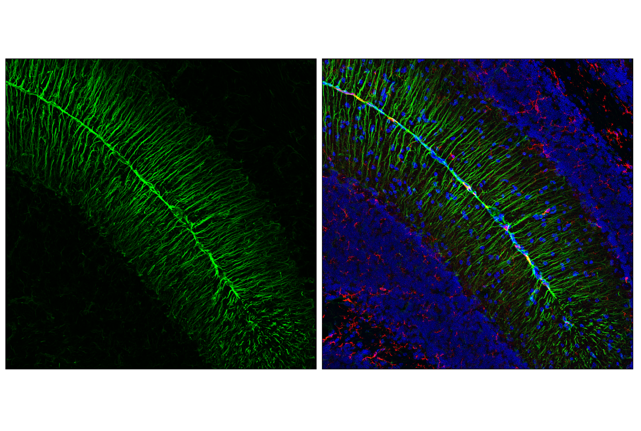
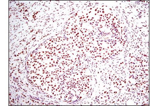
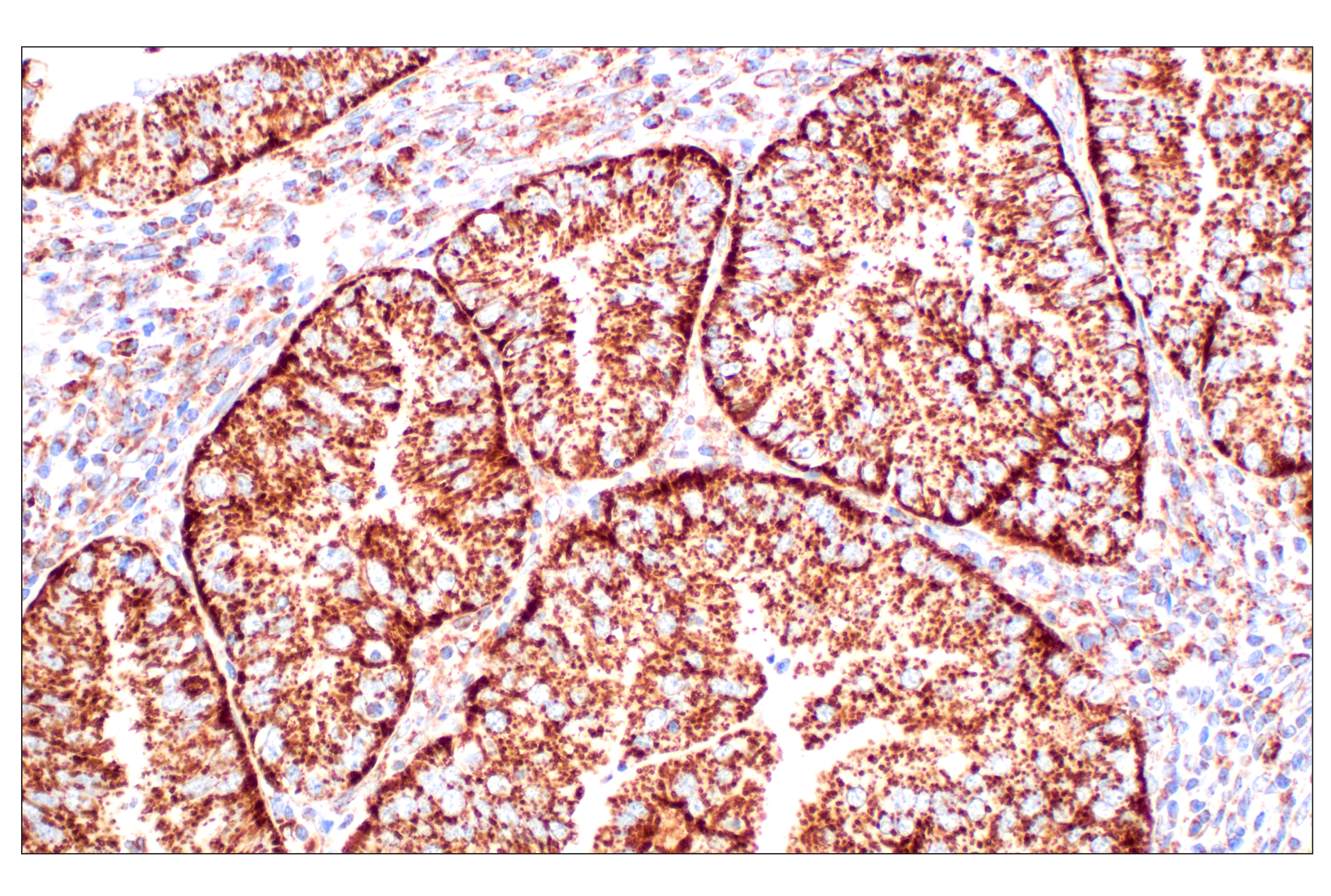
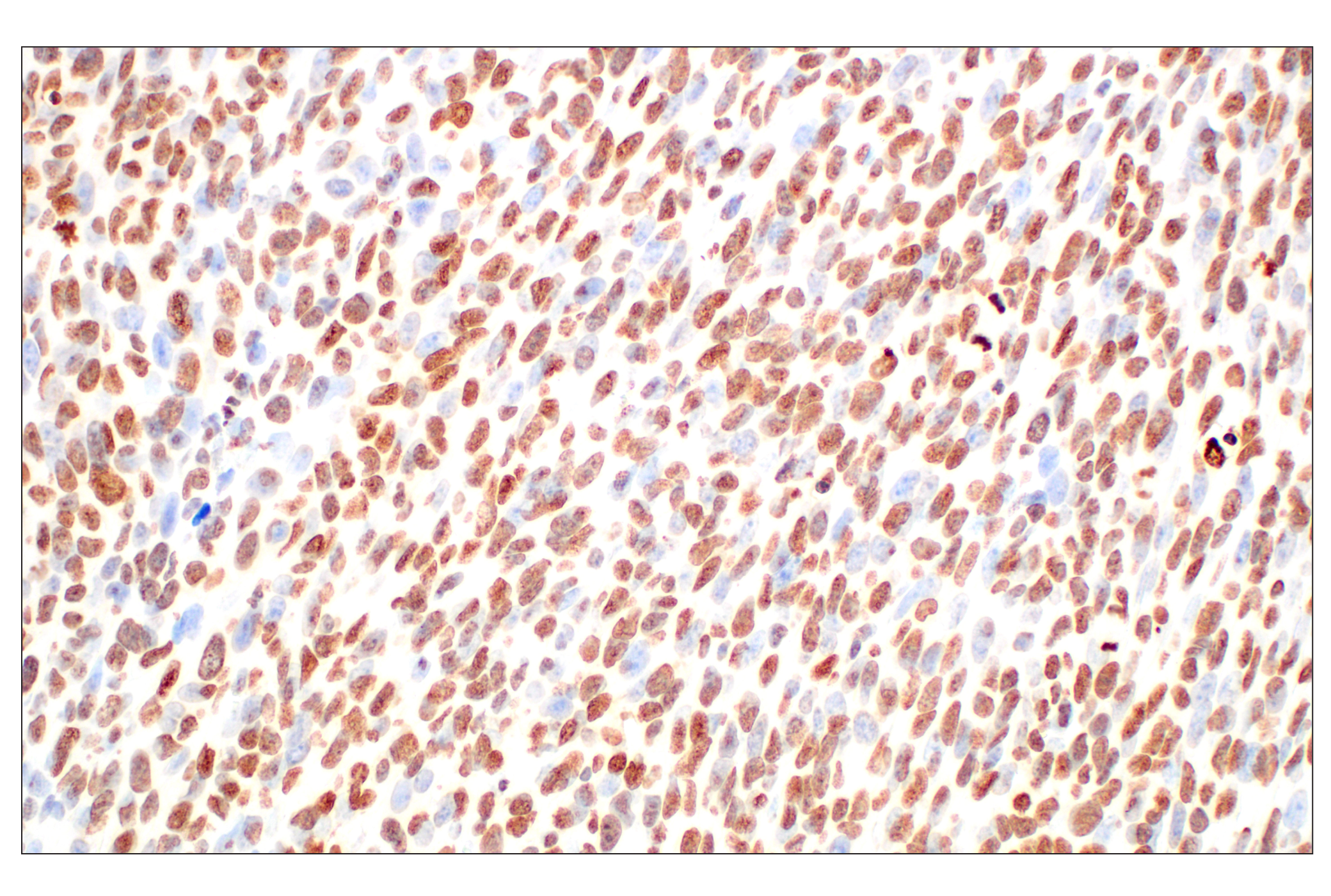
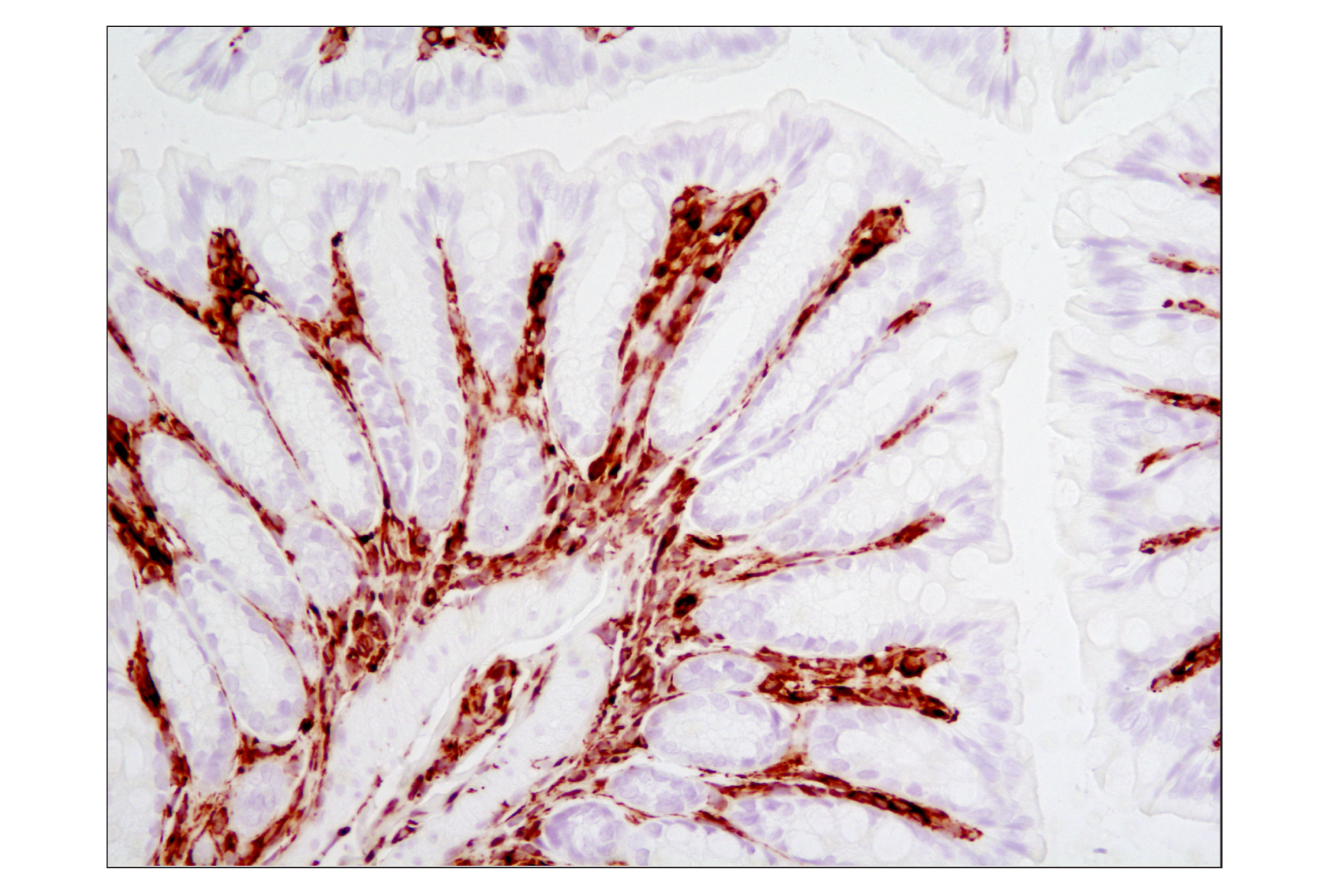
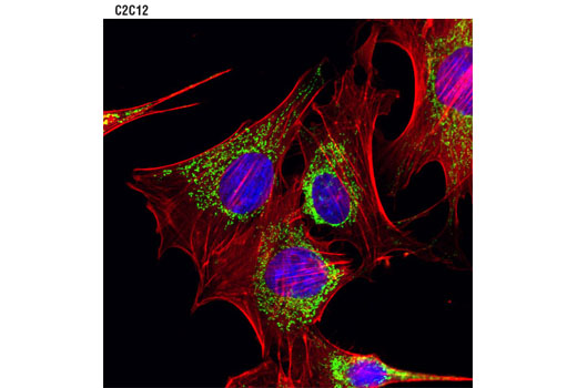
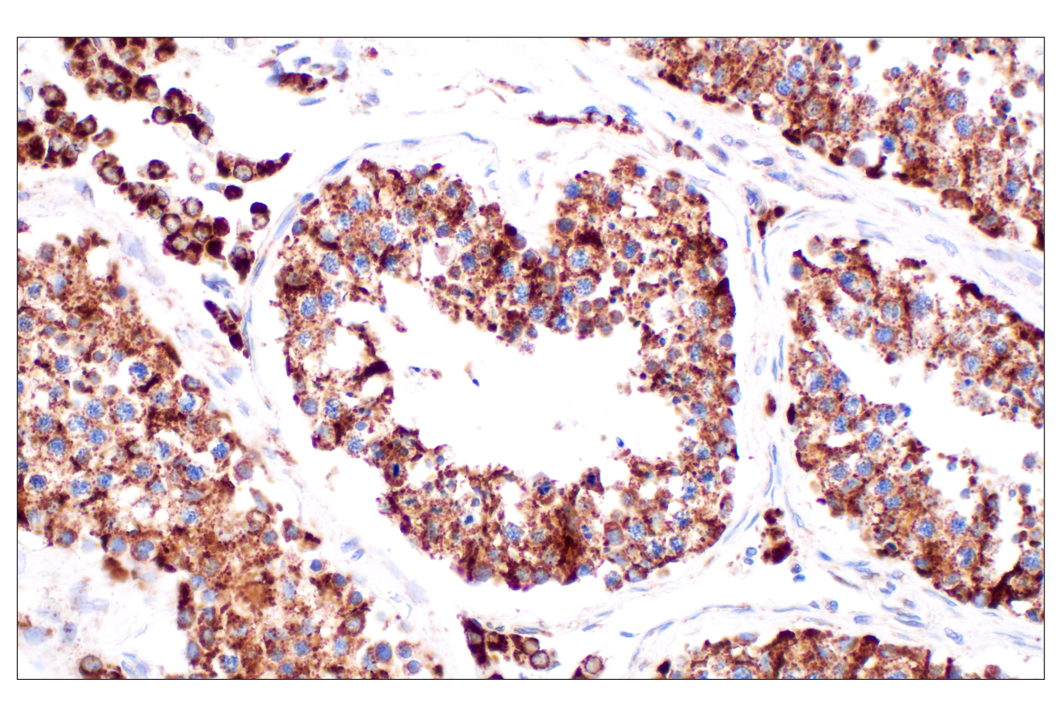
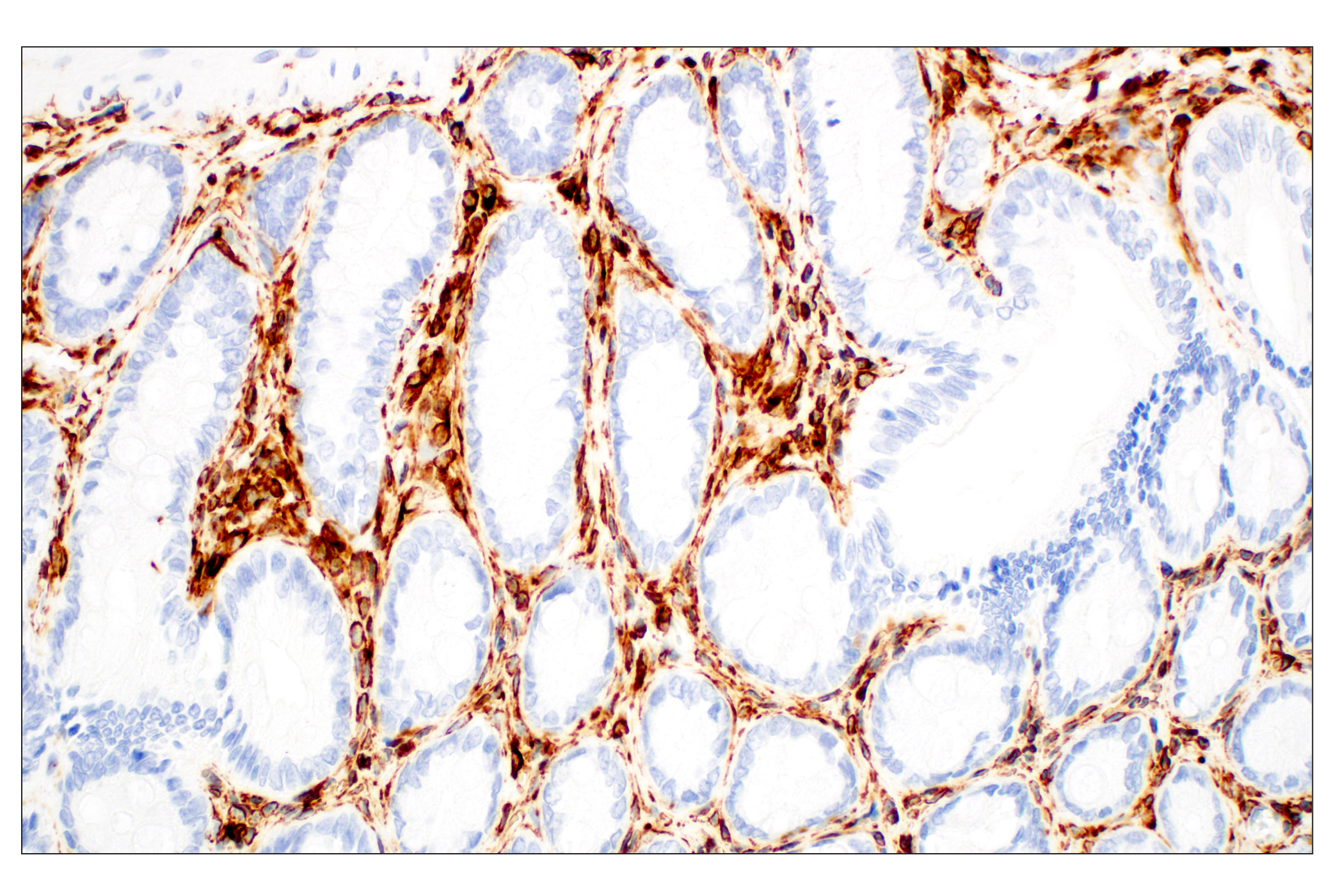
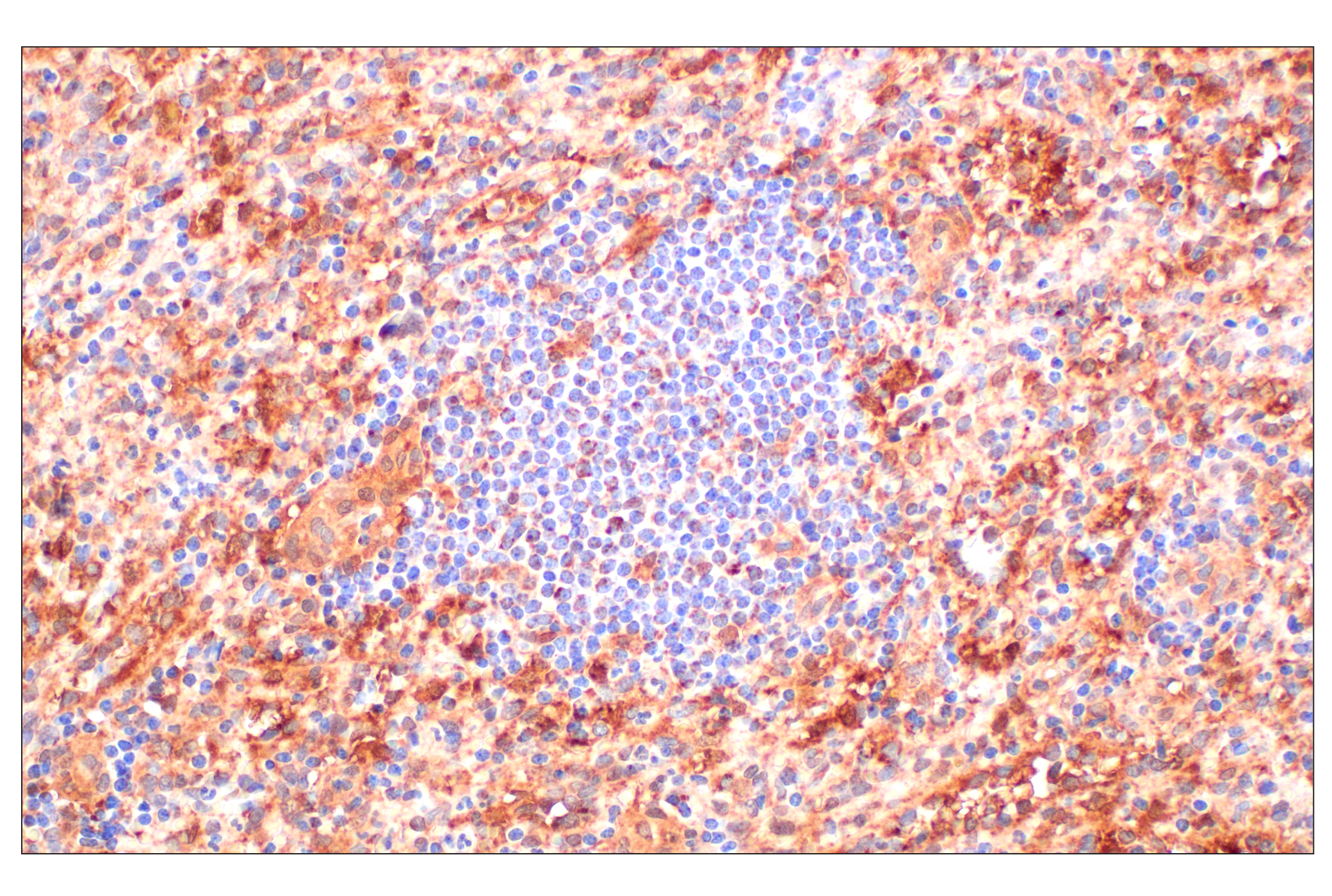
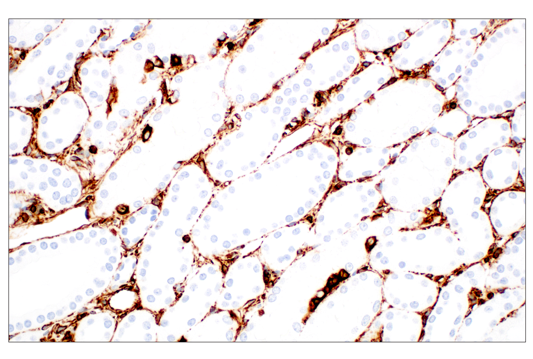
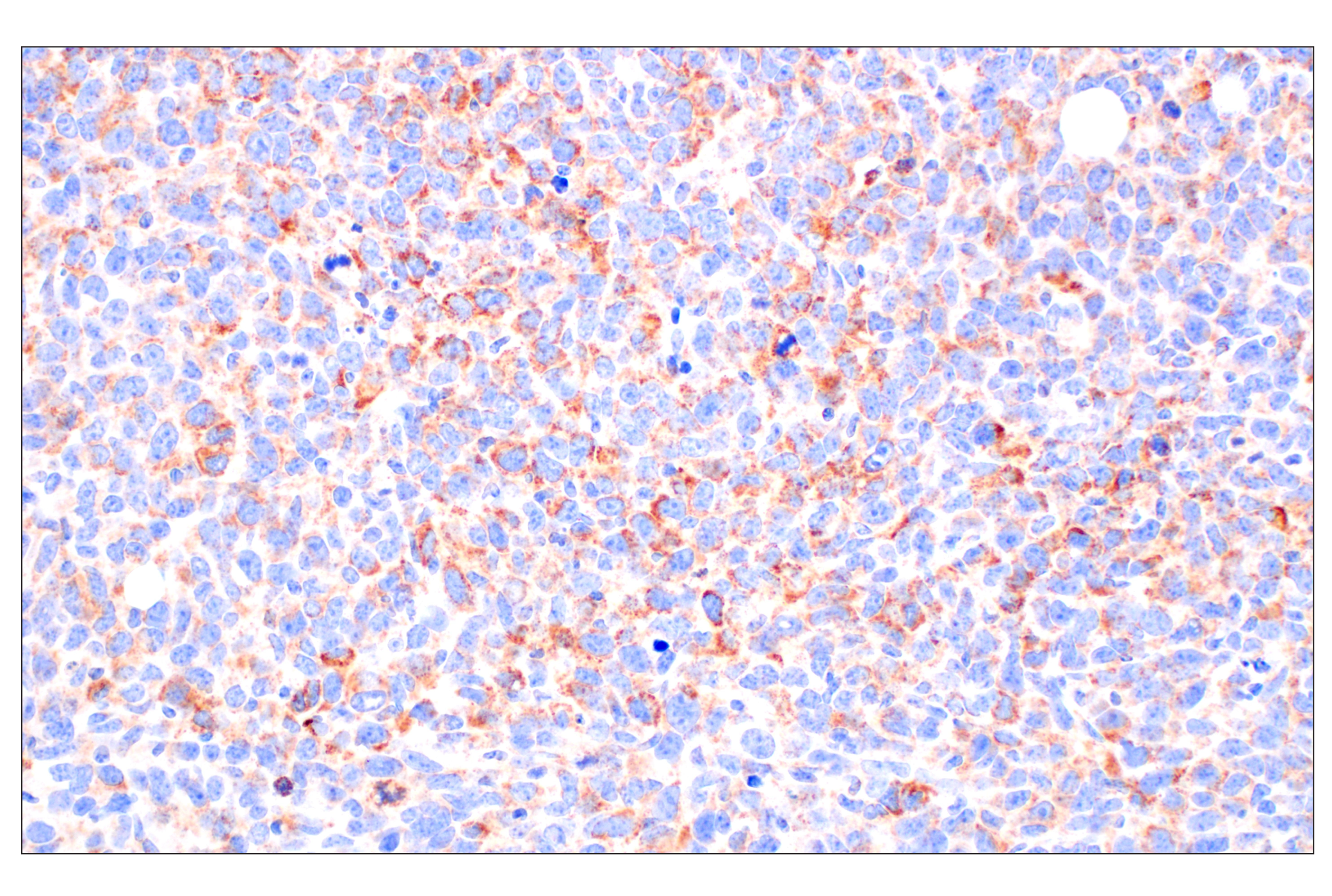
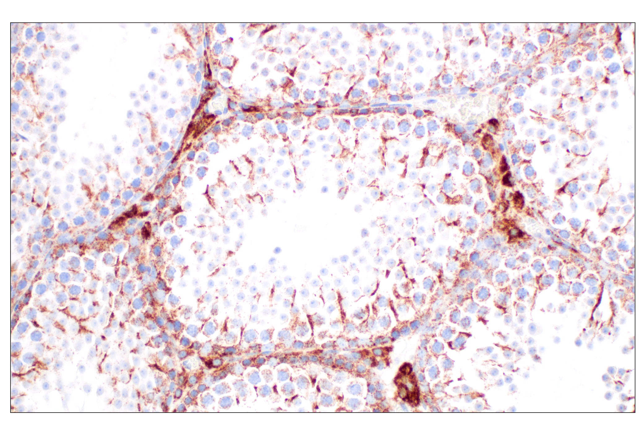
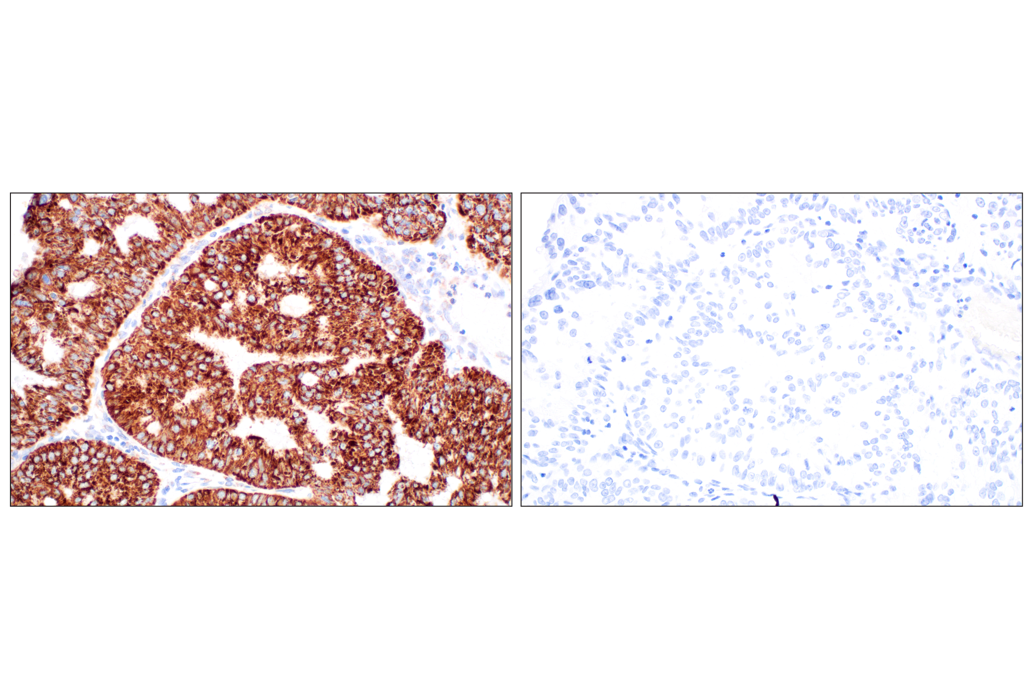
Knowledge of the subcellular location of a protein may reveal the potential role it plays in a variety of cellular processes. Antibodies in the Cell Fractionation Antibody Sampler Kit can be used as a marker to ensure that each subcellular fraction is efficiently separated from the next. MEK1 and MEK2, also called MAPK or Erk Kinases, are dual-specificity protein kinases that function in a mitogen activated protein kinase cascade (1). Apoptosis-inducing factor (AIF) is a ubiquitously expressed flavoprotein that plays a critical role in caspase-independent apoptosis (2). Core histone protein, H3 is one of the primary building blocks of chromatin that makes up the nucleosome (3). Vimentin is a cell-specific intermediate filament with mesenchyme origin that contributes to making up of the cytoskeleton (4). MEK1/2, AIF, histone H3, and vimentin localize to the cytoplasm, mitochondria, nucleus, and cytoskeleton, respectively.
Explore pathways related to this product.
STRING - Known and Predicted Protein-Protein Interactions.
Except as otherwise expressly agreed in a writing signed by a legally authorized representative of CST, the following terms apply to Products provided by CST, its affiliates or its distributors. Any Customer's terms and conditions that are in addition to, or different from, those contained herein, unless separately accepted in writing by a legally authorized representative of CST, are rejected and are of no force or effect.
Products are labeled with For Research Use Only or a similar labeling statement and have not been approved, cleared, or licensed by the FDA or other regulatory foreign or domestic entity, for any purpose. Customer shall not use any Product for any diagnostic or therapeutic purpose, or otherwise in any manner that conflicts with its labeling statement. Products sold or licensed by CST are provided for Customer as the end-user and solely for research and development uses. Any use of Product for diagnostic, prophylactic or therapeutic purposes, or any purchase of Product for resale (alone or as a component) or other commercial purpose, requires a separate license from CST. Customer shall (a) not sell, license, loan, donate or otherwise transfer or make available any Product to any third party, whether alone or in combination with other materials, or use the Products to manufacture any commercial products, (b) not copy, modify, reverse engineer, decompile, disassemble or otherwise attempt to discover the underlying structure or technology of the Products, or use the Products for the purpose of developing any products or services that would compete with CST products or services, (c) not alter or remove from the Products any trademarks, trade names, logos, patent or copyright notices or markings, (d) use the Products solely in accordance with CST Product Terms of Sale and any applicable documentation, and (e) comply with any license, terms of service or similar agreement with respect to any third party products or services used by Customer in connection with the Products.
View in English?