View in English?
View in English?
View in English?

| Cat. # | Size | Qty. | Price | Inventory |
|---|---|---|---|---|
| 9789T | 1 Kit (8 x 20 microliters) |
|
| Product Includes | Quantity | Applications | Reactivity | MW(kDa) | Isotype |
|---|---|---|---|---|---|
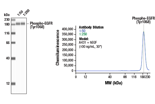
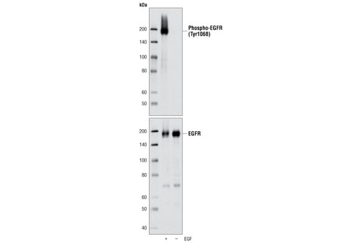
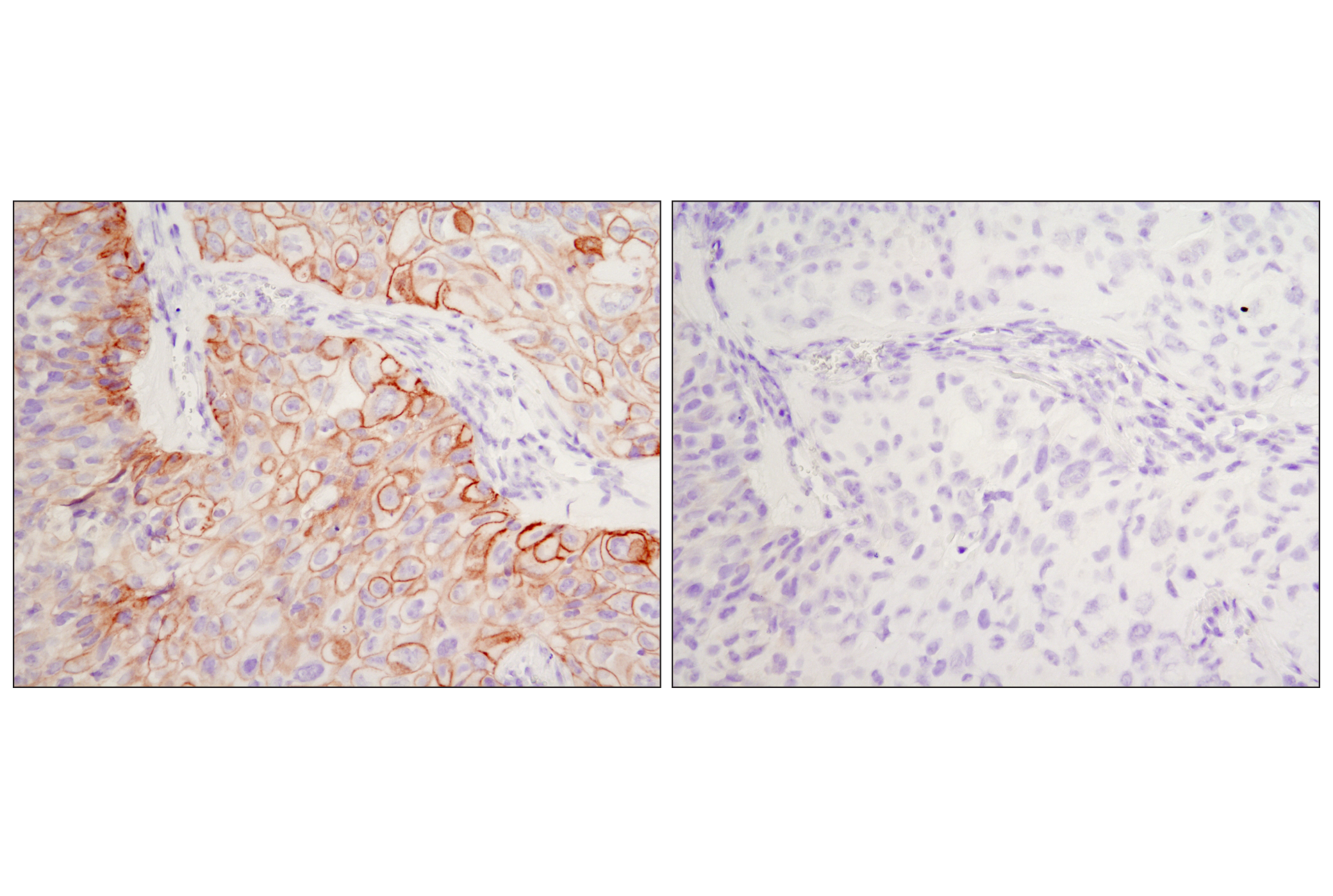
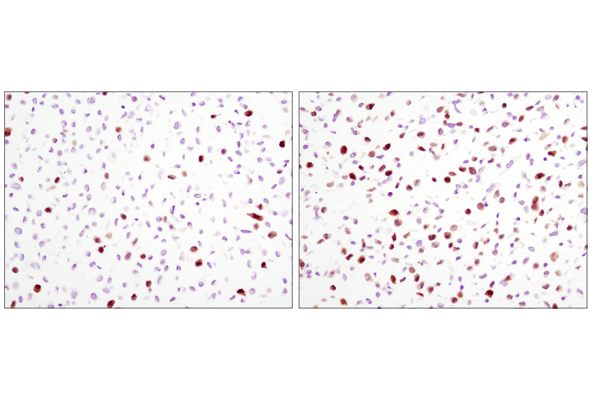
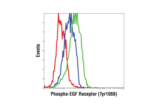
| Phospho-EGF Receptor (Tyr1068) (D7A5) XP® Rabbit mAb 3777 | 20 µl |
|
H M R Mk | 175 | Rabbit IgG |
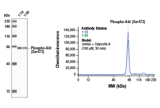
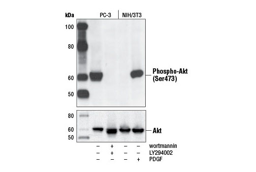
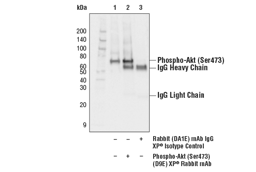
| Phospho-Akt (Ser473) (D9E) XP® Rabbit mAb 4060 | 20 µl |
|
H M R Hm Mk Dm Z B | 60 | Rabbit IgG |
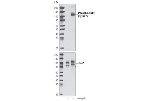
| Phospho-Gab1 (Tyr627) (C32H2) Rabbit mAb 3233 | 20 µl |
|
H | 110 | Rabbit IgG |
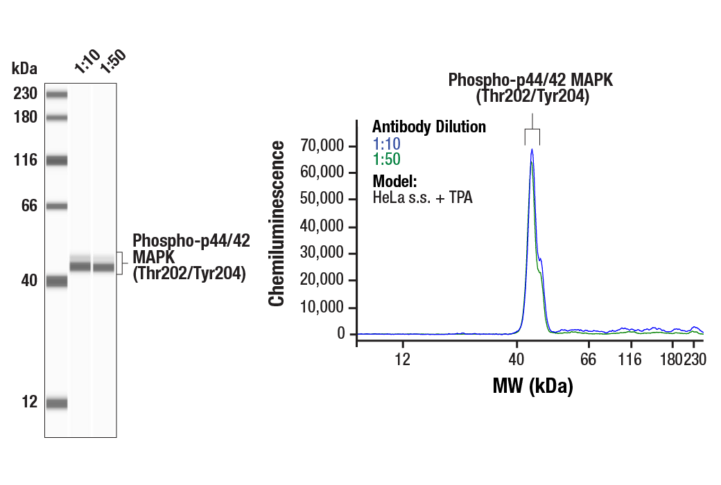
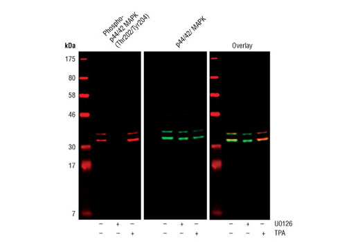
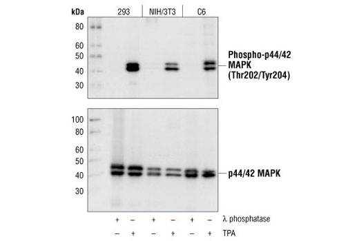
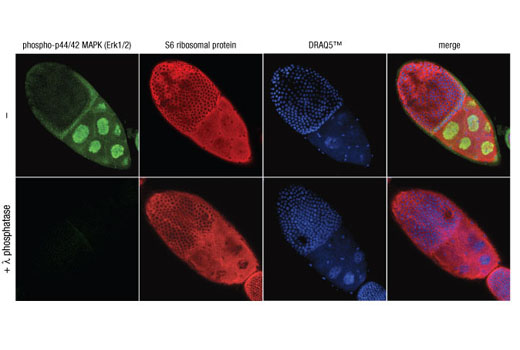
| Phospho-p44/42 MAPK (Erk1/2) (Thr202/Tyr204) (D13.14.4E) XP® Rabbit mAb 4370 | 20 µl |
|
H M R Hm Mk Mi Dm Z B Dg Pg Sc | 44, 42 | Rabbit IgG |
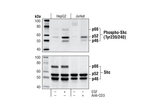
| Phospho-Shc (Tyr239/240) Antibody 2434 | 20 µl |
|
H M R | 50, 55, 70 | Rabbit |
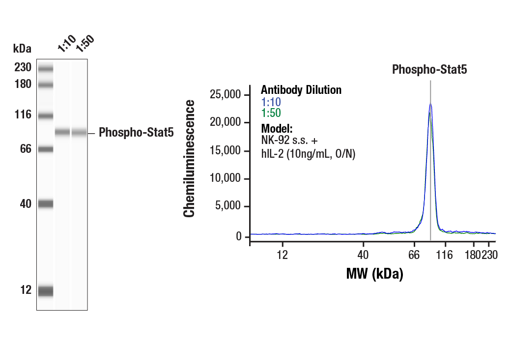
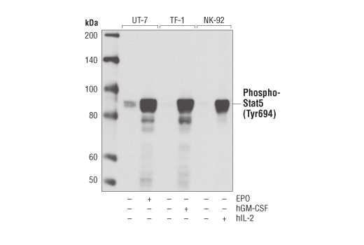
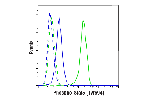
| Phospho-Stat5 (Tyr694) (D47E7) XP® Rabbit mAb 4322 | 20 µl |
|
H M | 90 | Rabbit IgG |
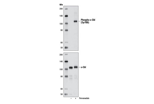
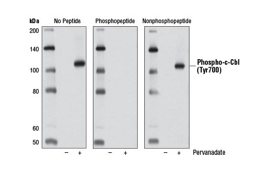
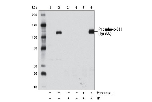
| Phospho-c-Cbl (Tyr700) (D16D7) Rabbit mAb 8869 | 20 µl |
|
H | 120 | Rabbit IgG |
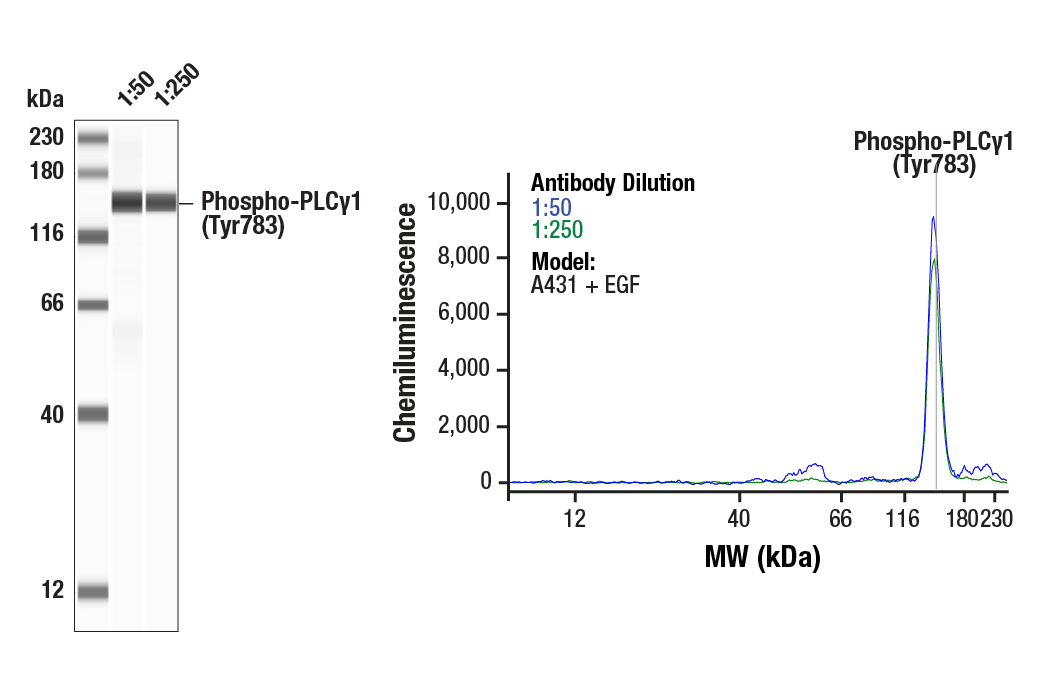
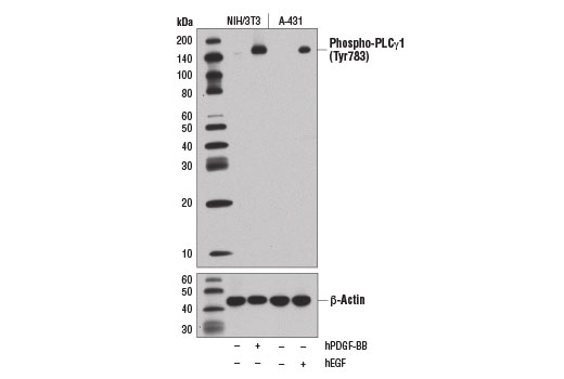
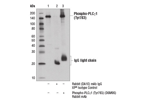
| Phospho-PLCγ1 (Tyr783) (D6M9S) Rabbit mAb 14008 | 20 µl |
|
H M | 155 | Rabbit IgG |
| Anti-rabbit IgG, HRP-linked Antibody 7074 | 100 µl |
|
Goat |
Product Information
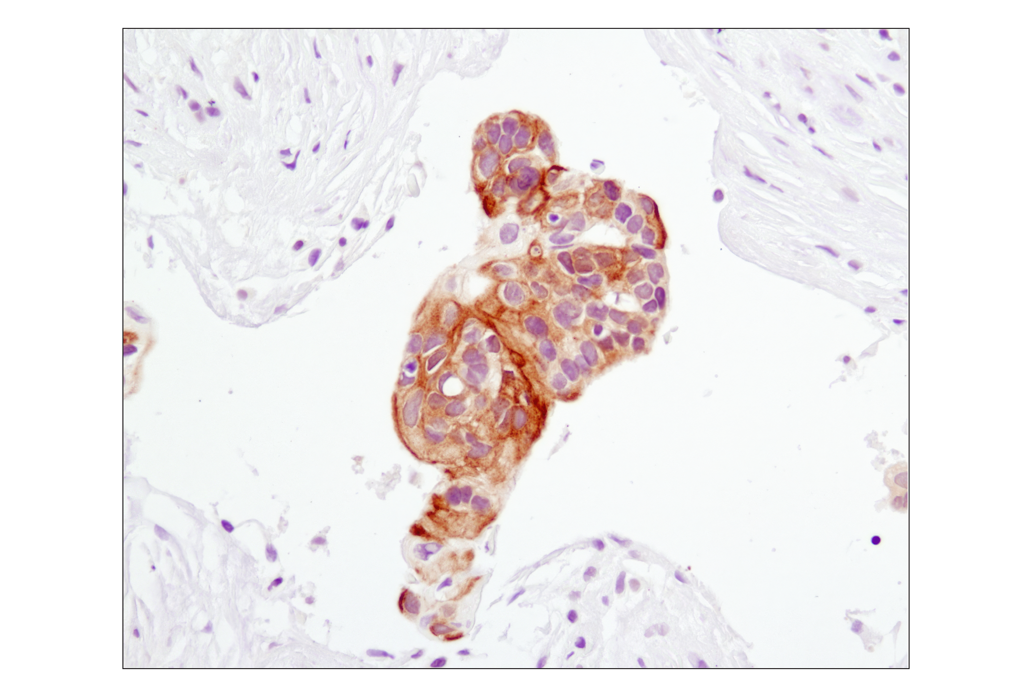
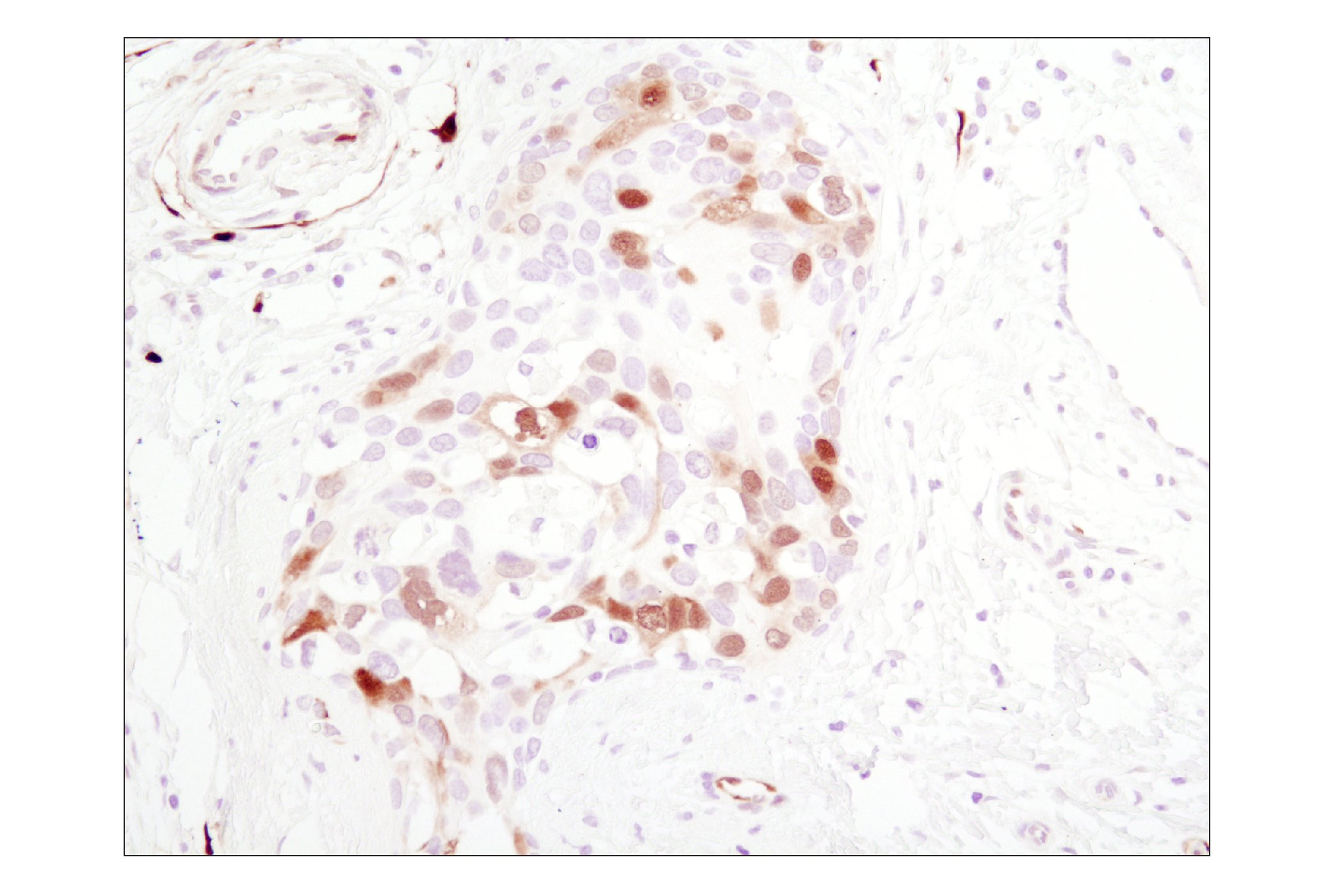
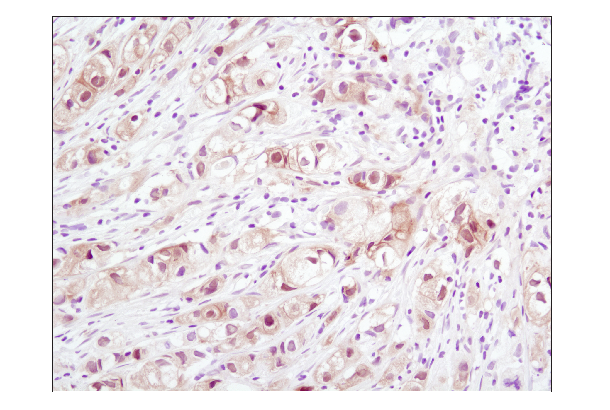
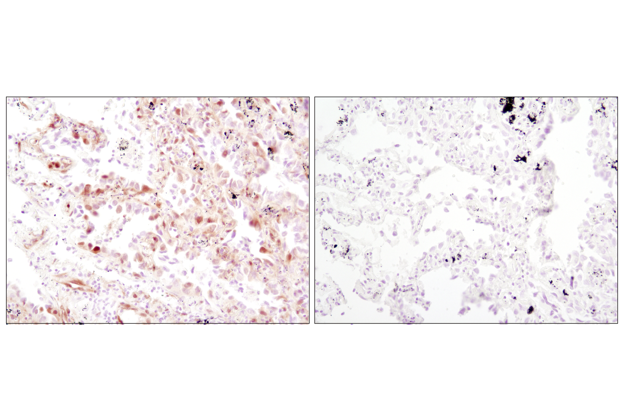
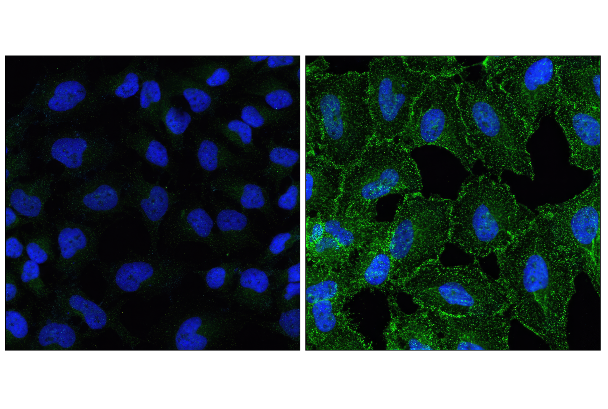
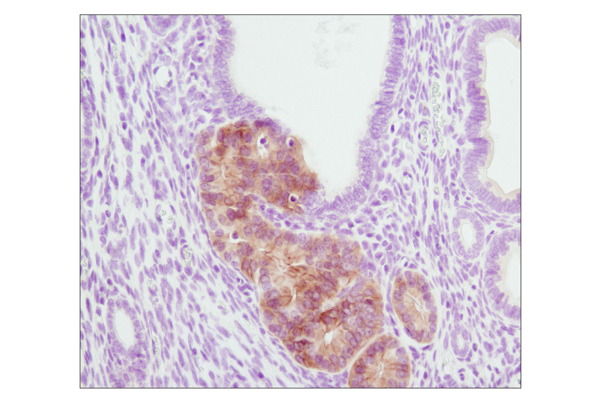
Activation state polyclonal antibodies are produced by immunizing animals with synthetic phosphopeptides corresponding to residues surrounding Tyr239/240 of human Shc. Polyclonal antibodies are purified by protein A and peptide affinity chromatography. Rabbit monoclonal antibodies are produced by immunizing animals with synthetic phosphopeptides corresponding to residues surrounding Tyr700 of c-Cbl, Tyr1068 of human EGF receptor, Tyr694 of Stat5a, Tyr627 of human Gab1, Ser473 of human Akt, Thr202/Tyr204 of human p44 MAP kinase, or Tyr783 of human PLCγ1 protein.
The epidermal growth factor (EGF) receptor is a transmembrane tyrosine kinase that belongs to the HER/ErbB protein family. Ligand binding results in receptor dimerization, autophosphorylation, activation of downstream signaling, internalization, and lysosomal degradation (1,2). Phosphorylation of EGF receptor (EGFR) at Tyr845 in the kinase domain is implicated in stabilizing the activation loop, maintaining the active state enzyme, and providing a binding surface for substrate proteins (3,4). c-Src is involved in phosphorylation of EGFR at Tyr845 (5). The SH2 domain of PLCγ binds at phospho-Tyr992, resulting in activation of PLCγ-mediated downstream signaling (6). Phosphorylation of EGFR at Tyr1045 creates a major docking site for the adaptor protein c-Cbl, leading to receptor ubiquitination and degradation following EGFR activation (7,8). The GRB2 adaptor protein binds activated EGFR at phospho-Tyr1068 (9). A pair of phosphorylated EGFR residues (Tyr1148 and Tyr1173) provide a docking site for the Shc scaffold protein, with both sites involved in MAP kinase signaling activation (2). Phosphorylation of EGFR at specific serine and threonine residues attenuates EGFR kinase activity. EGFR carboxy-terminal residues Ser1046 and Ser1047 are phosphorylated by CaM kinase II; mutation of either of these serines results in upregulated EGFR tyrosine autophosphorylation (10).
Explore pathways related to this product.
STRING - Known and Predicted Protein-Protein Interactions.
Except as otherwise expressly agreed in a writing signed by a legally authorized representative of CST, the following terms apply to Products provided by CST, its affiliates or its distributors. Any Customer's terms and conditions that are in addition to, or different from, those contained herein, unless separately accepted in writing by a legally authorized representative of CST, are rejected and are of no force or effect.
Products are labeled with For Research Use Only or a similar labeling statement and have not been approved, cleared, or licensed by the FDA or other regulatory foreign or domestic entity, for any purpose. Customer shall not use any Product for any diagnostic or therapeutic purpose, or otherwise in any manner that conflicts with its labeling statement. Products sold or licensed by CST are provided for Customer as the end-user and solely for research and development uses. Any use of Product for diagnostic, prophylactic or therapeutic purposes, or any purchase of Product for resale (alone or as a component) or other commercial purpose, requires a separate license from CST. Customer shall (a) not sell, license, loan, donate or otherwise transfer or make available any Product to any third party, whether alone or in combination with other materials, or use the Products to manufacture any commercial products, (b) not copy, modify, reverse engineer, decompile, disassemble or otherwise attempt to discover the underlying structure or technology of the Products, or use the Products for the purpose of developing any products or services that would compete with CST products or services, (c) not alter or remove from the Products any trademarks, trade names, logos, patent or copyright notices or markings, (d) use the Products solely in accordance with CST Product Terms of Sale and any applicable documentation, and (e) comply with any license, terms of service or similar agreement with respect to any third party products or services used by Customer in connection with the Products.
View in English?