View in English?
View in English?
View in English?

| Cat. # | Size | Qty. | Price | Inventory |
|---|---|---|---|---|
| 4487T | 1 Kit (5 x 20 microliters) |
|
| Product Includes | Quantity | Applications | Reactivity | MW(kDa) | Isotype |
|---|---|---|---|---|---|
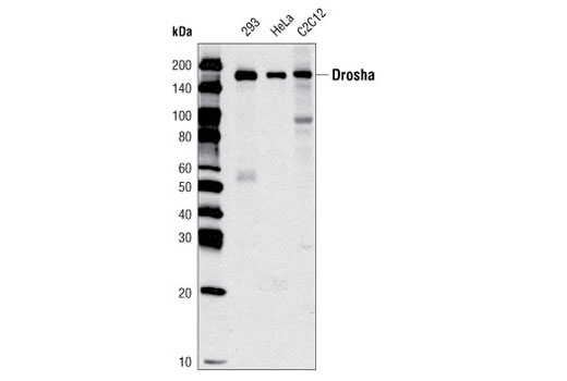
| Drosha (D28B1) Rabbit mAb 3364 | 20 µl |
|
H M | 160 | Rabbit IgG |
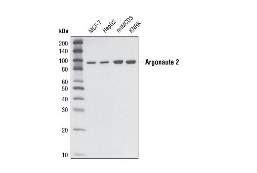
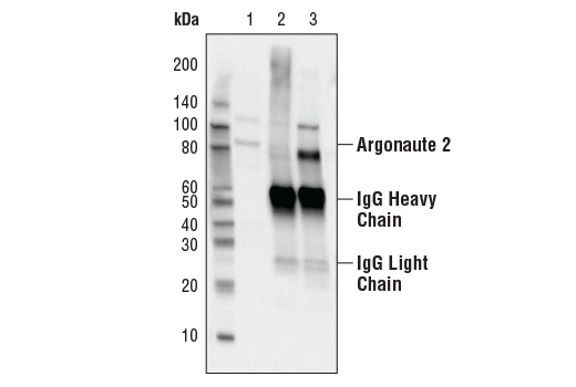
| Argonaute 2 (C34C6) Rabbit mAb 2897 | 20 µl |
|
H M R Mk | 97 | Rabbit IgG |
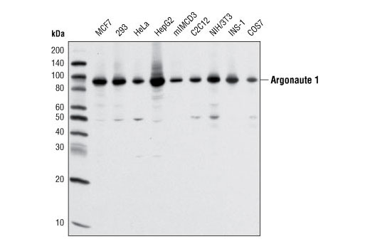
| Argonaute 1 (D84G10) XP® Rabbit mAb 5053 | 20 µl |
|
H M R Mk | 97 | Rabbit IgG |
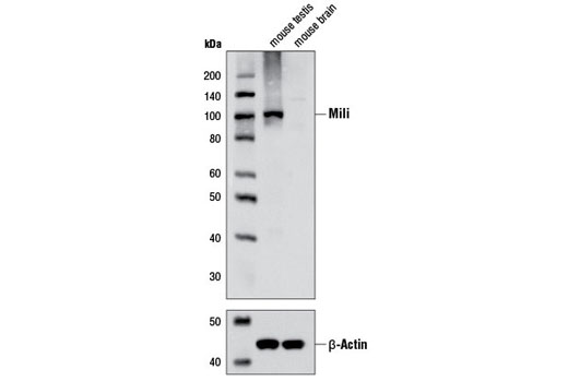
| Mili (D14F5) XP® Rabbit mAb 5940 | 20 µl |
|
M | 110 | Rabbit IgG |
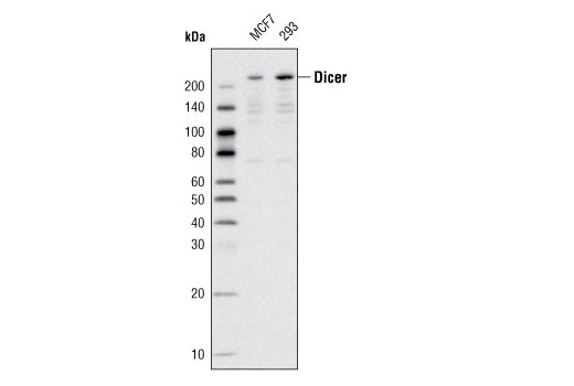
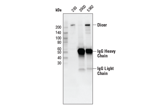
| Dicer (D38E7) Rabbit mAb 5362 | 20 µl |
|
H | 220 | Rabbit IgG |
| Anti-rabbit IgG, HRP-linked Antibody 7074 | 100 µl |
|
Goat |
Product Information
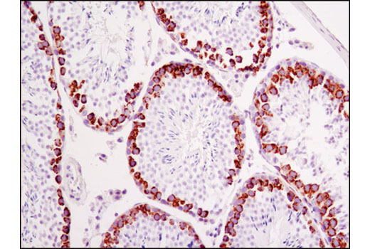
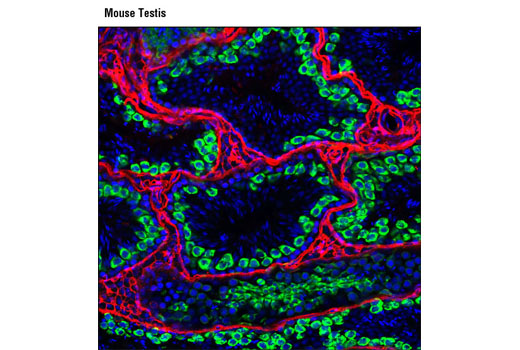
Polyclonal antibodies are produced by immunizing animals with synthetic peptides corresponding to the residues surrounding Ala1123 of human Dicer or residues surrounding Leu118 of mouse Mili protein. Polyclonal antibodies are purified by protein A and peptide affinity chromatography. Monoclonal antibody is produced by immunizing animals with synthetic peptides corresponding to the sequence of mouse Argonaute 2 or around Gly953 of human Drosha proteins.
RNA interference (RNAi) serves as a global mechanism of gene regulation in eukaryotes. Through interactions with Dicer, Drosha, Argonaute 2 (Ago2) and Miwi/Mili proteins, microRNA (miRNA) is processed within the nucleus and utilized for gene silencing and down regulation of gene expression. Dicer is a member of the RNase III family that specifically cleaves double-stranded RNA to generate microRNA (miRNA) (1). Long, primary transcripts (pri-miRNAs) are processed to stem-looped pre-miRNAs by the nuclear RNase III Drosha (2) and are then transported to the cytoplasm for further processing by Dicer to produce mature, 22-nucleotide miRNAs (3). The mature miRNA then becomes a part of the RNA-Induced Silencing Complex (RISC) and can bind to the 3' UTR of the target mRNA (3). Interference of Drosha pri-miRNA processing results in the increase of pri-miRNAs and the decrease of pre-miRNAs (2). Drosha forms part of a multiprotein complex called the Microprocessor along with other components, such as DGCR8 (4). Both Drosha and DGCR8 are necessary for miRNA biogenesis (5). Argonaute protein family members participate in various steps of miRNA-mediated gene silencing such as repression of translation and mRNA turnover (6). The Drosophila piwi gene was identified as being required for the self-renewal of germ-line stem cells, and its homologues are well conserved among various species including Arabidopsis, C. elegans and human (7). Miwi and Mili proteins are both mouse homologs of Piwi and contain a carboxy-terminal Piwi domain that binds to Piwi-interacting RNAs (piRNAs) in male germ cells and are essential for spermatogenesis in mouse (8-11).
Except as otherwise expressly agreed in a writing signed by a legally authorized representative of CST, the following terms apply to Products provided by CST, its affiliates or its distributors. Any Customer's terms and conditions that are in addition to, or different from, those contained herein, unless separately accepted in writing by a legally authorized representative of CST, are rejected and are of no force or effect.
Products are labeled with For Research Use Only or a similar labeling statement and have not been approved, cleared, or licensed by the FDA or other regulatory foreign or domestic entity, for any purpose. Customer shall not use any Product for any diagnostic or therapeutic purpose, or otherwise in any manner that conflicts with its labeling statement. Products sold or licensed by CST are provided for Customer as the end-user and solely for research and development uses. Any use of Product for diagnostic, prophylactic or therapeutic purposes, or any purchase of Product for resale (alone or as a component) or other commercial purpose, requires a separate license from CST. Customer shall (a) not sell, license, loan, donate or otherwise transfer or make available any Product to any third party, whether alone or in combination with other materials, or use the Products to manufacture any commercial products, (b) not copy, modify, reverse engineer, decompile, disassemble or otherwise attempt to discover the underlying structure or technology of the Products, or use the Products for the purpose of developing any products or services that would compete with CST products or services, (c) not alter or remove from the Products any trademarks, trade names, logos, patent or copyright notices or markings, (d) use the Products solely in accordance with CST Product Terms of Sale and any applicable documentation, and (e) comply with any license, terms of service or similar agreement with respect to any third party products or services used by Customer in connection with the Products.
View in English?