View in English?
View in English?
View in English?

| Cat. # | Size | Qty. | Price | Inventory |
|---|---|---|---|---|
| 47767T | 1 Kit (9 x 20 microliters) |
|
| Product Includes | Quantity | Applications | Reactivity | MW(kDa) | Isotype |
|---|---|---|---|---|---|
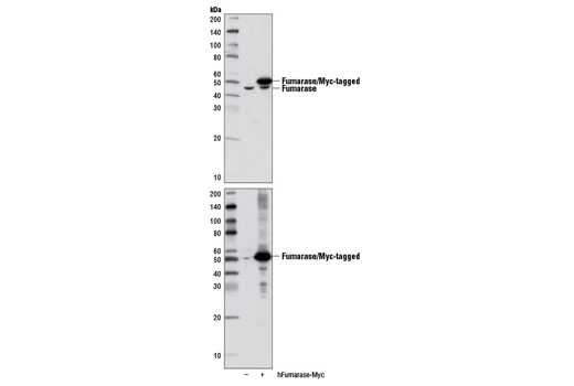
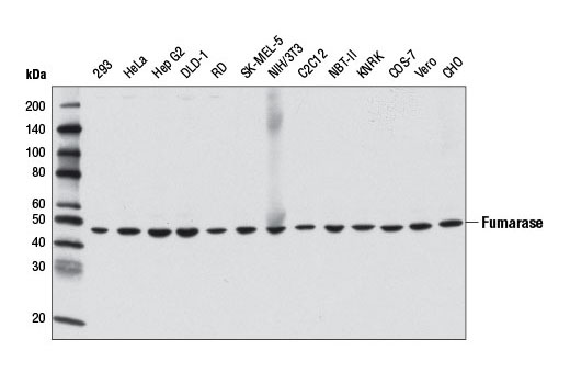
| Fumarase (D9C5) Rabbit mAb 4567 | 20 µl |
|
H M R Hm Mk | 49 | Rabbit IgG |
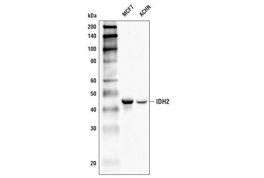
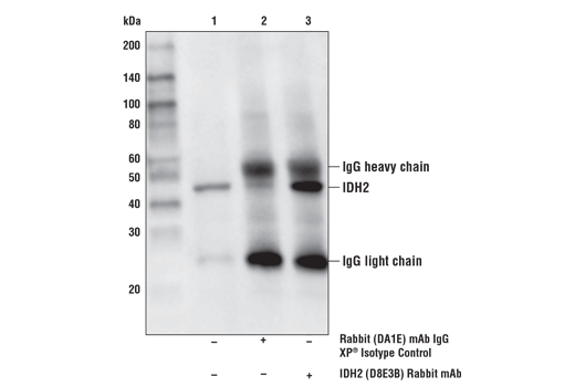
| IDH2 (D8E3B) Rabbit mAb 56439 | 20 µl |
|
H M R | 43 | Rabbit IgG |
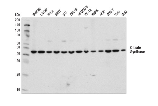
| Citrate Synthase (D7V8B) Rabbit mAb 14309 | 20 µl |
|
H M R Hm Mk | 45 | Rabbit IgG |
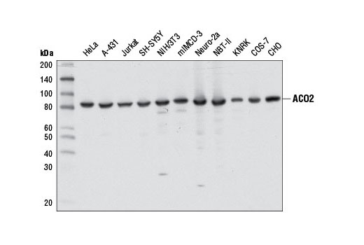
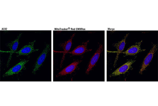
| ACO2 (D6D9) XP® Rabbit mAb 6571 | 20 µl |
|
H M R Hm Mk | 85 | Rabbit IgG |
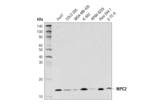
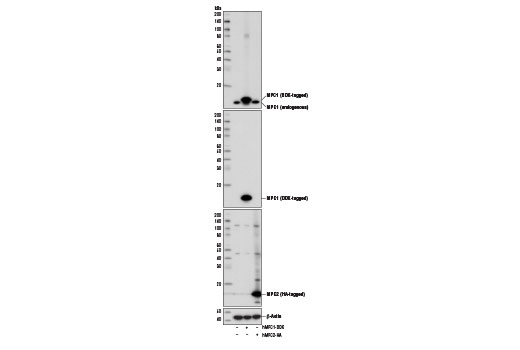
| MPC2 (D4I7G) Rabbit mAb 46141 | 20 µl |
|
H M R Mk | 14 | Rabbit IgG |
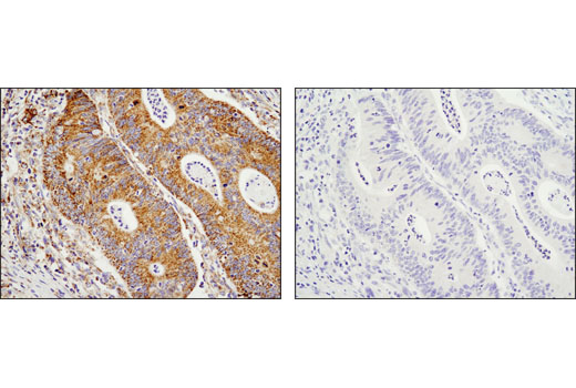
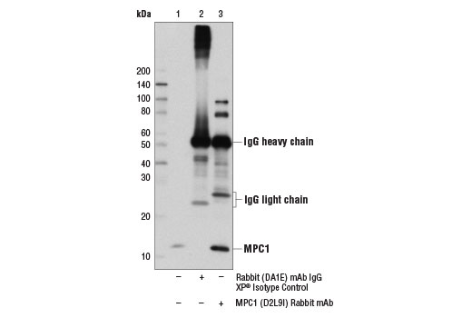
| MPC1 (D2L9I) Rabbit mAb 14462 | 20 µl |
|
H M R Mk | 12 | Rabbit IgG |
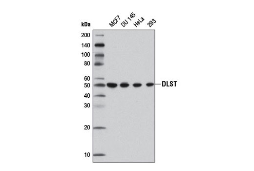
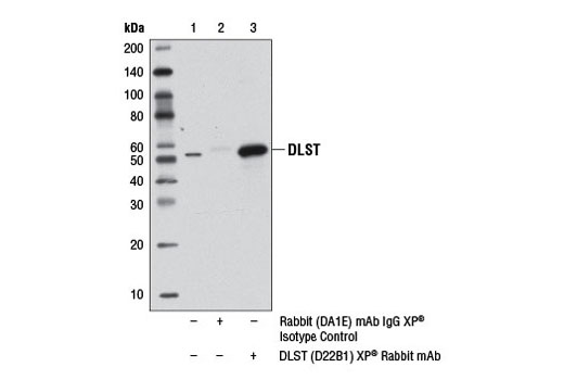
| DLST (D22B1) XP® Rabbit mAb 11954 | 20 µl |
|
H | 50 | Rabbit IgG |
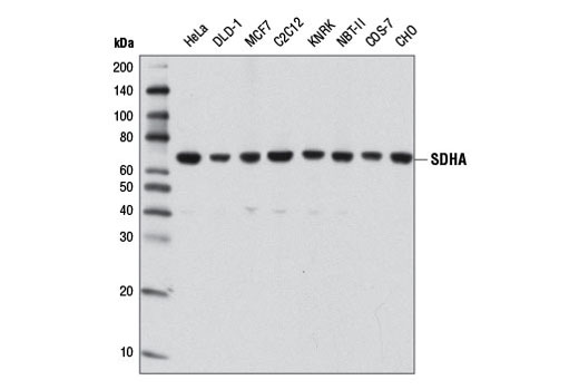
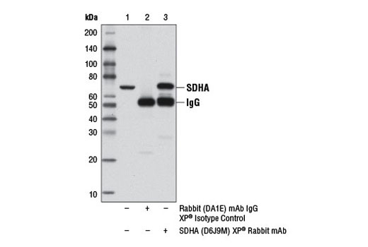
| SDHA (D6J9M) XP® Rabbit mAb 11998 | 20 µl |
|
H M R Hm Mk | 70 | Rabbit IgG |
| Anti-rabbit IgG, HRP-linked Antibody 7074 | 100 µl |
|
Rab | Goat | |
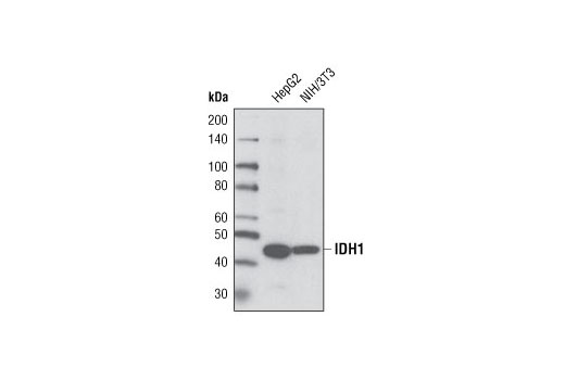
| IDH1 Antibody 3997 | 20 µl |
|
H M | 46 | Rabbit |
Product Information
Monoclonal antibodies are produced by immunizing animals with a synthetic peptide corresponding to residues surrounding Gly540 of human ACO2 protein, residues near the carboxy terminus of human citrate synthase protein, residues surrounding Pro188 of human DLST protein, residues surrounding Gly354 of human fumarase protein, residues surrounding Arg222 of human IDH1 protein, residues surrounding Val195 of human IDH2 protein, residues near the carboxy terminus of human MPC1 protein, residues surrounding Asn33 of human MPC2 protein and residues surrounding Gly166 of human SDHA protein, respectively.
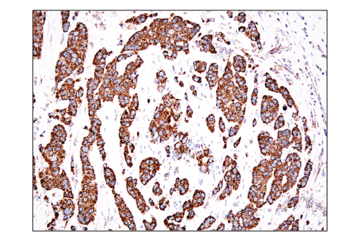
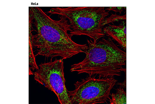
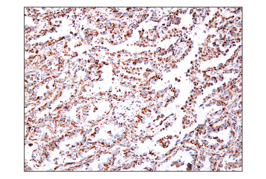
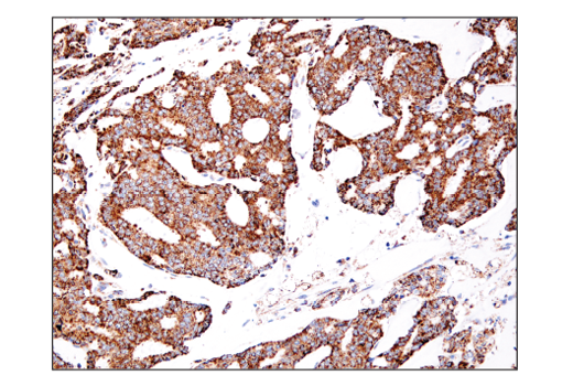
The tricarboxylic acid (TCA) cycle includes various enzymatic reactions that constitute a key part of cellular aerobic respiration. The transport of the glycolytic end product pyruvate into mitochondria and the decarboxylation of pyruvate in the TCA cycle generate energy through oxidative phosphorylation under aerobic conditions (1,2). Two inner mitochondrial membrane proteins, mitochondrial pyruvate carrier 1 (MPC1) and mitochondrial pyruvate carrier 2 (MPC2), form a 150 kDa complex and are essential proteins in the facilitated transport of pyruvate into mitochondria (1,2). Citrate synthase catalyzes the first and rate-limiting reaction of the TCA cycle (3). Mitochondrial aconitase 2 (ACO2) catalyzes the conversion of citrate to isocitrate via cis-aconitate (4). IDH1 and IDH2 are two of the three isocitrate dehydrogenases that catalyze oxidative decarboxylation of isocitrate to α-ketoglutarate (α-KG) (5). IDH1 functions as a tumor suppressor in the cytoplasm and peroxisomes, whereas IDH2 is in mitochondria and is involved in the TCA cycle (5). Mutations in IDH2 have also been identified in malignant gliomas (6). Dihydrolipoamide succinyltransferase (DLST) is a subunit of the α-ketoglutarate dehydrogenase complex, a key enzymatic complex in the TCA cycle (7). Succinate dehydrogenase subunit A (SDHA) is a component of the TCA cycle and the electron transport chain and is involved in the oxidation of succinate (8). Fumarase catalyzes the conversion of fumarate to malate (9). Fumarase deficiency leads to the accumulation of fumarate, an oncometabolite that has been shown to promote epithelial-to-mesenchymal-transition (EMT), a developmental process that has been implicated in oncogenesis (10).
Except as otherwise expressly agreed in a writing signed by a legally authorized representative of CST, the following terms apply to Products provided by CST, its affiliates or its distributors. Any Customer's terms and conditions that are in addition to, or different from, those contained herein, unless separately accepted in writing by a legally authorized representative of CST, are rejected and are of no force or effect.
Products are labeled with For Research Use Only or a similar labeling statement and have not been approved, cleared, or licensed by the FDA or other regulatory foreign or domestic entity, for any purpose. Customer shall not use any Product for any diagnostic or therapeutic purpose, or otherwise in any manner that conflicts with its labeling statement. Products sold or licensed by CST are provided for Customer as the end-user and solely for research and development uses. Any use of Product for diagnostic, prophylactic or therapeutic purposes, or any purchase of Product for resale (alone or as a component) or other commercial purpose, requires a separate license from CST. Customer shall (a) not sell, license, loan, donate or otherwise transfer or make available any Product to any third party, whether alone or in combination with other materials, or use the Products to manufacture any commercial products, (b) not copy, modify, reverse engineer, decompile, disassemble or otherwise attempt to discover the underlying structure or technology of the Products, or use the Products for the purpose of developing any products or services that would compete with CST products or services, (c) not alter or remove from the Products any trademarks, trade names, logos, patent or copyright notices or markings, (d) use the Products solely in accordance with CST Product Terms of Sale and any applicable documentation, and (e) comply with any license, terms of service or similar agreement with respect to any third party products or services used by Customer in connection with the Products.
View in English?