注目情報はこちら >>
93702
Mouse Anti-Rabbit IgG (Light-Chain Specific) (D4W3E) mAb (HRP Conjugate)
Secondary Antibodies
Secondary Antibody

Mouse Anti-Rabbit IgG (Light-Chain Specific) (D4W3E) mAb (HRP Conjugate) #93702

Citations (74)
Filter:
  1. WB
Immunoprecipitation of β-catenin from HeLa cell extracts. Lane 1 is 10% input, lane 2 is Rabbit (DA1E) mAb IgG XP® Isotype Control (Sepharose® Bead Conjugate) #3423, and lane 3 is β-Catenin (D10A8) XP® Rabbit mAb (Sepharose® Bead Conjugate) #12475. Western blot analysis was performed using β-Catenin (D10A8) XP® Rabbit mAb #8480 as the primary antibody and Mouse Anti-Rabbit IgG (Light-Chain Specific) (D4W3E) mAb (HRP Conjugate) as the secondary antibody.
To Purchase # 93702
Cat. # Size Qty. Price Inventory
93702S
100 µl

Supporting Data

REACTIVITY Rab
SENSITIVITY Endogenous
MW (kDa)
Source/Isotype Mouse 

Application Key:

  • WB-Western Blot
  • IP-Immunoprecipitation
  • IHC-Immunohistochemistry
  • ChIP-Chromatin Immunoprecipitation
  • C&R-CUT&RUN
  • C&T-CUT&Tag
  • DB-Dot Blot
  • eCLIP-eCLIP
  • IF-Immunofluorescence
  • F-Flow Cytometry

Species Cross-Reactivity Key:

  • H-Human
  • M-Mouse
  • R-Rat
  • Hm-Hamster
  • Mk-Monkey
  • Vir-Virus
  • Mi-Mink
  • C-Chicken
  • Dm-D. melanogaster
  • X-Xenopus
  • Z-Zebrafish
  • B-Bovine
  • Dg-Dog
  • Pg-Pig
  • Sc-S. cerevisiae
  • Ce-C. elegans
  • Hr-Horse
  • GP-Guinea Pig
  • Rab-Rabbit
  • All-All Species Expected

Product Description

This Cell Signaling Technology antibody is conjugated to the carbohydrate groups of horseradish peroxidase (HRP) via its amine groups.

Product Usage Information

Recommended Antibody Dilutions:

1:1000-1:3000

Storage

Supplied in 136 mM NaCl, 2.6 mM KCl, 12 mM sodium phosphate (pH 7.4) dibasic, 2 mg/ml BSA, and 50% glycerol. Store at –20°C. Do not aliquot the antibody.

Protocol

PRINT

View >Collapse >

Western Immunoblotting Protocol (for Light-Chain Specific Secondary Antibodies)

For western blots, incubate membrane with diluted primary antibody in either 5% w/v BSA or nonfat dry milk, 1X TBS, 0.1% Tween-20 at 4°C with gentle shaking, overnight.

NOTE: Please refer to primary antibody product webpage for recommended Primary Antibody Dilution Buffer and recommended antibody dilution.

A. Solutions and Reagents

NOTE: Prepare solutions with reverse osmosis deionized (RODI) or equivalent grade water.

  1. 20X Phosphate Buffered Saline (PBS): (#9808) To prepare 1 L 1X PBS: add 50 ml 20X PBS to 950 ml dH2O, mix.
  2. 10X Tris Buffered Saline (TBS): (#12498) To prepare 1 L 1X TBS: add 100 ml 10X to 900 ml dH2O, mix.
  3. 1X SDS Sample Buffer: Blue Loading Pack (#7722) or Red Loading Pack (#7723) Prepare fresh 3X reducing loading buffer by adding 1/10 volume 30X DTT to 1 volume of 3X SDS loading buffer. Dilute to 1X with dH2O.
  4. 10X Tris-Glycine SDS Running Buffer: (#4050) To prepare 1 L 1X running buffer: add 100 ml 10X running buffer to 900 ml dH2O, mix.
  5. 10X Tris-Glycine Transfer Buffer: (#12539) To prepare 1 L 1X Transfer Buffer: add 100 ml 10X Transfer Buffer to 200 ml methanol + 700 ml dH2O, mix.
  6. 10X Tris Buffered Saline with Tween® 20 (TBST): (#9997) To prepare 1 L 1X TBST: add 100 ml 10X TBST to 900 ml dH2O, mix.
  7. Nonfat Dry Milk: (#9999).
  8. Blocking Buffer: 1X TBST with 5% w/v nonfat dry milk; for 150 ml, add 7.5 g nonfat dry milk to 150 ml 1X TBST and mix well.
  9. Wash Buffer: (#9997) 1X TBST.
  10. Bovine Serum Albumin (BSA): (#9998).
  11. Primary Antibody Dilution Buffer: 1X TBST with 5% BSA; for 20 ml, add 1.0 g BSA to 20 ml 1X TBST and mix well.
  12. Biotinylated Protein Ladder Detection Pack: (#7727).
  13. Blue Prestained Protein Marker, Broad Range (11-250 kDa): (#59329).
  14. Blotting Membrane and Paper: (#12369) This protocol has been optimized for nitrocellulose membranes. Pore size 0.2 µm is generally recommended.
  15. Mouse Anti-Rabbit IgG (Light-Chain Specific) mAb.
  16. Secondary Antibody Conjugated to HRP: Anti-mouse IgG, HRP-linked Antibody (#7076).
  17. Detection Reagent: SignalFire™ ECL Reagent (#6883).

B. Protein Blotting

A general protocol for sample preparation is described below.

  1. Treat cells by adding fresh media containing regulator for desired time.
  2. Aspirate media from cultures; wash cells with 1X PBS; aspirate.
  3. Lyse cells by adding 1X SDS sample buffer (100 μl per well of 6-well plate or 500 μl per plate of 10 cm diameter plate). Immediately scrape the cells off the plate and transfer the extract to a microcentrifuge tube. Keep on ice.
  4. Sonicate for 10–15 seconds for complete cell lysis and to shear DNA (to reduce sample viscosity).
  5. Heat a 20 μl sample to 95–100°C for 5 minutes; cool on ice.
  6. Microcentrifuge for 5 minutes.
  7. Load 20 μl onto SDS-PAGE gel (10 cm x 10 cm).

    NOTE: Loading of prestained molecular weight markers (#59329, 10 µl/lane) to verify electrotransfer and biotinylated protein ladder (#7727, 10 µl/lane) to determine molecular weights are recommended.

  8. Electrotransfer to nitrocellulose or PVDF membrane.

C. Membrane Blocking and Antibody Incubations

NOTE: Volumes are for 10 cm x 10 cm (100 cm2) of membrane; for different sized membranes, adjust volumes accordingly.

I. Membrane Blocking

  1. (Optional) After transfer, wash nitrocellulose membrane with 25 ml TBS for 5 minutes at room temperature.
  2. Incubate membrane in 25 ml of blocking buffer for one hour at room temperature.
  3. Wash three times for 5 minutes each with 15 ml of TBS/T.

II. Primary Antibody Incubation

  1. Incubate membrane and primary antibody (at the appropriate dilution as recommended in the product webpage) in 10 ml primary antibody dilution buffer with gentle agitation overnight at 4°C.
  2. Wash three times for 5 minutes each with 15 ml of TBS/T.
  3. Incubate membrane with Mouse anti-Rabbit IgG (Light-Chain Specific) mAb (1:2000) in 10 ml of blocking buffer with gentle agitation for 1 hour at room temperature.
  4. Wash three times for 5 minutes each with 15 ml of TBS/T.
  5. Incubate membrane with Anti-mouse IgG, HRP-linked antibody (#7076) (1:2000) and Anti-biotin, HRP-linked Antibody (#7075) (1:1000) to detect biotinylated protein markers in 10 ml of blocking buffer with gentle agitation for one hour at room temperature.
  6. Wash three times for 5 minutes each with 15 ml of TBS/T.
  7. Proceed to detection step in section D.

D. Detection of Proteins

Directions for Use:

  1. Wash membrane-bound HRP (antibody conjugate) three times for 5 minutes in TBST.
  2. Prepare 1X SignalFire™ ECL Reagent (#6883) by diluting one part 2X Reagent A and one part 2X Reagent B (e.g. for 10 ml, add 5 ml Reagent A and 5 ml Reagent B). Mix well.
  3. Incubate substrate with membrane for 1 minute, remove excess solution (membrane remains wet), wrap in plastic and expose to X-ray film.

* Avoid repeated exposure to skin.

posted June 2009

revised June 2020

Protocol Id: 29

Specificity / Sensitivity

Mouse Anti-rabbit IgG (Light-Chain Specific) (D4W3E) mAb (HRP Conjugate) recognizes the light chain of rabbit IgG, either in its native conformation or in its denatured and reduced form. It does not bind to the denatured and reduced rabbit IgG heavy chain (about 50 kDa) on western blot.

Species Reactivity:

Rabbit

Source / Purification

Monoclonal antibody is produced by immunizing animals with native total rabbit IgG.

Background

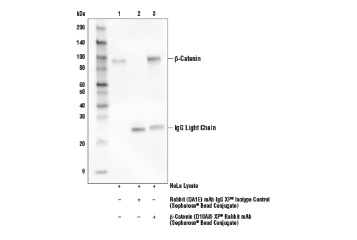
Mouse Anti-rabbit IgG (Light-Chain Specific) (D4W3E) mAb (HRP Conjugate) only reacts with the light chain of rabbit IgG and does not bind to the denatured and reduced rabbit IgG heavy chain. When performing immunoprecipitation (IP) followed by western blotting, the denatured rabbit IgG heavy chain of the primary antibody used for IP runs at approximately 50 kDa on the subsequent western blot and can often obscure bands of proteins that have a similar molecular weight. Using Mouse Anti-rabbit IgG (Light-Chain Specific) (D4W3E) mAb (HRP Conjugate) as a secondary antibody will eliminate this problem.

Limited Uses

Except as otherwise expressly agreed in a writing signed by a legally authorized representative of CST, the following terms apply to Products provided by CST, its affiliates or its distributors. Any Customer's terms and conditions that are in addition to, or different from, those contained herein, unless separately accepted in writing by a legally authorized representative of CST, are rejected and are of no force or effect.

Products are labeled with For Research Use Only or a similar labeling statement and have not been approved, cleared, or licensed by the FDA or other regulatory foreign or domestic entity, for any purpose. Customer shall not use any Product for any diagnostic or therapeutic purpose, or otherwise in any manner that conflicts with its labeling statement. Products sold or licensed by CST are provided for Customer as the end-user and solely for research and development uses. Any use of Product for diagnostic, prophylactic or therapeutic purposes, or any purchase of Product for resale (alone or as a component) or other commercial purpose, requires a separate license from CST. Customer shall (a) not sell, license, loan, donate or otherwise transfer or make available any Product to any third party, whether alone or in combination with other materials, or use the Products to manufacture any commercial products, (b) not copy, modify, reverse engineer, decompile, disassemble or otherwise attempt to discover the underlying structure or technology of the Products, or use the Products for the purpose of developing any products or services that would compete with CST products or services, (c) not alter or remove from the Products any trademarks, trade names, logos, patent or copyright notices or markings, (d) use the Products solely in accordance with CST Product Terms of Sale and any applicable documentation, and (e) comply with any license, terms of service or similar agreement with respect to any third party products or services used by Customer in connection with the Products.

For Research Use Only. Not for Use in Diagnostic Procedures.
Cell Signaling Technology is a trademark of Cell Signaling Technology, Inc.
XP is a registered trademark of Cell Signaling Technology, Inc.
All other trademarks are the property of their respective owners. Visit our Trademark Information page.