View in English?
View in English?
View in English?

| Cat. # | Size | Qty. | Price | Inventory |
|---|---|---|---|---|
| 26768T | 1 Kit (4 x 20 microliters) |
|
| Product Includes | Quantity | Applications | Reactivity | MW(kDa) | Isotype |
|---|---|---|---|---|---|
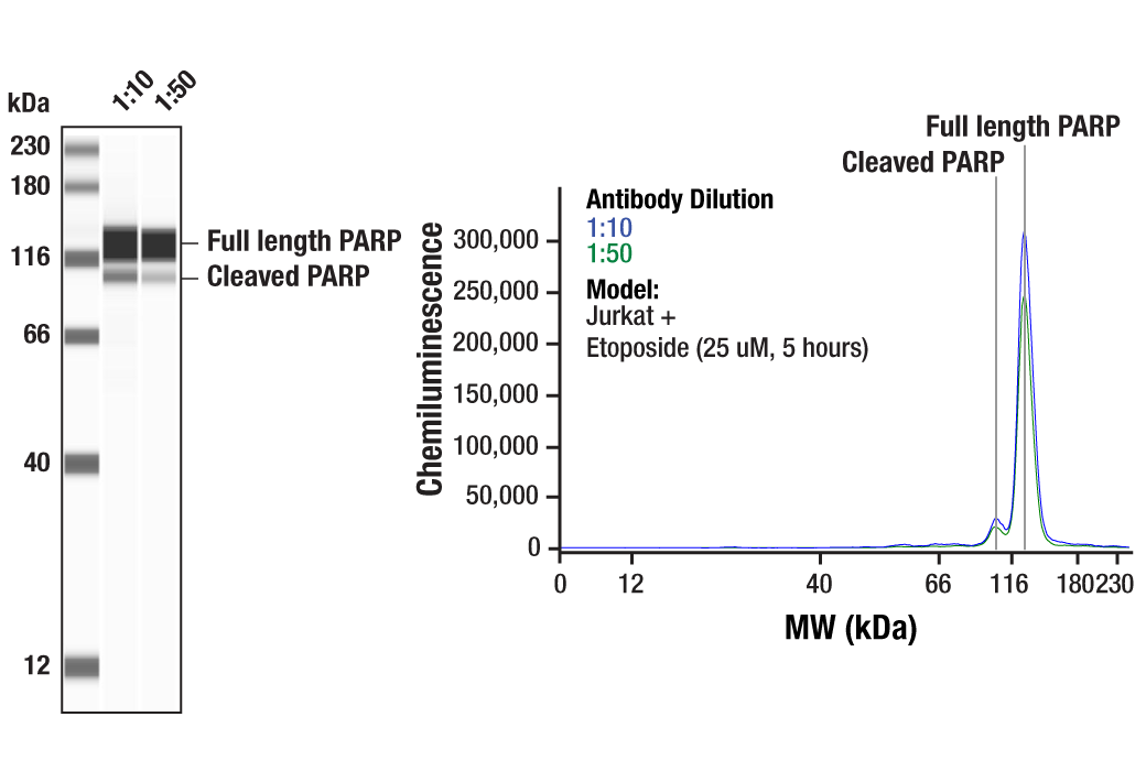
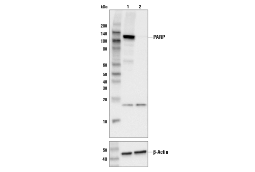
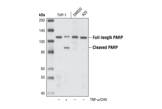
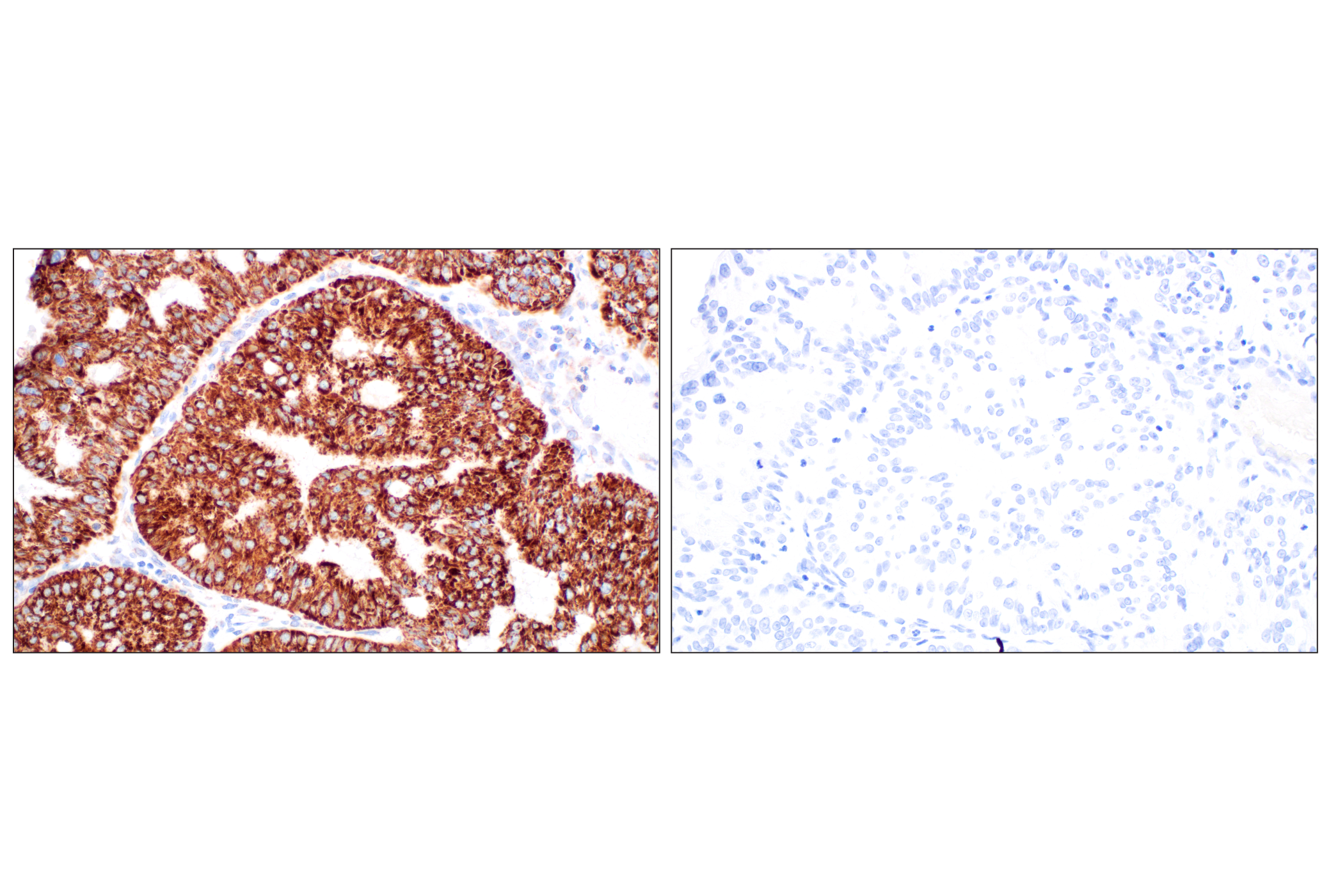
| PARP (46D11) Rabbit mAb 9532 | 20 µl |
|
H M R Mk | 116, 89 | Rabbit |
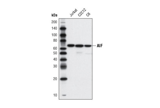
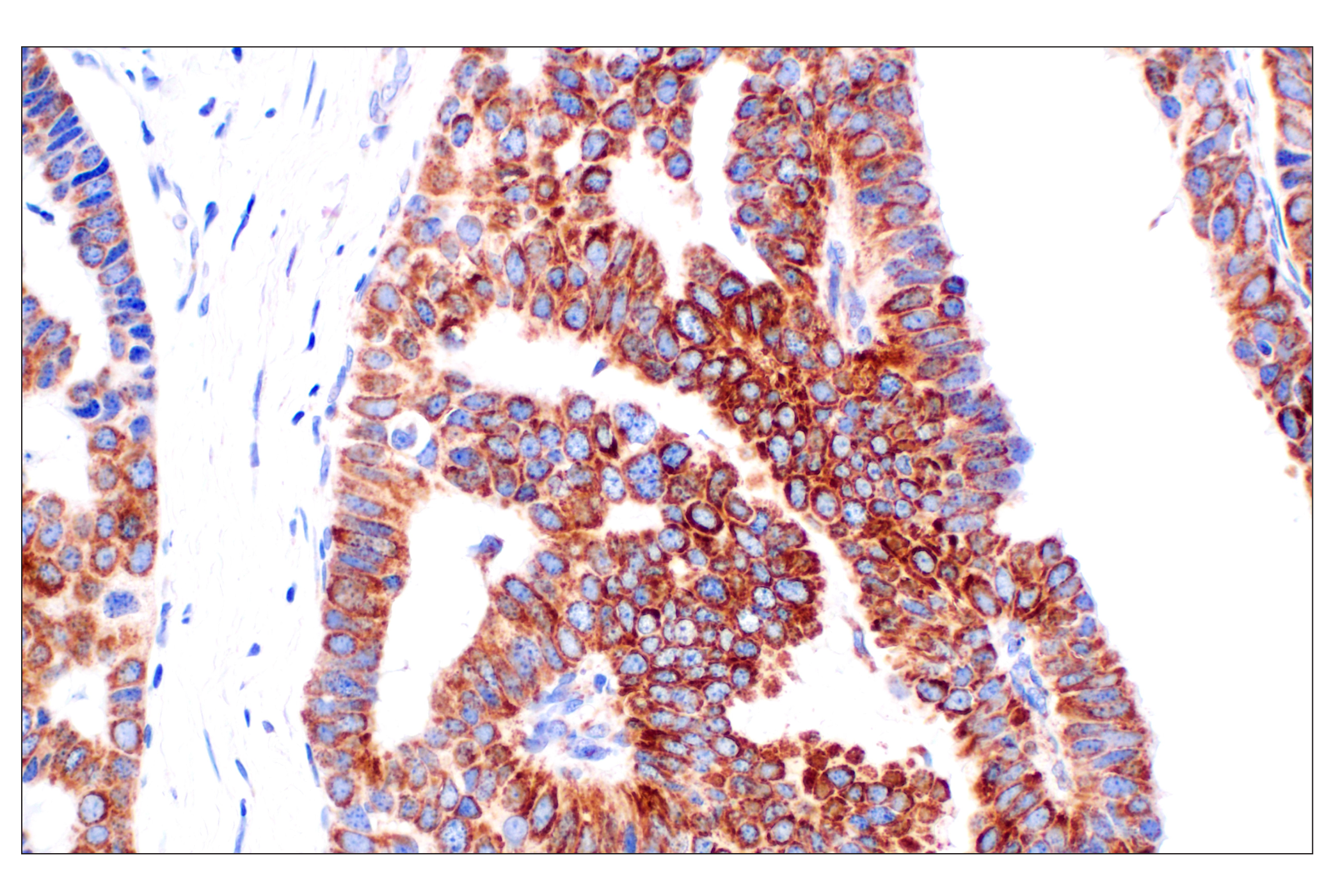
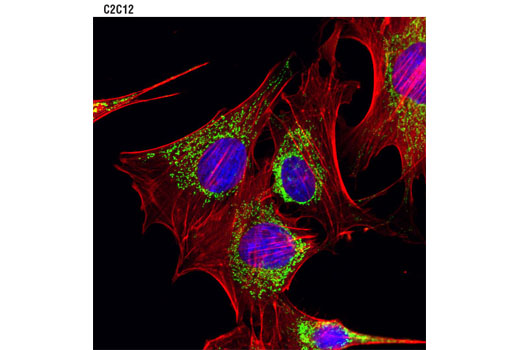
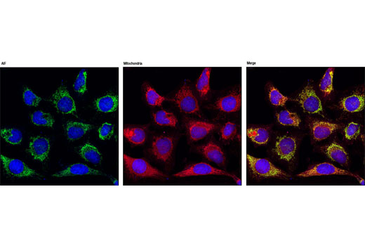
| AIF (D39D2) XP® Rabbit mAb 5318 | 20 µl |
|
H M R Mk | 67 | Rabbit IgG |
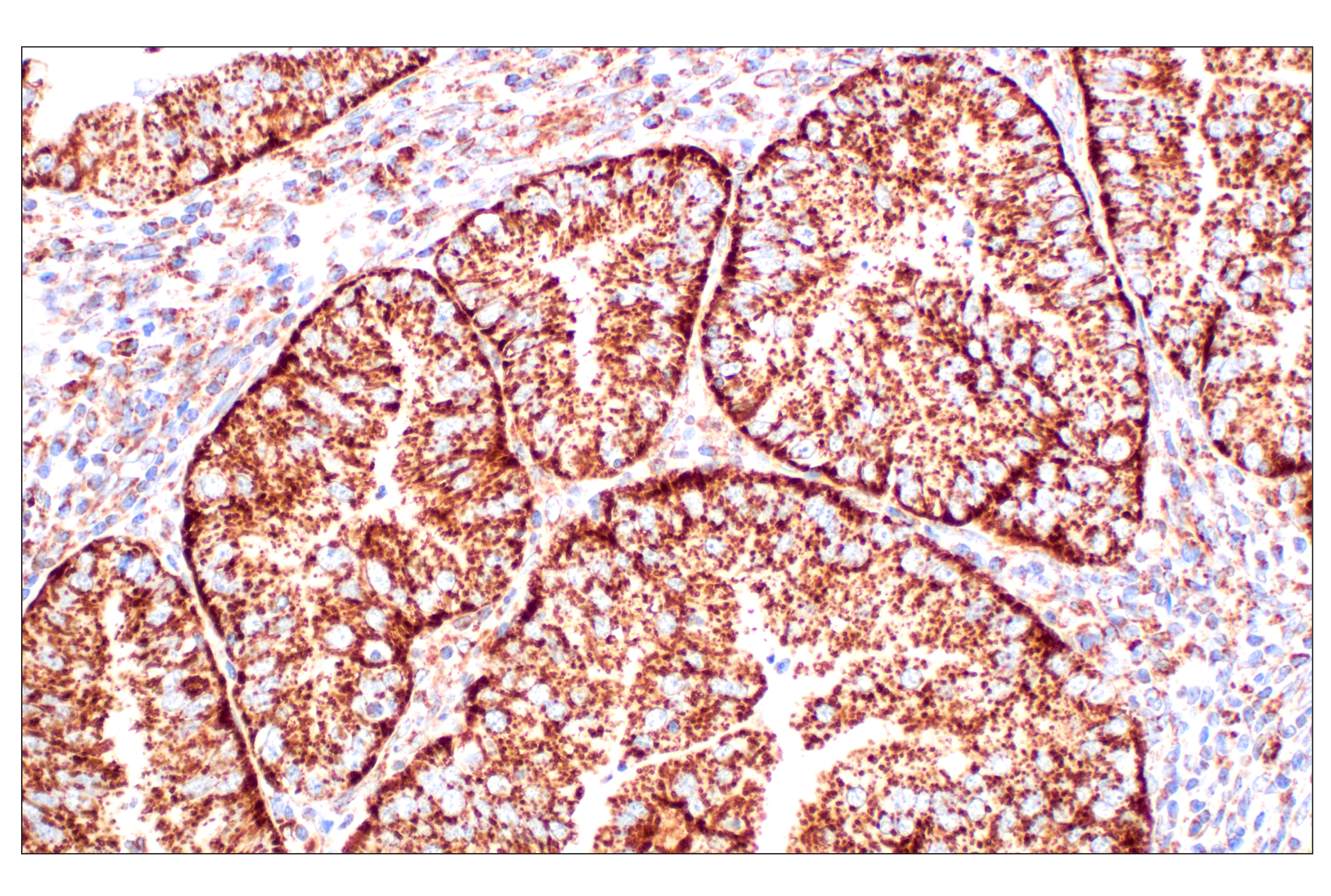
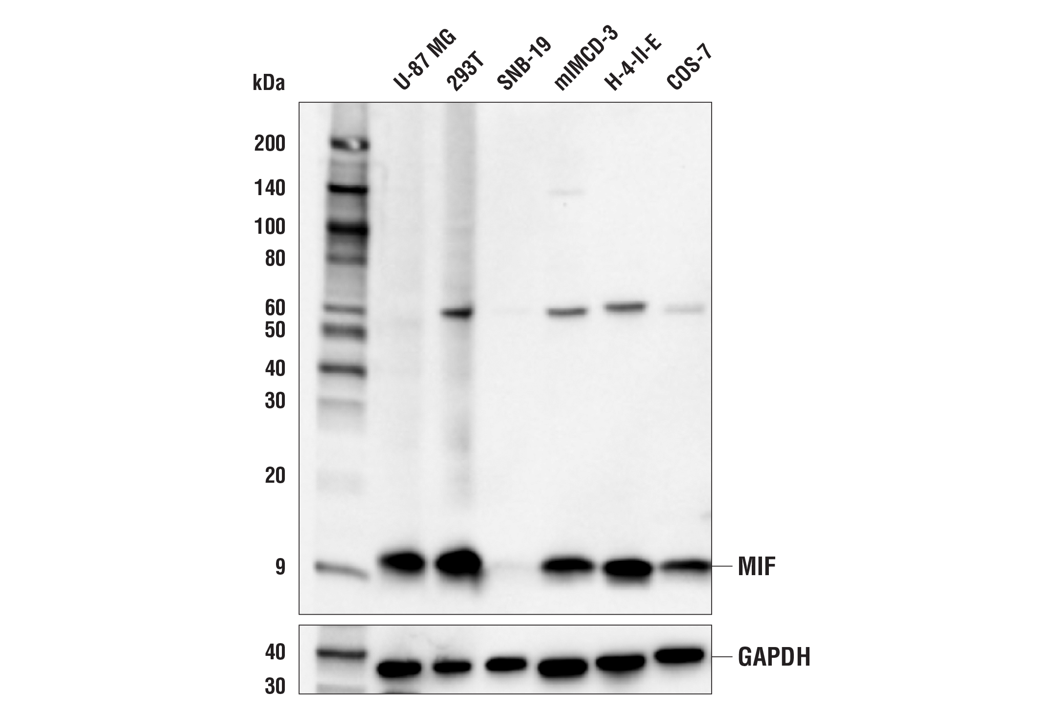
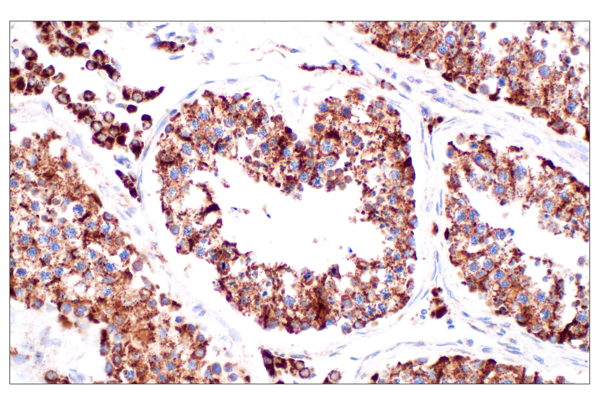
| MIF (E8S8H) Rabbit mAb 75038 | 20 µl |
|
H M R Hm | 12 | Rabbit IgG |
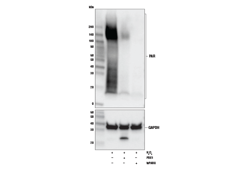
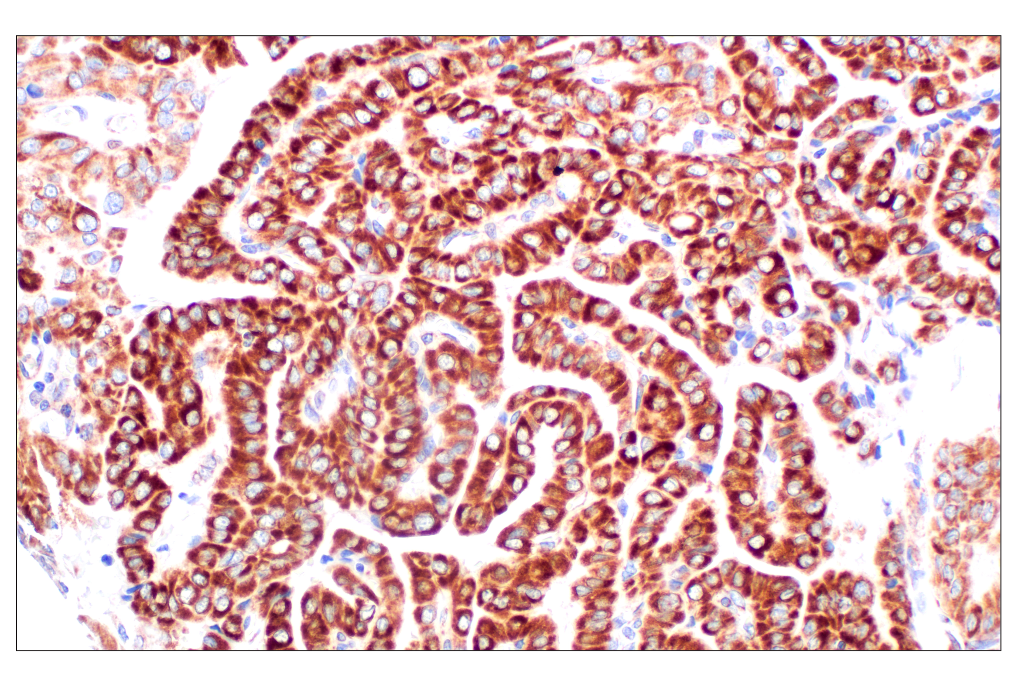
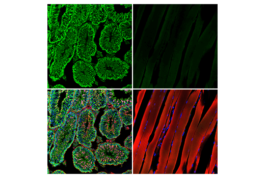
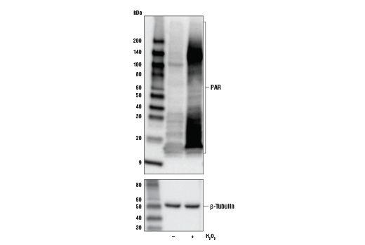
| Poly/Mono-ADP Ribose (E6F6A) Rabbit mAb 83732 | 20 µl |
|
All | Rabbit IgG | |
| Anti-rabbit IgG, HRP-linked Antibody 7074 | 100 µl |
|
Goat |
Product Information
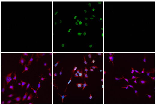
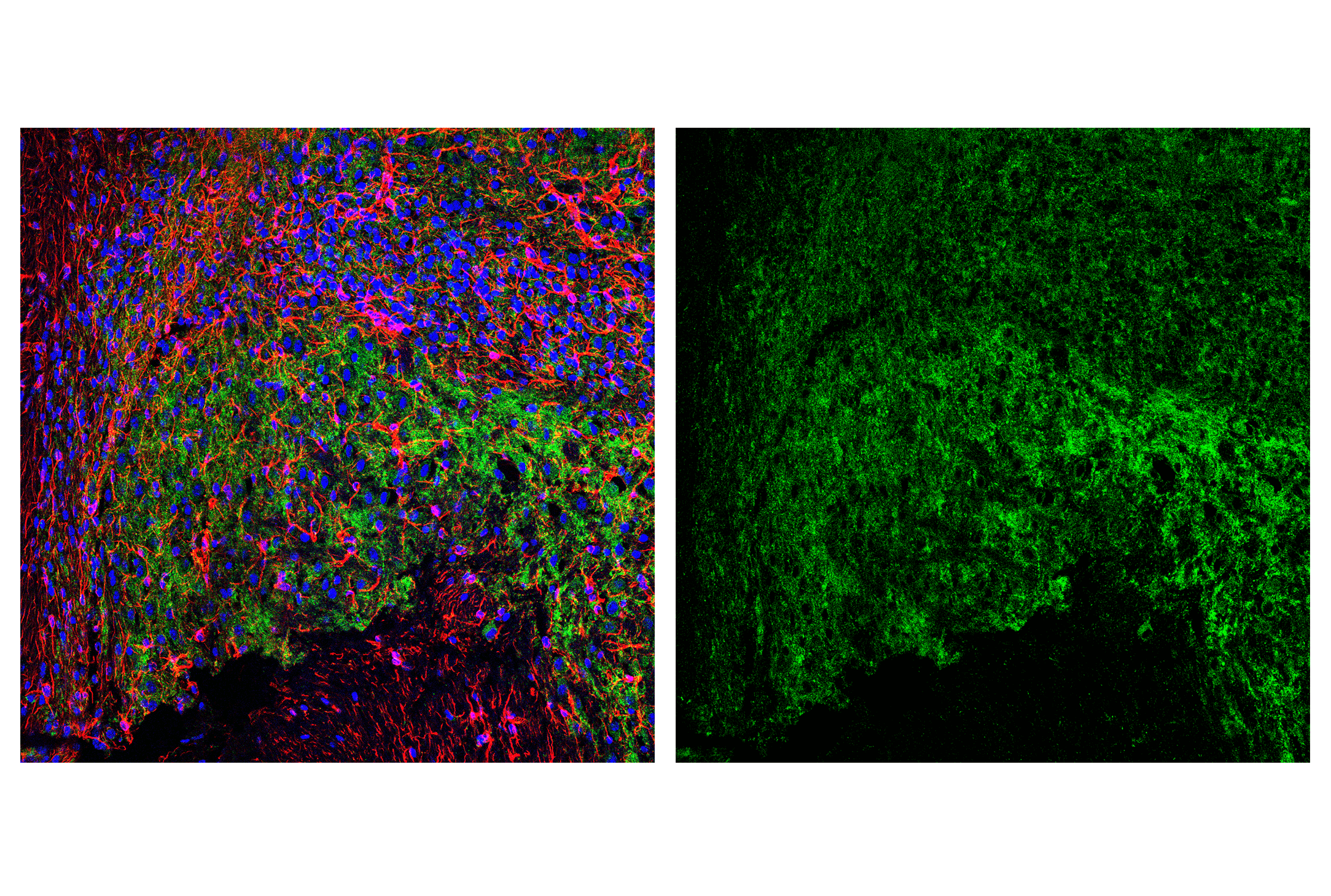
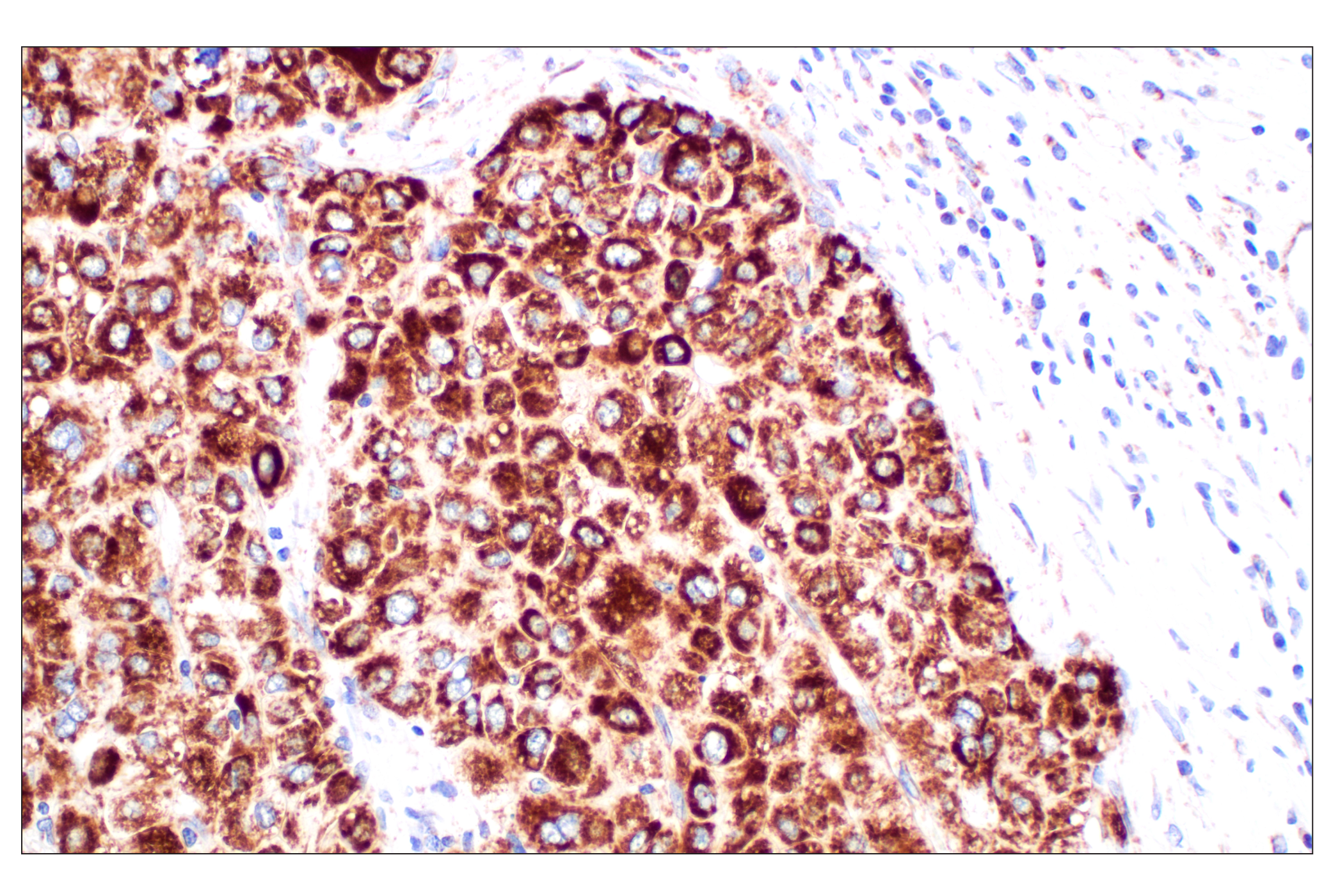
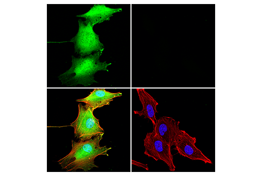
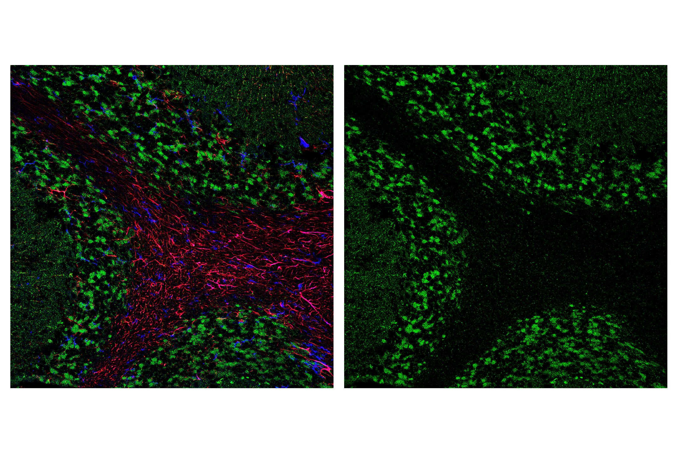
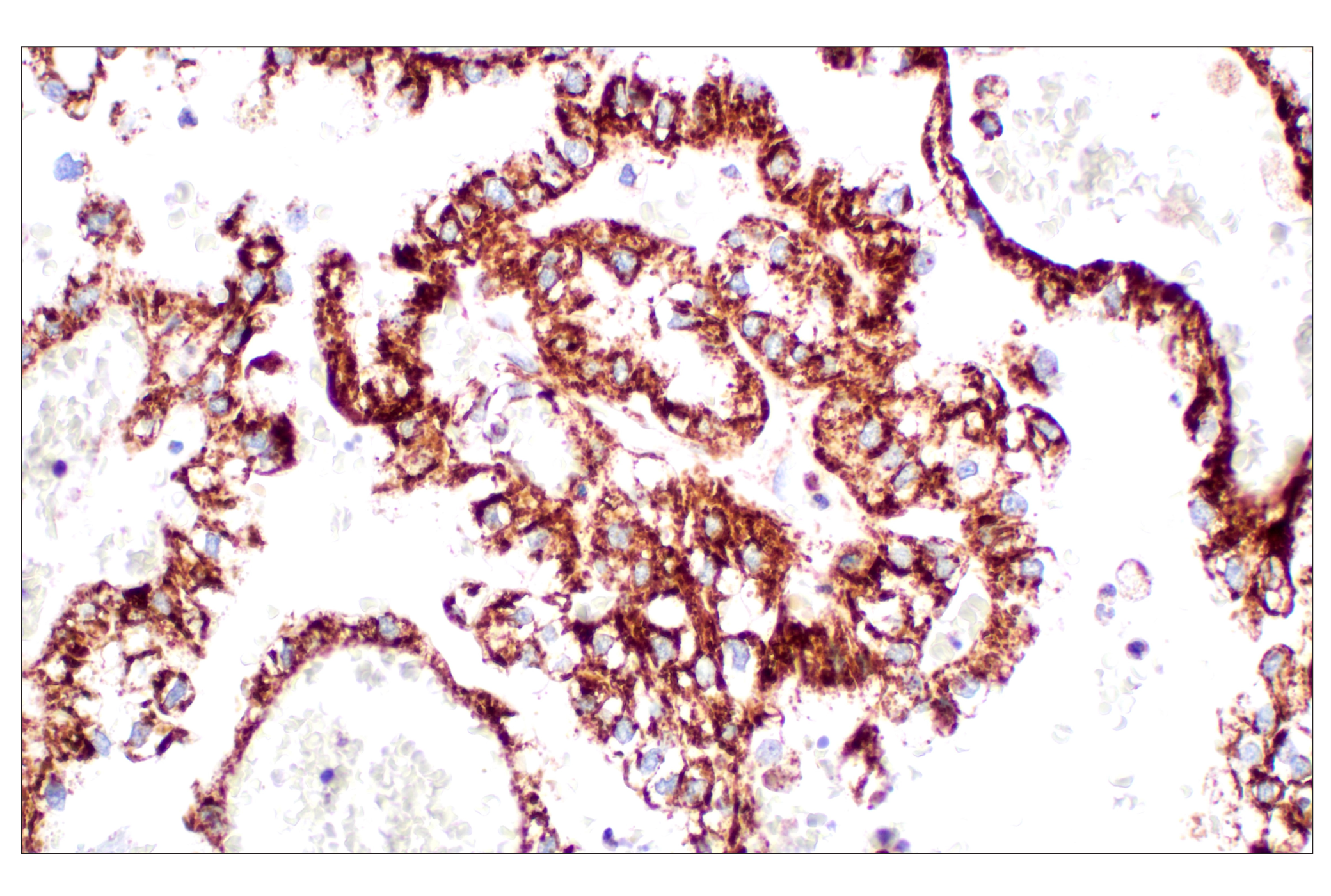
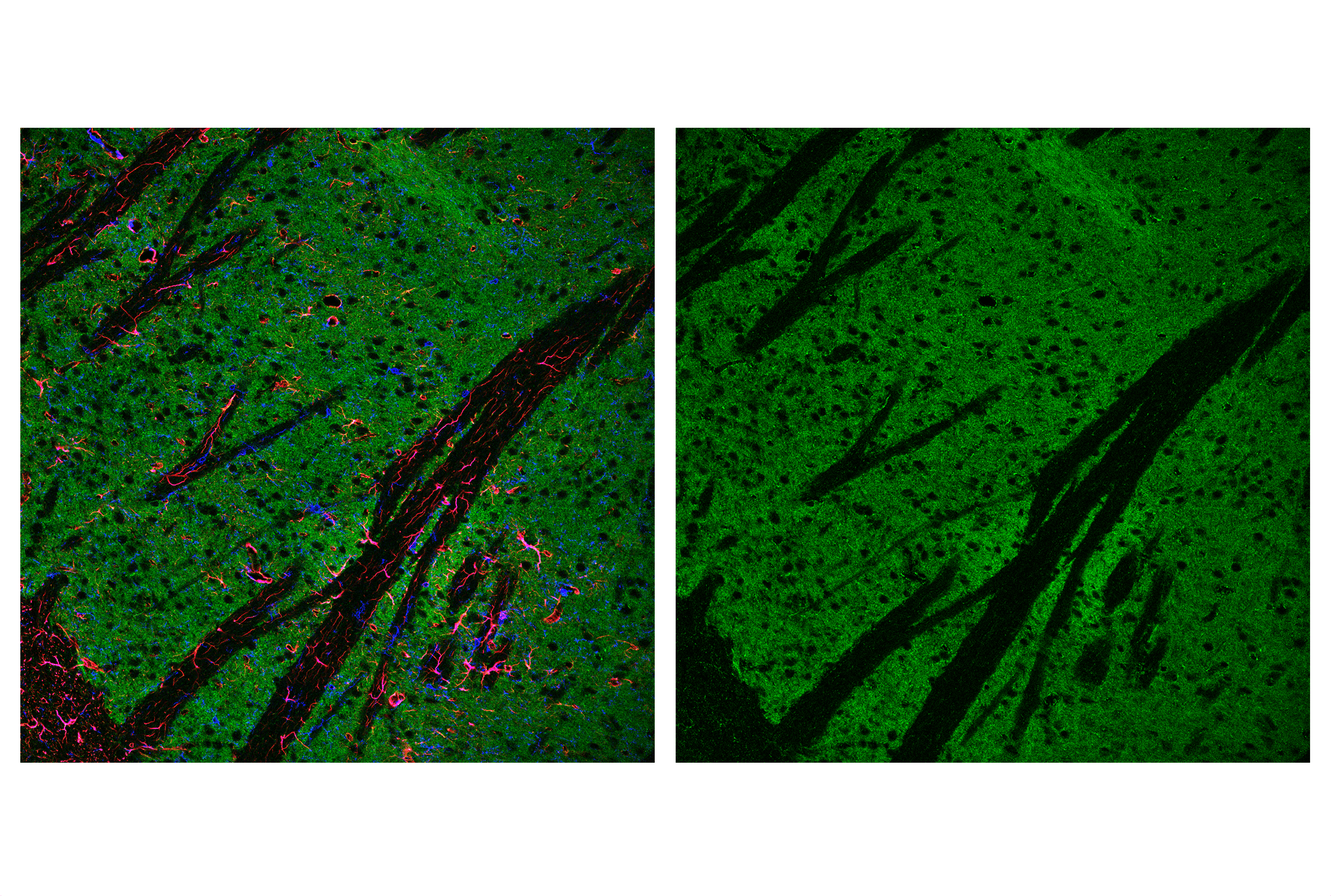
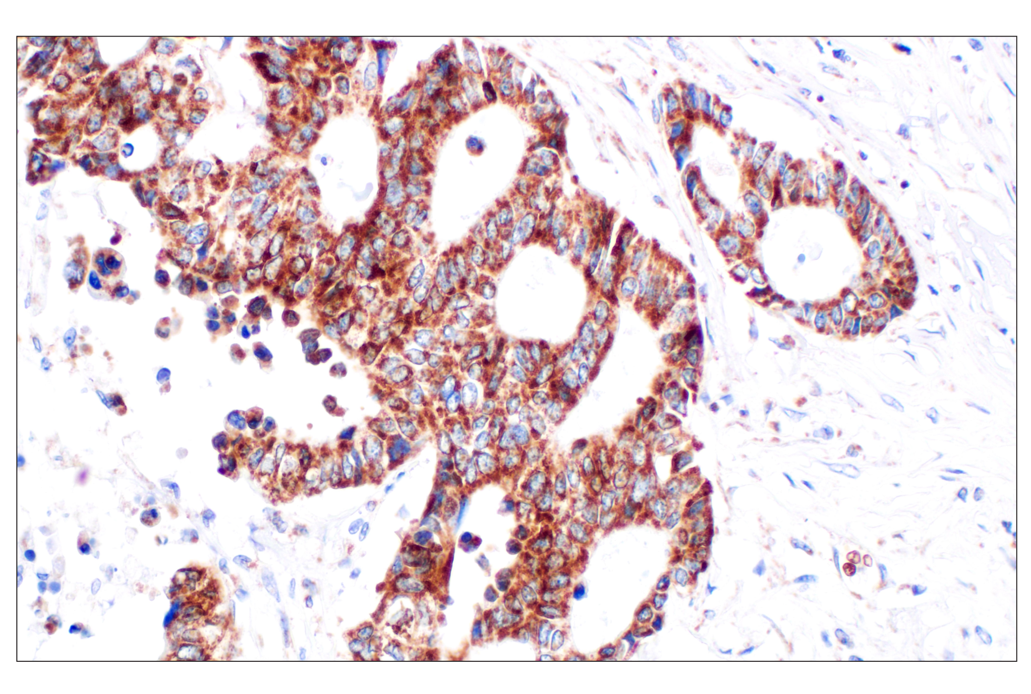
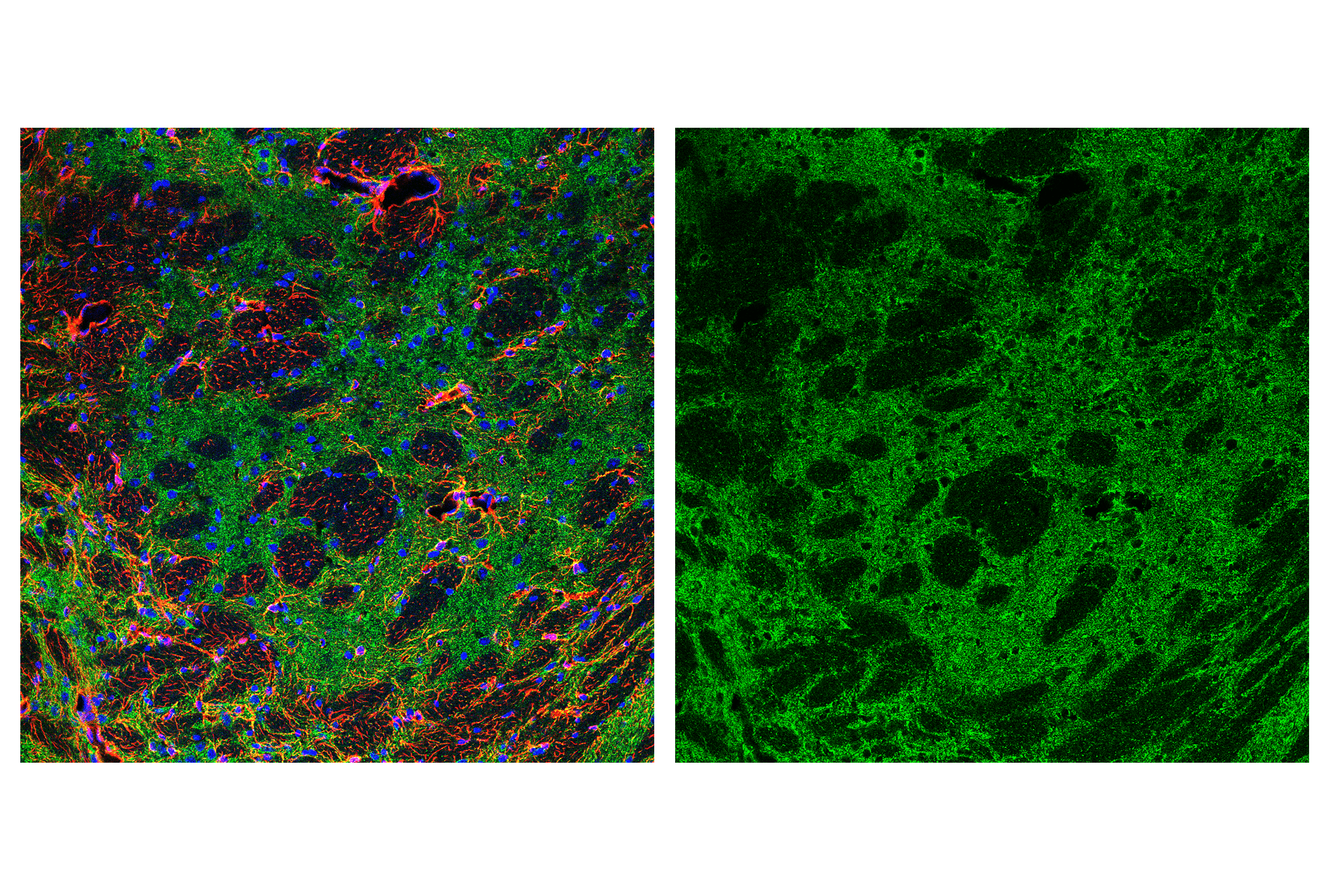
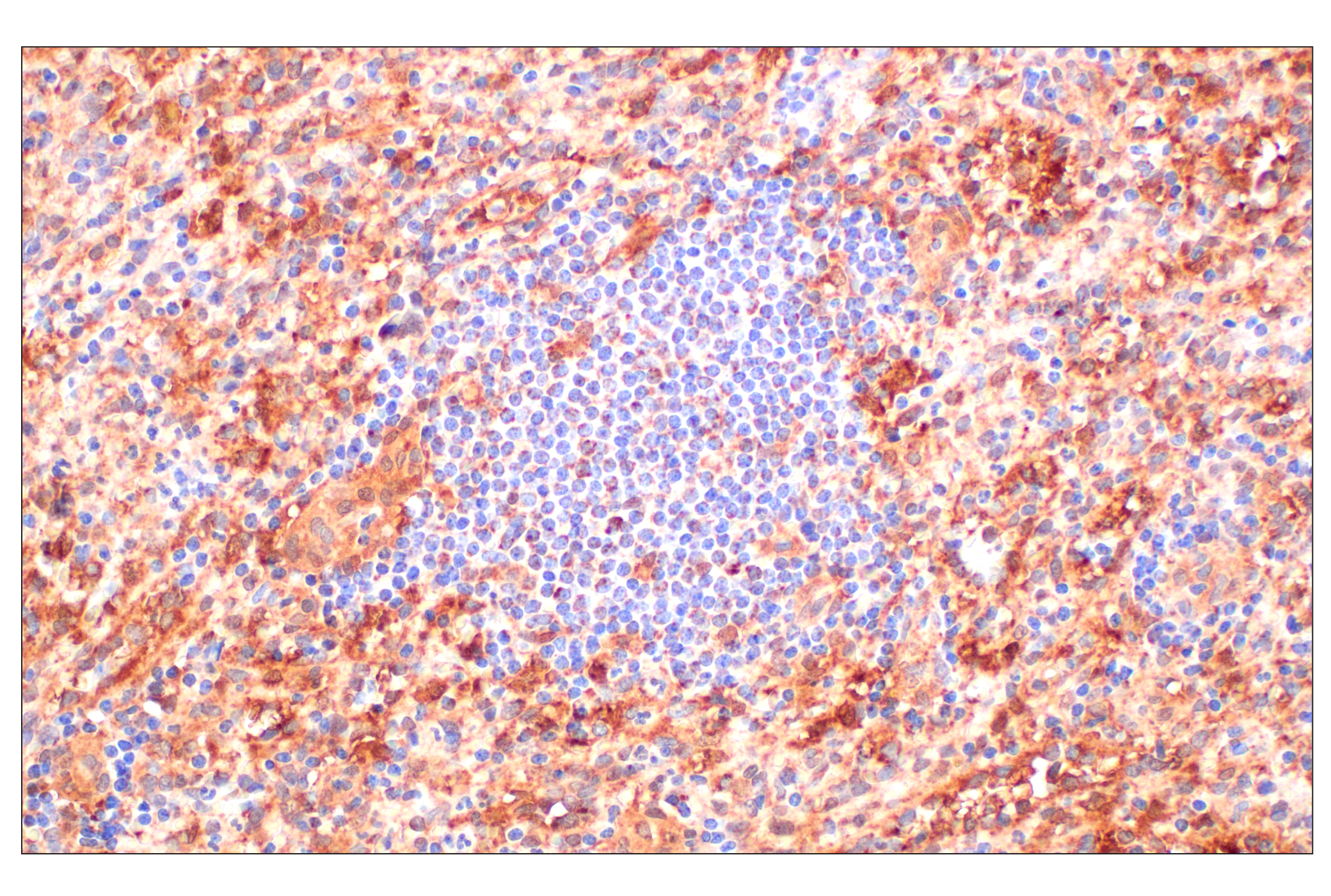
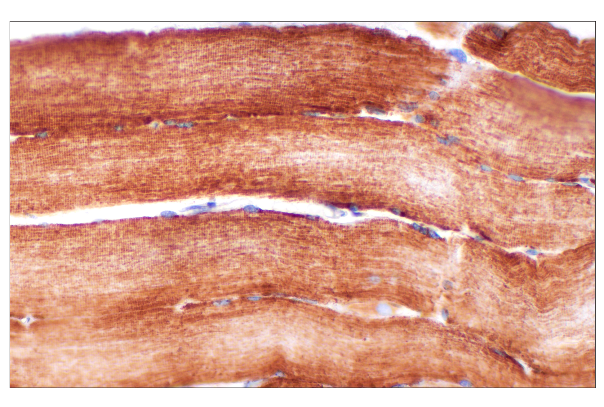
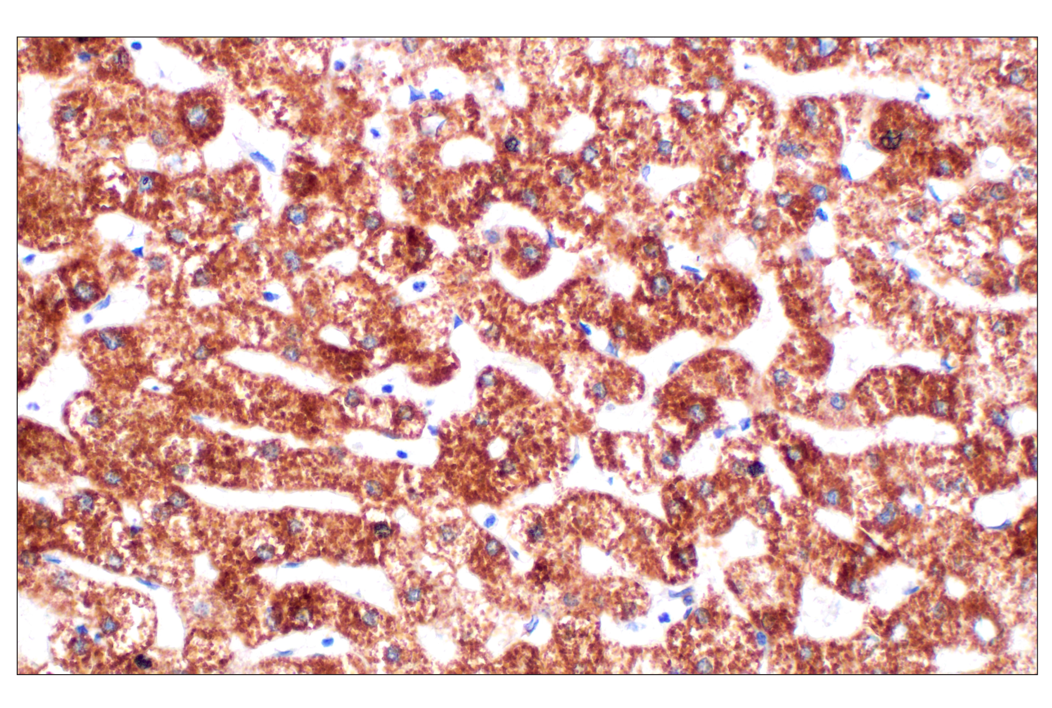
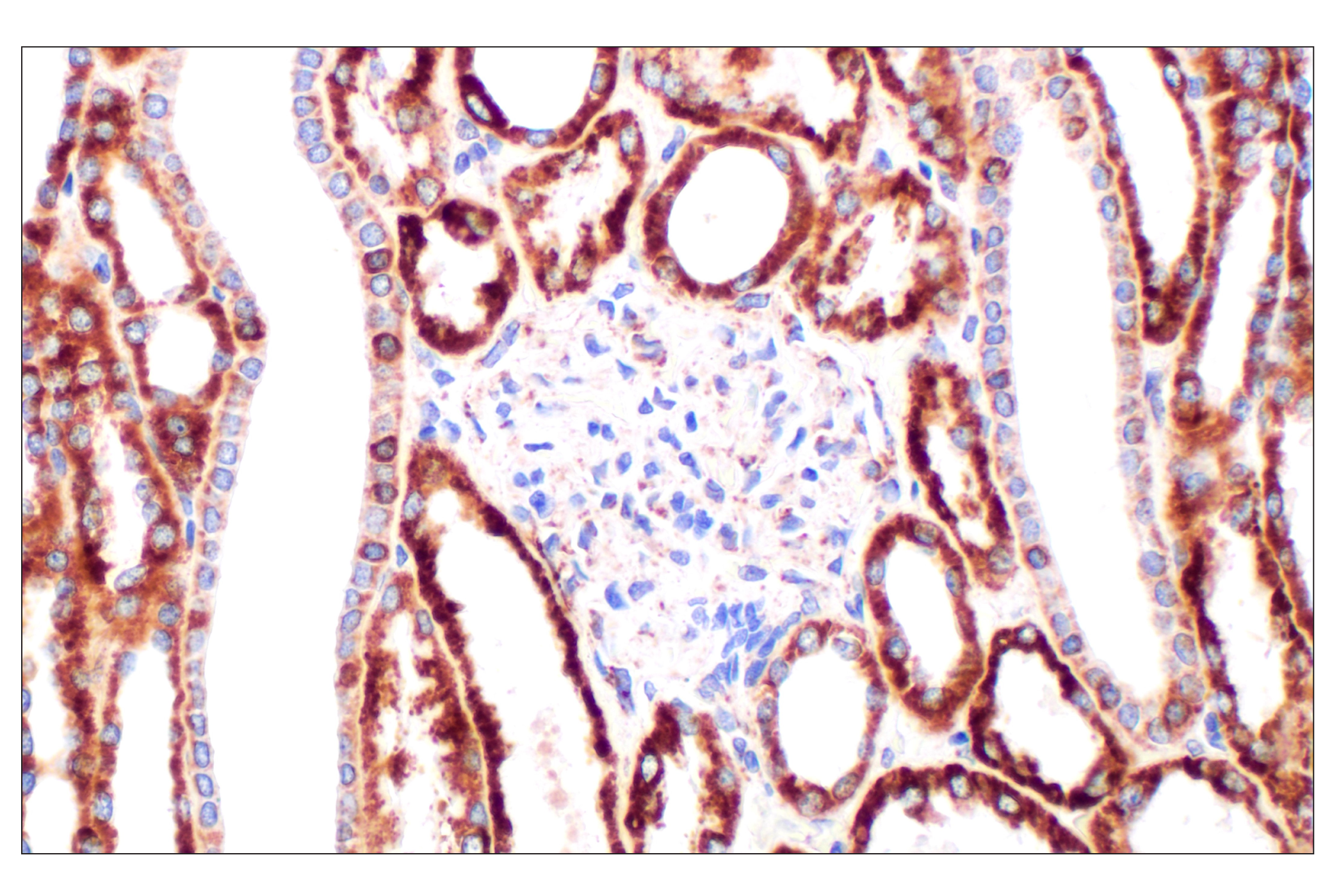
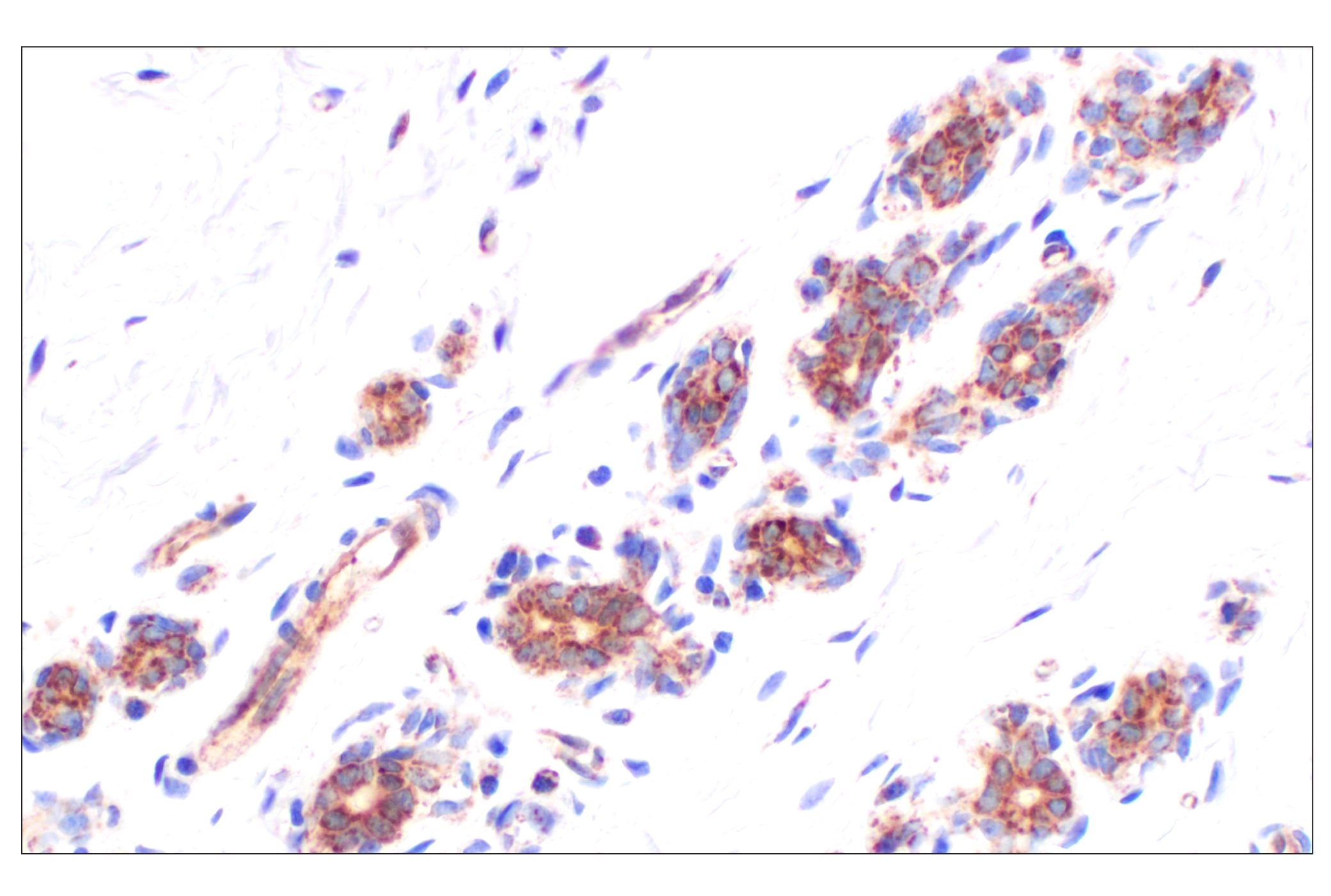
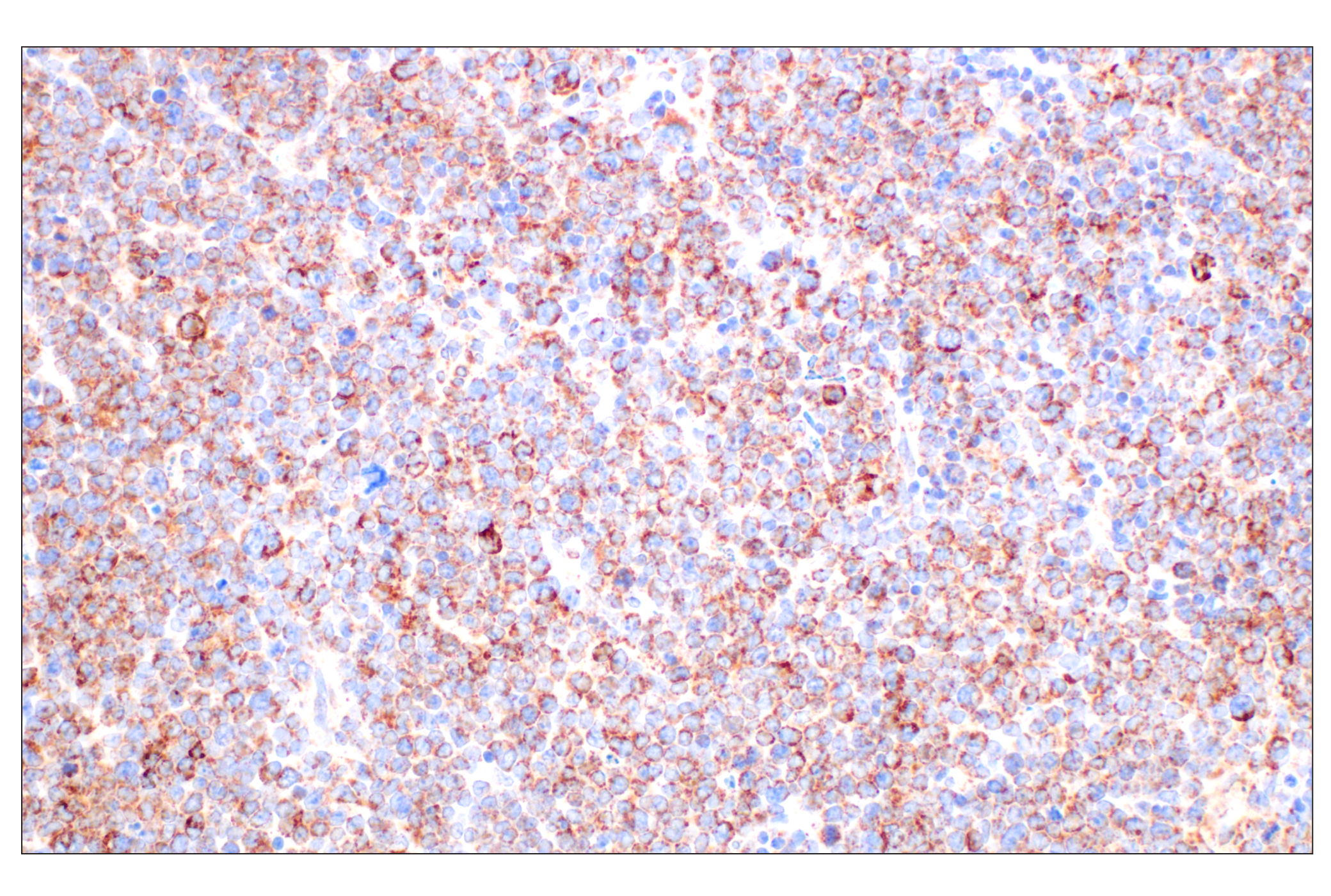
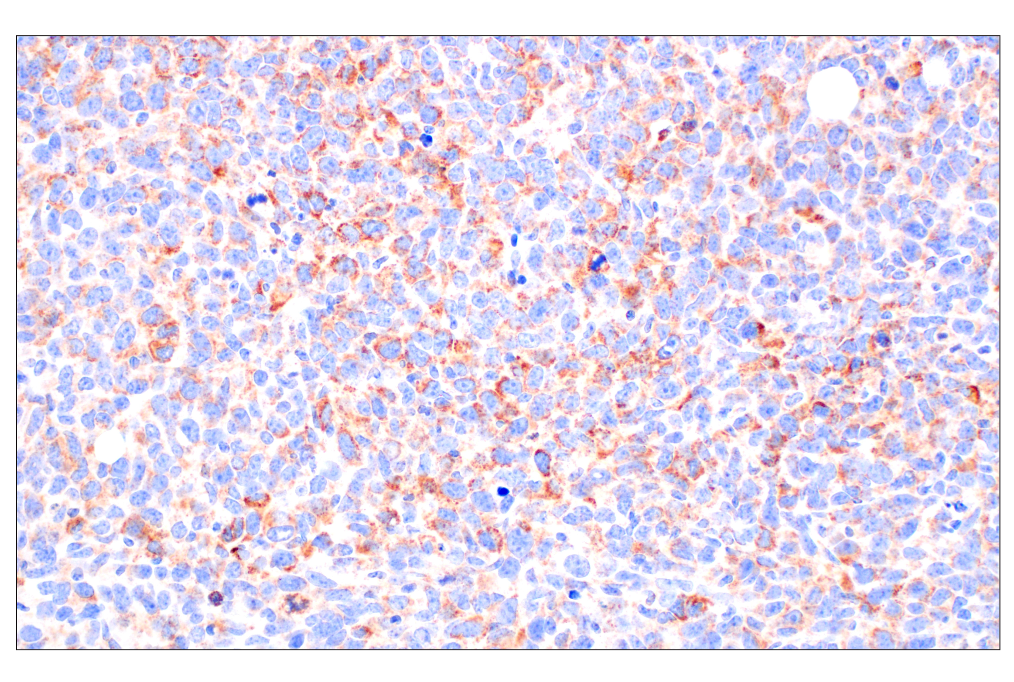
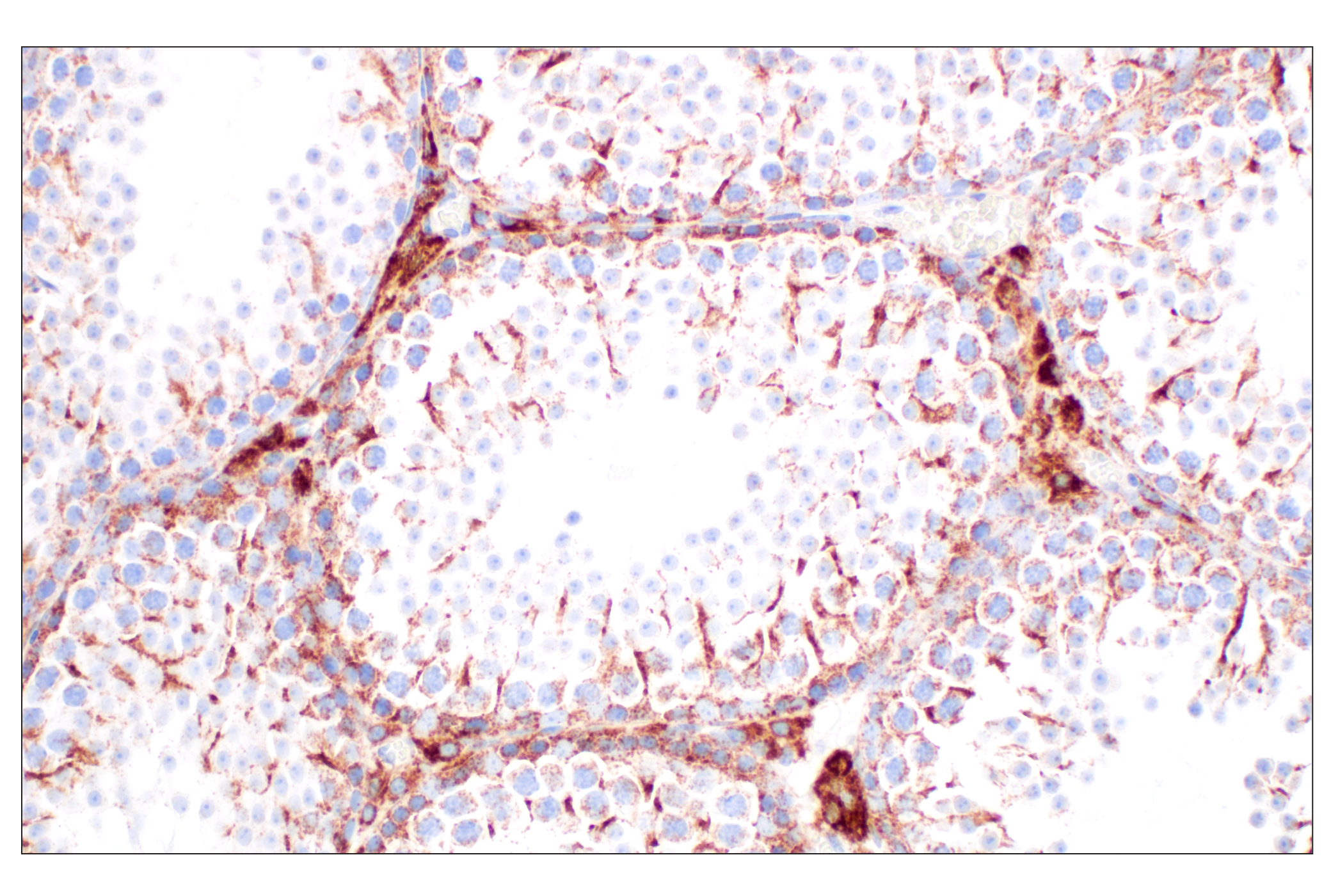
Monoclonal antibodies are produced by immunizing animals with synthetic peptides corresponding to residues surrounding Gly623 of human PARP-1, Ala520 of human AIF, and Tyr100 of human MIF protein, or with KLH modified on lysines with ADP ribose.
Parthanatos is a form of regulated cell death that follows a multistep cascade and is triggered by the accumulation of poly (ADP-ribose) (PAR). When PAR polymerase-1 (PARP-1) is overactivated under certain conditions, excessive PAR is produced and binds to apoptosis-inducing factor (AIF). As a result, AIF is released from the mitochondria and forms a complex with macrophage migration inhibitory factor (MIF). Subsequently, the AIF/MIF complex is translocated to the nucleus where MIF cleaves genomic DNA into large fragments, and cell death follows (1-3). Studies have found that parthanatos is involved in the pathogenesis of many diseases, particularly neurodegenerative disorders, such as Alzheimer's disease, Huntington's disease, Parkinson's disease, and amyotrophic lateral sclerosis (ALS) (4-7).
Explore pathways related to this product.
STRING - Known and Predicted Protein-Protein Interactions.
Except as otherwise expressly agreed in a writing signed by a legally authorized representative of CST, the following terms apply to Products provided by CST, its affiliates or its distributors. Any Customer's terms and conditions that are in addition to, or different from, those contained herein, unless separately accepted in writing by a legally authorized representative of CST, are rejected and are of no force or effect.
Products are labeled with For Research Use Only or a similar labeling statement and have not been approved, cleared, or licensed by the FDA or other regulatory foreign or domestic entity, for any purpose. Customer shall not use any Product for any diagnostic or therapeutic purpose, or otherwise in any manner that conflicts with its labeling statement. Products sold or licensed by CST are provided for Customer as the end-user and solely for research and development uses. Any use of Product for diagnostic, prophylactic or therapeutic purposes, or any purchase of Product for resale (alone or as a component) or other commercial purpose, requires a separate license from CST. Customer shall (a) not sell, license, loan, donate or otherwise transfer or make available any Product to any third party, whether alone or in combination with other materials, or use the Products to manufacture any commercial products, (b) not copy, modify, reverse engineer, decompile, disassemble or otherwise attempt to discover the underlying structure or technology of the Products, or use the Products for the purpose of developing any products or services that would compete with CST products or services, (c) not alter or remove from the Products any trademarks, trade names, logos, patent or copyright notices or markings, (d) use the Products solely in accordance with CST Product Terms of Sale and any applicable documentation, and (e) comply with any license, terms of service or similar agreement with respect to any third party products or services used by Customer in connection with the Products.
View in English?